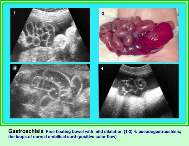
Gastroschisis
บทนำ
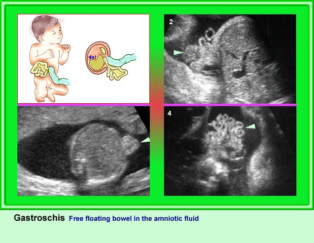
เป็นภาวะที่อวัยวะในช่องท้องออกมาอยู่ในน้ำคร่ำ ซึ่งเกิดจากความบกพร่องของผนังหน้าท้องทุกชั้น มักมีพยาธิสภาพขนาดค่อนข้างเล็ก เกือบทุกรายเกิดที่ตำแหน่งข้างขวาต่อสะดือ แต่ข้างซ้ายก็พบได้ พบได้ประมาณ 1 ต่อ 12,000 ไม่สัมพันธ์กับพันธุกรรม และไม่เพิ่มโอกาสเป็นซ้ำในครรภ์ต่อไป ไม่ขึ้นกับอายุมารดา และเนื่องจากอวัยวะในช่องท้องสัมผัสโดยตรงกับน้ำคร่ำ จึงระดับ alpha-fetoprotein ในน้ำคร่ำได้สูงบ่อยกว่า omphalocele ไม่เพิ่มอัตราความผิดปกติทางโครโมโซม ความผิดปกติอื่น ๆ นอกจากลำไส้พบได้น้อยมาก ถ้าได้รับการผ่าตัดแก้ไขอย่างเหมาะสมจะมีพยากรณ์ที่ดี
ลักษณะทางคลื่นเสียงความถี่สูง
- ลักษณะทางคลื่นเสียงความถี่สูงที่ช่วยในการวินิจฉัยก่อนคลอดที่ให้ความเชื่อถือสูง ได้แก่
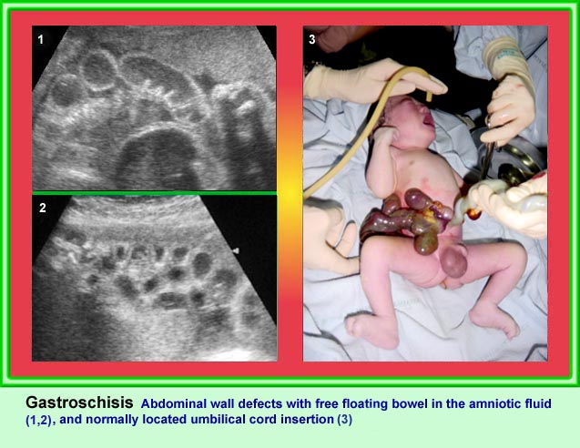
- ตำแหน่งบกพร่องอยู่ที่ข้างขวาต่อสะดือ โดยเห็นสะดืออยู่ในตำแหน่งปกติ
- เห็นลำไส้ผ่านรูดังกล่าวไปลอยอยู่ในน้ำคร่ำ
- ลำไส้ที่ออกไปอาจเห็นปริมาณมาก เมื่อเทียบกับรูโหว่ขนาดค่อนข้างเล็ก
- ไม่เห็นเยื่อหุ้ม (membrane) คลุมส่วนลำไส้ที่ออกมา
- ลำไส้เล็กเป็นส่วนที่ออกมาด้วยเสมอ แต่บางทีอาจมีลำไส้ใหญ่ หรือกระเพาะอาหารร่วมด้วย แต่น้อยรายมากที่จะมีตับออกมาด้วย
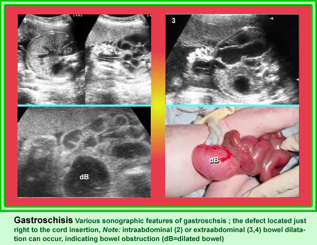
ครรภ์แฝดน้ำพบได้เพียงส่วนน้อยของราย gastroschisis ซึ่งถ้ามีจะบ่งชี้ว่าอาจมีการตีบตันของลำไส้เกิดขึ้น
ลำไส้ที่อยู่ในน้ำคร่ำอาจมีผนังหนาขึ้น หรือมีแผ่นไฟบรัสหุ้ม และจับกลุ่มเป็นก้อน เชื่อว่ามีการอักเสบทางเคมีเกิดขึ้น
บ่อยครั้งที่ลำไส้ในน้ำคร่ำโป่งขยายขึ้นบ้าง แต่ถ้าโตขยายมากผิดปกติอาจแสดงถึงการขาดเลือด หรืออุดตัน และอาจทำให้ลำไส้มีเนื้อตาย หรือทะลุตามมาได้ ในรายเช่นนี้อาจต้องให้รีบคลอด
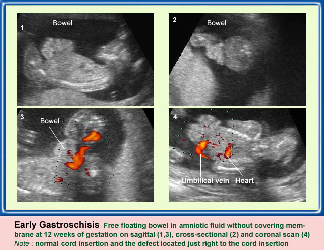
การเห็นลำไส้ออกมาอยู่หน้าท้องในไตรมาสแรก มักจะเป็น physiologic hernia ไม่ควรวินิจฉัย gastroschisis แต่ควรตรวจซ้ำ ซึ่งทารกปกติลำไส้จะกลับสู่ช่องท้องหมดทุกรายหลัง 12 สัปดาห์ไป
ข้อแตกต่างสำคัญระหว่าง gastroschisis กับ omphalocele
| Gastroschisis | Omphalocele |
| โหว่ด้านขวา | โหว่ที่สะดือ |
| ลำไส้ลอยอิสระ | ลำไส้มีเยื่อบาง ๆ คลุม |
| ตับออกมาน้อยรายมาก | ตับออกมาได้บ่อย |
| ลำไส้อุดตันบ่อย | ลำไส้อุดตันน้อย |
| ความผิดปกติอื่น ๆ พบร่วมน้อยมาก | ความผิดปกติอื่น ๆ พบร่วมได้บ่อย โดยเฉพาะหัวใจ |
| ความผิดปกติของโครโมโซมไม่เพิ่มขึ้น | ความผิดปกติของโครโมโซมเพิ่มขึ้นมาก |
| สัมพันธ์กับกลุ่มอาการอื่น ๆ น้อย | สัมพันธ์กลุ่มอาการอื่น ๆ ได้บ่อย |
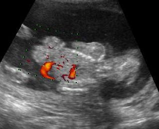
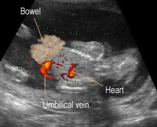

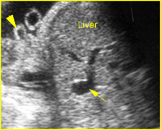
Gastroschisis
Coronal scan : bowel protruding through the defect on the right-side of the umbilical vein (the same side as fetal heart)
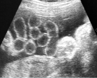
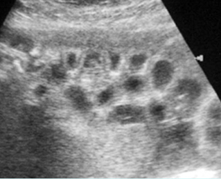
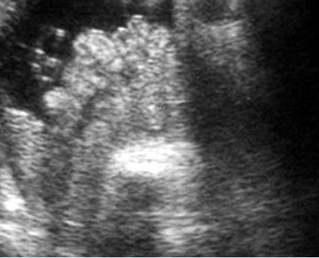
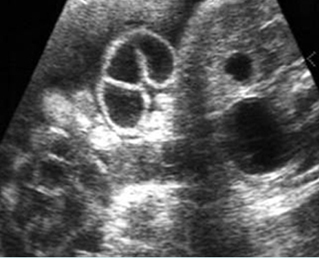
Gastroschisis
Scan of the free-floating bowel loops in the amniotic fluid
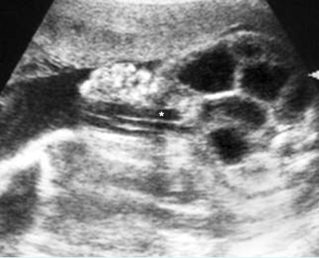
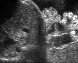
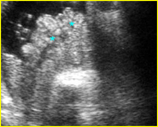
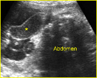
Gastroschisis
Cross-sectional scan of the abdomen: Free floating echogenic bowel loops (arrowhead), protruding through the abdominal wall defect on the right-side of the umbilical vessels (*)
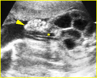
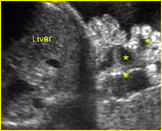
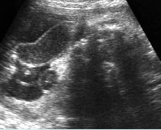
Gastroschisis
Cross-sectional scan of the abdomen: Echogenic bowel (arrowhead), protruding through the defect on the right-side of the umbilical vein, with intra-abdominal bowel dilatation (*), (solid circle = intra-abdominal stomach)

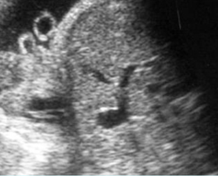
Gastroschisis
Free floating bowel loops in the amniotic fluid with dilatation and wall thickening
Gastroschisis
Cross-sectional scan of the abdomen: free floating bowel in the amniotic fluid (*) protruding through the abdominal wall defect
Gastroschisis
Cross-sectional scan of the abdomen: bowel (arrowhead) protruding through the abdominal wall defect on the right-side of the umbilicus (arrow = umbilical vein)
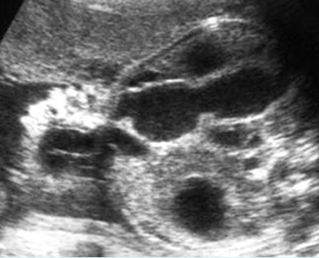
Gastroschisis
Free floating bowel loops (*) in the amniotic fluid
Gastroschisis
Free floating dilated bowel loops (*) in the amniotic fluid
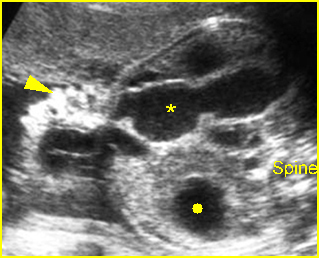
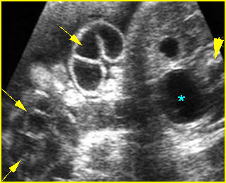
Gastroschisis
Cross-sectional scan of the abdomen: Free floating dilated bowel loops (arrow) in the amniotic fluid (arrowhead = spine, * = intra-abdominal stomach)
Classic Images