Absent Stomach
The stomach should be visualized on the standard axial views of the abdominal circumference. Failure to visualize the stomach in the normal location after 14 weeks of gestation is suggestive of abnormalities. However, absent stomach bubble can be identified in 1.4% of all fetal sonographic surveys and it may be found in up to 1% of normal fetuses on an initial scan.
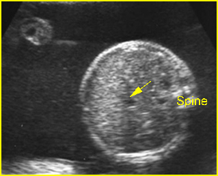
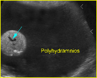
Fig 1, Fig 2
Differential diagnosis of absent stomach bubble
- Transient non-visualization of the stomach in normal fetuses
- Esophageal atresia + tracheoesophageal fistula
- Diaphragmatic hernia
- Situs inversus
- Oligohydramnios from any cause, i.e. renal agenesis
- Facial clefts (ineffective swallowing)
- Central nervous system disorders.
A three-sectional view of the neck and upper chest is useful for in utero detection of an esophageal pouch that may enhance the prenatal diagnosis of early amniocentesis (EA)

Fig 1: Esophageal atresia Cross-sectional scan of the abdomen: absent stomach with marked polyhydramnios (arrow = umbilical vein)

Fig 2: Esophageal atresia Cross-sectional scan of the abdomen: absent stomach at the level of umbilical vein complex (arrow) with marked polyhydramnios
Video clips of absent stomach

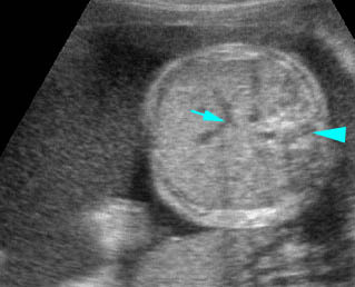
Esophageal atresia: Cross-sectional scan of the abdomen: persistently absent stomach (arrow = umbilical vein, arrowhead = spine)

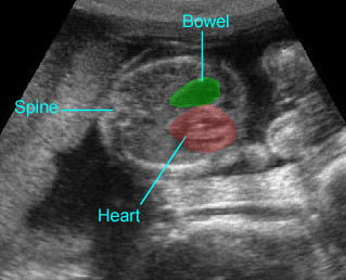
Diaphragmatic hernia: Cross-sectional scan: The stomach is herniated into the chest, not seen in the abdomen, with cardiac displacement

