Bladder Outlet Obstruction
Bladder outlet obstruction occurs almost exclusively in males, and is most often related to the posterior urethral valve, resulting in failure of complete disintegration of the urogenital membrane leaving membranous tissue within the posterior urethra; it is rarely secondary to a nearby lesion such as cloacal dysgenesis or ureterocele. It is the most common cause of severe obstructive uropathy in childhood.
Incidence: 1 in 5000-8000 boys.
Sonographic findings:
- Only males are affected.
- The urinary bladder is dilated (megacystis) regardless of the cause. Fig 1, Fig 2
- The bladder wall is commonly thickened.
- The posterior urethra may be dilated and appears as a projection from the bladder base, giving a keyhole appearance. Fig 3, Fig 4
- The ureters are usually dilated (megaureter). Fig 5, Fig 6, Fig 7
- Asymmetric hydronephrosis, with massive hydronephrosis on one side and minimal hydronephrosis on the other with relative sparing.
- The kidneys have a variable appearance depending on the presence and extent of dysplasia including hydronephrosis, cystic renal parenchyma, or small shrunken kidneys with echogenic parenchyma.
- Oligohydramnios with pulmonary hypoplasia.
- Prolonged distention of the bladder with deficiency of the abdominal musculature development, called Prune belly syndrome, is commonly seen.
- Usually presenting at 18-22 weeks but possibly as early as 11 weeks.

Fig 1: Megacystis Sagittal scan of the trunk: marked dilatation of the bladder (solid circle) with protrusion of the lower abdomen (arrow = diaphragm)
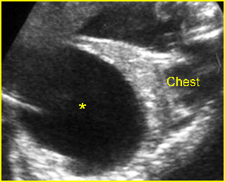
Fig 2: Megacystis Sagittal scan of the trunk: marked dilatation of the bladder (*), occupying nearly total abdomen
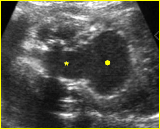
Fig 3: Megacystis Coronal scan of the lower abdomen: dilatation of the bladder (solid circle) with marked dilatation of the proximal urethra giving the key-hole appearance (*)
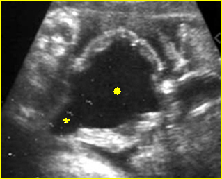
Fig 4: Megacystis Coronal scan of the lower abdomen: dilatation of the bladder (solid circle) with thickened wall; dilatation of the proximal urethra giving the key-hole appearance (*)
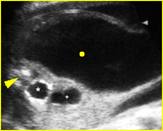
Fig 5: Megacystis Cross-sectional scan of the abdomen: marked dilatation of the bladder (solid circle) and ureter (+) and renal pelvis (*) with echogenic thin parenchyma (arrow = spine)
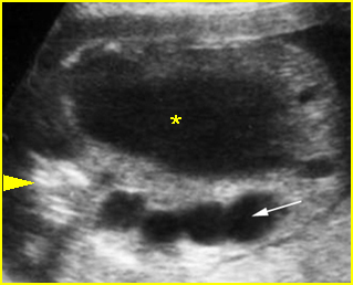
Fig 6: Megaureter and megacystis Oblique scan of the abdomen: marked dilatation of the ureter (arrow), simulating bowel loop, and bladder (*) (arrowhead = spine)
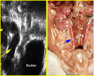
Fig 7: Megacystis and megaureter Coronal scan of the abdomen: marked dilatation of the ureter (arrow) with dilated renal pelvis and dilated bladder
Video clips of bladder outlet obstruction
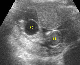
Bladder outlet obstruction: Sagittal scan: Megacystis (C) at 11 weeks of gestation (H = head)
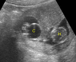
Bladder outlet obstruction: Coronal scan: Megacystis (C) at 11 weeks of gestation (H = head)
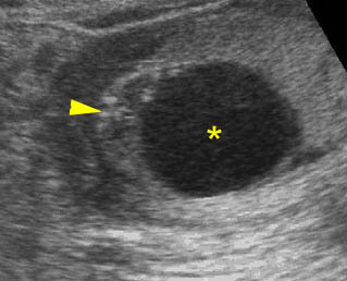
Megacystis: Cross-sectional scan of the fetal abdomen: markedly enlarged bladder (*) with oligohydramnios (arrowhead = spine)
Associations: Most are isolated and are rarely related to urethral atresia or caudal regression, and aneuploidy (trisomy 21, 18 and 13) in some cases.
Management: Termination of pregnancy can be offered when diagnosed before viability when oligohydramnios or renal insufficiency is present. In utero decompression can be considered in some selected cases. Cytogenetic analysis or FISH can be done using fetal urine specimens. The cause of obstruction may be corrected in some cases, such as ureterocele.
Prognosis: Variable, but most are poor, especially when combined with severe oligohydramnios, lung hypoplasia and renal dysplasia.
Recurrence risk: Sporadic (rare recurrence) with rare familial inheritance.

