Small Bowel Obstruction
Small bowel obstructions are non-duodenal bowel atresias with congenital obliteration of the lumen of segments of the large or small intestine, usually caused by in utero vascular accident. There are several types of atresias, the most common of which results in blind ends of bowel separated from each other by a gap.
Incidence: About 1 in 3000-5000 births.
Sonographic findings:
- Multiple dilated loops of small bowel (more than 7 mm in diameter or 15 mm in length), often with increased peristalsis.
Fig 1, Fig 2, Fig 3, Fig 4, Fig 5, Fig 6, Fig 7, Fig 8
- Polyhydramnios usually appears in the third trimester and more likely with a proximal atresia.
- The abdominal circumference may be large.
- Differentiating small bowel from large bowel obstruction is usually possible by the location of the loops, the absence of haustra in small bowel, and the presence of polyhydramnios. The dilated ureter can easily be differentiated from bowel, as urine is echogenic-free, whereas the bowel contains low-level echoes.
- It is usually difficult to differentiate jejunal atresia from ileal atresia or volvulus, although the extent of bowel dilatation is often a clue (more loops are present with ileal abnormalities).
- MRI is helpful in determining the site of obstruction.
- Pitfalls:
- Cystic masses, such as bowel duplication, mesenteric cyst or urinary tract cyst including hydronephrosis as well as multicystic kidneys can also be confused with fluid-filled dilated bowel. These anomalies, however, are rarely related to polyhydramnios.
- Occasionally, small bowel atresia can present as a cyst-like mass.
- Usually detected after 24 weeks
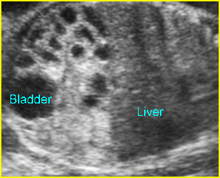
Fig 1: Small bowel obstruction Coronal scan of the abdomen: Multiple dilated loops of small bowel
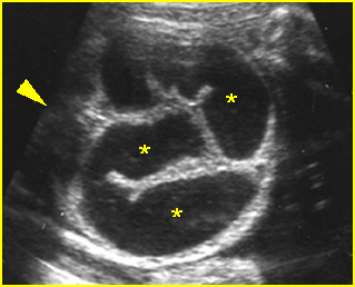
Fig 2: Small bowel obstruction Cross-sectional scan of the abdomen: Multiple loops of small bowel with marked dilatation (arrowhead = spine)
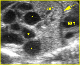
Fig 3: Small bowel obstruction Coronal scan of the abdomen: Multiple loop of small bowel loops with marked dilatation (*) (arrow = diaphragm)
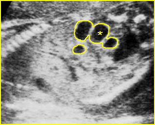
Fig 4: Small bowel obstruction Coronal scan of the abdomen: Multiple dilated loops of small bowel (*)
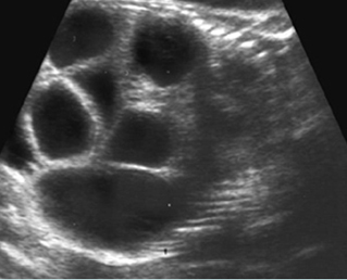
Fig 5: Small bowel obstruction Coronal scan of the fetal abdomen: markedly dilated loops of the small bowels
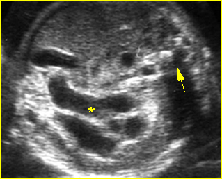
Fig 6: Small bowel obstruction Cross-sectional scan of the abdomen: multiple dilated small bowel loops (*) (arrow = spine)
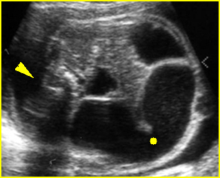
Fig 7: Small bowel obstruction Cross-sectional scan of the abdomen: Markedly dilated loops of small bowel (solid circle) (arrowhead = spine)
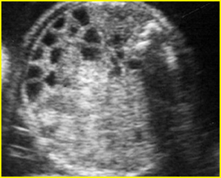
Fig 8: Small bowel obstruction Cross-sectional scan of the abdomen: Multiple dilated loops of small bowel (arrowhead = spine)
Video clips of small bowel obstruction
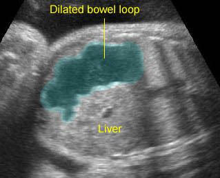
Proximal jejunal atresia: Dilated bowel loop at the upper abdomen representing the proximal jejunal loop separated from the stomach and large bowel
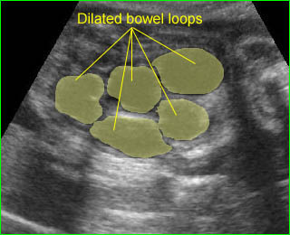
Small bowel obstruction: Multiple markedly dilated bowel loops of small bowels
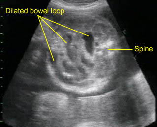
Small bowel obstruction: Multiple dilated bowel loops of small bowels
Associations: Unlike duodenal atresia, small bowel atresia is associated with a low rate of extragastrointestinal tract abnormalities and chromosomal abnormalities. However, additional GI anomalies, such as volvulus, malrotation, and duplication, are relatively common. Other complication such as intrauterine hemorrhage or bowel perforation may rarely be associated with intestinal atresia. Cystic fibrosis is commonly related to jejunal and ileal atresia.
Management: Delivery should be at term where appropriate personnel are available for surgical correction, however, early delivery might be beneficial in cases with massive bowel dilatation.
Prognosis: Depends on associated anomalies, the health of the remaining bowel and whether perforation or necrosis has occurred. The prognosis is good if it occurs as an isolated abnormality.
Recurrence risk: Sporadic (with rare autosomal recessive inheritance).

