Meconium Peritonitis
Meconium peritonitis is a condition resulting from a small bowel perforation, leading to an inflammatory reaction of the peritoneum and intra-abdominal calcifications.
Incidence: About 1 in 2000 births.
Sonographic findings:
- Calcifications (bright echogenic area) in the abdomen, often with ascites.
- The calcifications can occur anywhere within the peritoneal cavity but must be distinguished from intraluminal or intrahepatic calcifications. Fig1, Fig2
- Typically the calcifications are linear in nature, sometimes rimming a hypoechoic mass or pseudocyst. Fig3
- Pseudocyst, a hypoechoic mass representing extraluminal meconium, resulting from a contained bowel perforation.
- Usually associated with ascites and polyhydramnios.
- Typically diagnosed in the third trimester.
- Persistent ascites, pseudocyst or dilated bowel loop are most sensitive for predicting postnatal surgery.
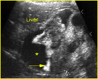
Fig 1: Meconium peritonitis Coronal scan of the abdomen: ascites (*) and calcification (arrow) outlining the visceral structures
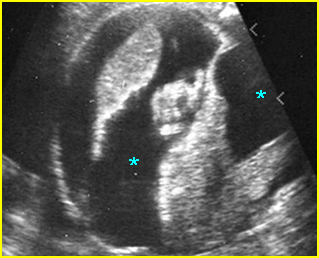
Fig 2: Meconium peritonitis Cross-sectional scan of the abdomen: ascites (*) and calcification outlining the visceral structures
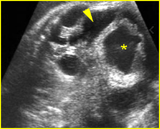
Fig 3: Meconium peritonitis Oblique coronal scan of the abdomen: dilated large bowel loops (*) with thickened wall and ascites (arrowhead)
Video clips of meconium peritonitis
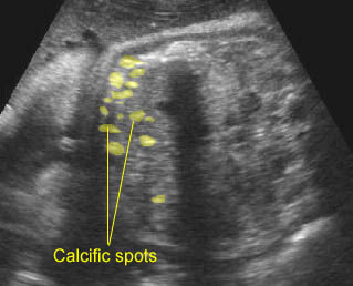
Intraperitoneal calcifications: Intraperitoneal calcifications; postnatally proven to be due to meconium peritonitis
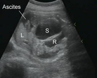
Anorectal atresia: Anorectal atresia with ruptured large bowel: Oblique cross-sectional scan of the abdomen shows ascites with dilated rectum (R) and sigmoid colon
Associations: Cystic fibrosis is found in 15% of cases with meconium peritonitis in neonatal series but is rarely found in prenatal series. Some cases are related to pavovirus B 19.
Management: Sonographic monitoring may be beneficial in assessing fluid volume, fetal growth and the degree of the severity. Early delivery may be appropriate if there is marked bowel dilatation. The pregnancy should be managed in a tertiary center.
Prognosis: Rather good in isolated cases, with most cysts disappearing spontaneously during pregnancy.
Recurrence risk: Rare for isolated cases, but may be as high as 25% when associated with cystic fibrosis.

