Bladder and Cloacal Exstrophy
This is a rare congenital anomaly characterized by the exteriorization of the viscera on the abdominal surface, low insertion of the umbilical cord, divergent pubic rami and abnormal exterior genitalia. Bladder and cloacal exstrophy share a common embryologic origin in abnormal cloacal development, but are different in terms of severity and extent of involvement. Bladder exstrophy is a lower abdominal wall defect with herniation of the bladder whereas cloacal exstrophy is much more complex, including omphalocele, bladder exstrophy, imperforate anus, spine malformation (also known as OEIS complex), spina bifida and intersex.
Incidence: Sporadic occurrence with a prevalence of 1 in 30,000 births with a male to female ratio of 2:1 for bladder exstrophy, 1 in 50,000 births with a higher frequency in twins for cloacal exstrophy, and 1 in 200,000-300,000 for OEIS complex.
Sonographic findings:
- Soft tissue mass at the lower abdominal wall
Fig 1, Fig 2, Fig 3, Fig 4 - Persistently absent fetal bladder with a large midline infraumbilical cystic or solid mass.
- A large cystic mass in the pelvis representing a persistent cloaca can be seen in some cases.
- Omphalocele is usually seen.
- Lumbosacral abnormalities.
- Associated anomalies such as renal agenesis, myelomeningocele, horseshoe kidney, and clubfeet.
- Umbilical arteries running alongside the mass suggestive of bladder exstrophy.
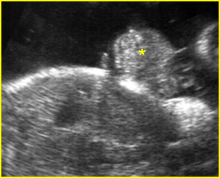
Fig 1: Bladder extrosphy Sagittal view of the fetal abdomen: complex extra-abdominal mass (*) below the umbilicus, finally proven to be bladder extrosphy
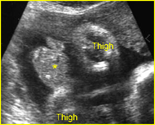
Fig 2: Cloacal extrosphy Free floating complex mass (*) connecting with fetal perineum
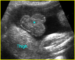
Fig 3: Cloacal extrosphy Free floating complex mass (*) connecting with fetal perineum
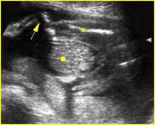
Fig 4: Cloacal extrosphy Free floating complex mass (solid circle) connecting with fetal perineum, abnormal fetal limb (arrow) (* = femur)
Video clips of bladder and cloacal exstrophy
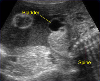
Bladder extrosphy : Bladder is located outside the abdomen, presented as a cystic mass below the cord insertion
Associations: Genital defects in both conditions; renal, skeletal, neural tube, intestinal, cardiovascular and omphaloceles defects in cloacal exstrophy.
Management: Termination of pregnancy can be offered when diagnosed before viability. Isolated bladder exstrophy requires postnatal surgery, either with initial repair using the staged approach or a complete primary repair technique with continent diversion. Neonatal assignment of genetic males to the female sex is often necessary in cloacal exstrophy because of severe phallic inadequacy resulting in unpredictable sexual identification.
Prognosis: Recently good in isolated bladder exstrophy, but poor in cloacal exstrophy, with a high rate of urinary tract infection in adult women with sexual problems in several cases.
Recurrence risk: Sporadic (rare recurrence) but there is a significant genetic predisposition in some familial cases.

