Abdominal Cysts
Sonographic differential diagnosis of abdominal cyst is often necessary to obtain the following critical information to narrow the diagnosis:
Fig 1, Fig 2, Fig 3, Fig 4, Fig 5, Fig 6, Fig 7, Fig 8, Fig 9
- Location
- close to the spine: usually related to renal abnormalities, etc.
- right upper quadrant: typically hepatic cyst or choledochal cyst
- close to the umbilicus: mesenteric cyst, umbilical vein varix, meconium pseudocyst, ovarian cyst
- lower abdomen: urachal cyst, ovarian cyst, hydrometrocolpos, megacystis
- Size and shape of the lesion
- unilocular, round: ovarian cyst
- multiple loops: bowel obstruction
- tubular: dilated ureter
- Fetal gender
- always female: ovarian cyst, hydrometrocolpos
- usually female: hepatic cyst, choledochal cyst
- usually male: bladder outlet obstruction, ureterocele, hydronephrosis
- The change in appearance over time
- dilated bowel
- partial obstruction of the urinary tract
- Other information is often helpful: the wall and contents of the cyst, the relationship to other abdominal organs, peristalsis activity, etc.
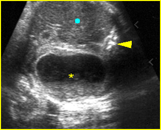
Fig 1: Fetal dermoid cyst Cross-section at the upper abdomen: cystic mass (*) containing echogenic debris or sebum (arrowhead = spine, circle = liver)
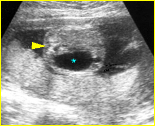
Fig 2: Fetal ovarian cyst Cross-section of the fetal upper abdomen: simple anechoic cyst (*) (arrowhead = spine)
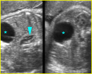
Fig 3: Fetal ovarian cyst Cross-sectional scan of the abdomen: Simple anechoic cyst (*) in the rightside of the abdomen, note normal kidney and adrenal gland (arrowhead)
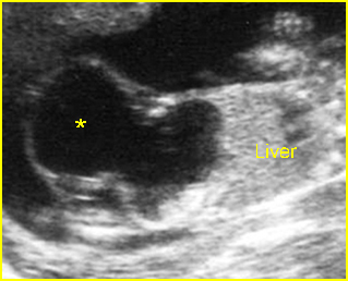
Fig 4: Large urachal cyst Sagittal scan of the fetal trunk: large cystic mass (*) connecting with the bladder, externally protruding from the lower abdomen
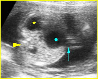
Fig 5: Large umbilical vein aneurysm Cross-sectional scan of the abdomen: cystic mass (solid circle) connecting with umbilical vein (arrow) with Doppler signal on flow study (* = stomach, arrowhead = spine)
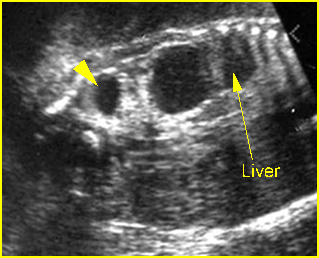
Fig 6: Choledocal duct cyst Coronal scan of the abdomen: anechoic cystic mass (*) located at the right upper abdomen with pressure effect on the liver (arrowhead = renal cystic dysplasia)
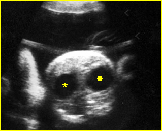
Fig 7: Duodenal atresia Cross-sectional scan of the abdomen: Double bubble sign: two cystic masses (* = proximal duodenum, solid circle = stomach) in the upper abdomen
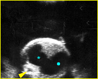
Fig 8: Proximal jejunal atresia Cross-sectional scan of the abdomen: marked dilatation of the bowel loop with continuation of stomach (solid circle), duodenum (*) and proximal jejunal
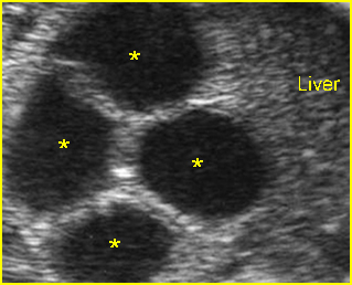
Fig 9: Small bowel obstruction Cross-sectional scan of the abdomen: Multiple loop of small bowel loops with marked dilatation (*)
Video clips of abdominal cysts
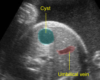
Hepatic cyst: Isolated cystic mass located at right upper abdomen
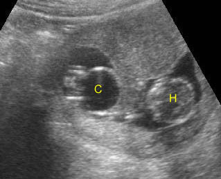
Bladder outlet obstruction: Coronal scan: Megacystis (C) at 11 weeks of gestation (H = head)
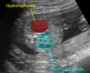
Renal cysts: Multicystic kidney and marked hydronephrosis of the contralateral kidney
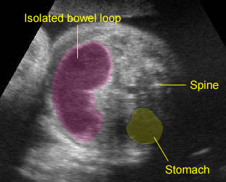
Bowel duplication: Sausage cystic mass secondary to bowel duplication; isolated bowel loop with no other visible bowel loops
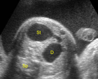
Duodenal atrestia: Double bubble sign with continuation representing stomach (St) and duodenum (D) (Sp = spine)
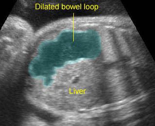
Proximal jejunal atresia: Dilated bowel loop at the upper abdomen representing the proximal jejunal loop separated from the stomach and large bowel
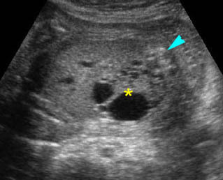
Renal cystic dysplasia: Cross-sectional scan of the abdomen: muliticystic kidney (*) with oligohydramnios and absent contralateral kidney
The main sonographic differential diagnoses include
Choledochal cyst/hepatic cyst:
- anechoic single cyst in liver, choledochal cyst located adjacent to hepatic ducts
- more common in females
- not associated with other anomalies
Multicystic kidneys:
- anechoic multicysts of variable size which are non-communicating
- located posteriorly
- unilateral or bilateral
- male or female
Hydronephrosis:
- anechoic single or often multiple communicating cysts within renal fossa
- more common in males
- probably associated with other GU anomalies
Hydroureter:
- anechoic tubular cyst in nature
- communicating with renal pelvis and bladder
- located posteriorly
- more common in males
- may be associated with megacystis
Meconium pseudocyst:
- echogenic cyst with calcified wall at mid-abdomen
- associated with bowel obstruction
- male or female
Mesenteric/omental cyst:
- isolated anechoic cyst, unilocular or less commonly septated
- often mobile
- located centrally
- male or female
Urachal cyst:
- isolated anechoic simple
- communicates with the bladder and also commonly with the umbilicus
- located anteriorly
- male or female
Ovarian cyst:
- isolated unilocular, sometimes septated, or daughter cyst
- usually unilateral
- located lower or laterally
- only in females
Duodenal atresia:
- double bubble sign in upper abdomen
- connection between the two bubbles
- polyhydramnios
- male or female
- commonly related to trisomy 21
Small bowel obstruction:
- multiple bowel loops with echogenic contents
- tubular in nature
- located centrally or throughout the abdomen
- peristalsis
- male or female
Hydrometrocolpos:
- anechoic to echogenic cyst
- located retrovesically
- only in females

