Masses at the Anterior Body Wall
Differential diagnoses of the masses at the anterior body wall include
Fig 1, Fig 2, Fig 3, Fig 4, Fig 5, Fig 6
- Physiologic omphalocele (8-12 weeks); size of <7 mm, no liver content
- Omphalocele
- Gastroschisis
- Ectopia cordis
- Limb-body wall defect
- Bladder exstrophy
- Cloacal exstrophy
- Stack of umbilical cord (positive Doppler signal or color flow)
- Cord edema or localized Wharton’s jelly near the umbilicus
- Cord cysts (omphalomesenteric cyst or allantoic cyst)
- Placental mass (chorioangioma) close to the anterior wall
- Artifacts: pseudo-omphalocele secondary to oblique plane
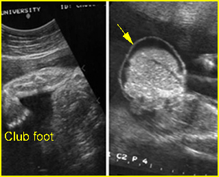
Fig 1: Omphalocele Clubfoot and free floating sac containing liver with covering membrane (arrow)
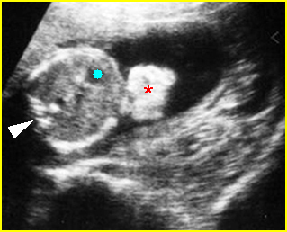
Fig 2: Gastroschisis Free floating echogenic bowel (*) in the amniotic fluid (arrowhead = spine, solid circle = intra-abdominal stomach)
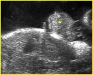
Fig 3: Bladder extrosphy Sagittal view of the fetal abdomen: complex extra-abdominal mass (*) below the umbilicus, finally proven to be bladder extrosphy
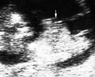
Fig 4: Physiologic omphalocele Sagittal scan of the fetus (10 weeks): bowel contents protruding into the proximal umbilical cord (arrow)
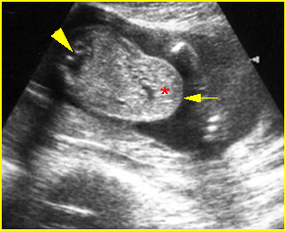
Fig 5: Pseudo-omphalocele Oblique cross-sectional scan of the fetus: the liver (*) covered by normal abdominal wall (arrow) protrudes anteriorly, may be mistaken for omphalocele (arrowhead = spine)
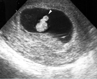
Fig 6: Physiologic omphalocele Cross-sectional scan of the embryo (8 weeks): bowel contents protruding into the proximal umbilical cord (arrowhead)
Video clips of masses at the anterior body wall
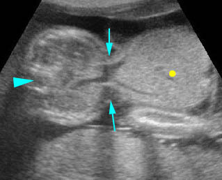
Omphalocele: Cross-sectional scan: large omphalocele with liver content (solid circle) (arrow = the defect, arrowhead = spine)
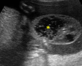
Umbilical cord cyst: Focus on the proximal cord: cord cyst with coarse particles (*) may be due to liquidfaction of the Wharton’s jelly
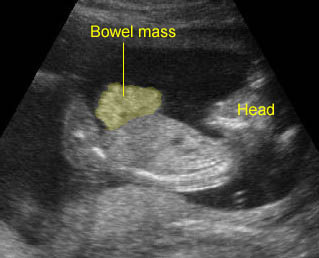
Gastroschisis: Free floating bowel mass anterior to abdominal wall
True anterior wall defect
- Omphalocele
- Gastroschisis
- Limb-body wall complex
- Amniotic band syndrome
- Bladder exstrophy
- Cloacal exstrophy
- Pentralogy of Cantrel
Cystic mass close to the umbilicus
- Allantoic cyst (urachal cyst)
- Omphalomesenteric cyst
- Pseudocyst (liquefaction of Wharton’s jelly)
- Umbilical vein varix
- Omphalocele
The approach of the anterior wall defects
- Relationship of the cord insertion to the defect: various sites of the defect suggest the nature of pathology as follows:
- above the umbilicus: pentalogy of Cantrell
- at the umbilicus: omphalocele
- paraumbilical area: gastroschisis
- below the umbilicus: bladder/cloacal exstrophy
- defects throughout the abdomen: limb-body wall complex
- severe and asymmetric defect: amniotic band syndrome
- Characteristics of herniated organs: a herniated organ can suggest the nature of pathology as follows:
- bowel: either a gastroschisis, omphalocele, or LBWC
- liver only: highly suggestive of omphalocele (but most omphaloceles also include portions of bowel), very unlikely to be gastroschisis
- bowel only (in omphalocele): more often related to chromosomal abnormalities
- solid mass at the lower abdomen: bladder or cloacal exstrophy
- Presence of covering membranes:
- presence: omphaloceles (be careful, the covering membrane may not always be seen or may be ruptured), limb-body wall complex (not always)
- absence: gastroschisis
- the presence of a herniated liver in a ventral wall defect without a covering membrane is more suggestive of a ruptured omphalocele
- Associated anomalies:
- multiple anomalies: omphalocele, LBWC
- extra-, intra-abdominal bowel obstruction: gastroschisis
- scoliosis: LBWC
- non-visualization of bladder: bladder or cloacal exstrophy
- amputation defects: amniotic band syndrome

