Hepatosplenomegaly
Enlargement of the liver or spleen is usually related to systemic abnormalities such as fetal hydrops, however it may occur secondary to an isolated finding such as a liver tumor (hemangioendothelioma).
Sonographic findings:
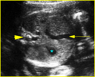
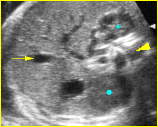
Fig 1, Fig 2
- Fetal liver measurements are usually obtained in a longitudinal plane, from the dome of the right hemidiaphragm to the tip of the right lobe.
- Commonly associated with ascites or other signs of hydrops fetalis.
- Tables of normal values are available for analyzing both fetal liver and spleen measurements.
- Hepatosplenomegaly could be seen in more than 90% of cases of hydrops fetalis from hemoglobin Bart’s disease.
- Splenic circumference is an excellent predictor of severe anemia.
- Hepatomegaly is constantly demonstrated in Beckwith-Wiedemann syndrome.
Differential diagnosis:
- Hydrops fetalis due to various causes, especially hemoglobin Bart’s disease.
- Severe anemia or myeloproliferative disorder.
- Congenital infection; for example, cytomegalovirus infection and syphilis.
- Beckwith-Wiedemann syndrome.
- Down’s syndrome.
Associations: Hydrops fetalis due to various causes.
Prognosis: Depends on underlying causes.

Fig 1: Splenomegaly Cross-sectional scan of the abdomen: enlarged spleen behind the stomach (*) (arrow= the umbilical vein, arrowhead = spine)

Fig 2: Splenomegaly
Cross-sectional scan of the upper abdomen: The enlarged spleen (solid circle) behind the stomach, Note adrenal gland (*) close to the spine (arrowhead) (arrow = umbilical vein)
Video clips of hepatosplenomegaly

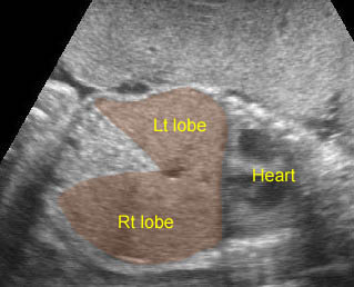
Hepatomegaly: Coronal scan of the upper abdomen shows the enlarged liver, both left and right lobe

