Ascites
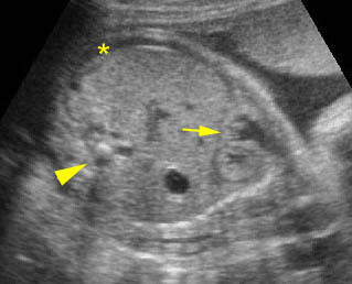
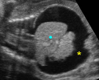
Ascites, fluid collection within the fetal abdomen, is always abnormal. In severe cases, ascites can be readily appreciated, however, mild ascites, often seen in early hydrops fetalis, may be confused with pseudoascites (described previously). Therefore, first of all, one must distinguish between pseudoascites (a symmetric hypoechoic rim of the anterior abdominal wall) and true ascites. The following findings are often related to true ascites but not to pseudoascites:
- Fluid between bowel loops or outlines the visceral structures (liver, omentum, umbilical vein, falciform ligament)
- Other signs of hydropic changes
- Polyhydramnios.
The major causes of ascites are as follows:
- Hydrops fetalis secondary to various causes (most common)
- Intrauterine infection
- GI or urinary tract obstruction with perforation
- Abdominal tumors (rare)
- Perforation of the common bile duct (very rare)
- Chyloperitoneum (very rare).
Note: Ascites associated with hydrops fetalis is usually accompanied by fluid collection in other spaces such as subcutaneous edema, pleural effusion or pericardial effusion. Isolated ascites is most often secondary to intra-abdominal disorders.

Mild ascites: Oblique cross-sectional scan: * = mild ascites, arrow = diaphragm, arrowhead = spine

Marked ascites: Coronal scan of the fetal trunk: marked fluid collection in the abdomen (*) (solid circle = liver)

