Double Bubbles
Differential diagnosis of the appearance of double bubbles at the upper abdomen includes:
- Duodenal obstruction (connection between proximal duodenum and stomach)
- duodenal atresia
- duodenal stenosis
- duodenal web
- Bowel duplication
- Choledocal cyst
- Hepatic cyst
- Ovarian cyst (with stomach bubble)
- Renal cyst (with stomach bubble)
Fig 1, Fig 2, Fig 3, Fig 4
Continuity with the stomach should be demonstrated to distinguish a distended duodenum from other cystic masses in the right upper quadrant, such as choledocal cyst, or duplication cyst. In the case of congenital pyloric atresia, an intermittent ‘double bubble’ sign can be visualized. The double bubble sign disappears during gastric peristalsis. However, a whole stomach configuration should be delineated by continuous observation covering periods when gastric peristalsis is active as well as quiet.
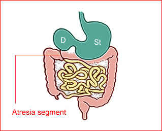
Fig 1: Schematic drawing of duodenal atresia, The fluid-filled stomach (St) and duodenum (D) cause double bubble sign on ultrasound examination
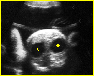
Fig 2: Duodenal atresia Cross-sectional scan of the abdomen: Double bubble sign: two cystic masses (* = proximal duodenum, solid circle = stomach) in the upper abdomen

Fig 3: Duodenal atresia Cross-sectional scan of the abdomen: Double bubble sign: two cystic masses (* = stomach, solid circle = proximal duodenum ) in the upper abdomen
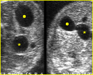
Fig 4: Duodenal atresia Cross-sectional scan of the abdomen: Double bubble sign: two cystic masses (* = proximal duodenum, solid circle = stomach) with connection together in the upper abdomen
Video clips of double bubbles
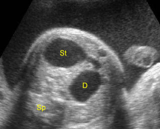
Duodenal atresia: Double bubble sign with continuation representing stomach (St) and duodenum (D) (Sp = spine)
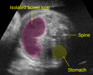
Bowel duplication: Sausage cystic mass secondary to bowel duplication; isolated bowel loop with no other visible bowel loops

