Skull Defects
Cranial defects vary from a small meningocele, which is very difficult to visualize, to complete absence of the cranium such as with anencephaly or exencephaly. In practice, there is no problem in the diagnosis of complete absence of the skull or a large defect, however, severe bone demineralization, such as osteogenesis imperfecta, may give the appearance of a large skull defect. Partial defects, or small cephaloceles are occasionally difficult to visualize.
Fig 1, Fig 2, Fig 3
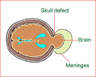
Fig1: Schematic drawing: Occipital meningocele, protrusion of the brain tissue and meninges through the skull defect
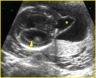
Fig 2: Occipital cephalocele Transverse scan of the skull: small defect of occipital bone with meningocele (*) (arrow = ventriculomegaly)
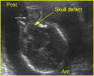
Fig 3: Occipital cephalocele Transverse scan of the skull: defect of occipital bone with meningoencephalocele (arrow)
Video clips of skull defects
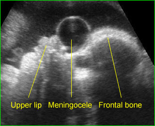
Anterior meningocele: Sagittal scan of the fetal face: frontal bone defect with midline cystic mass (meningocele)
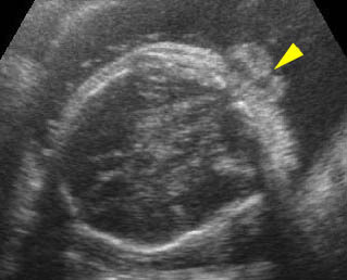
Occipital cephalocele: Transverse scan the level of cerebellum: small defect of occipital bone with encephalocele
Differential diagnosis
The differential diagnoses and some sonographic features of skull defects are as follows:
Anencephaly:
- absent bony calvarium above the orbits
- orbits well visualized
- absence of supratentorial brain
- residual brain
- possibly spina bifida
- polyhydramnios
Excencephaly
- calvarium absent
- disorganized supratentorial brain tissue
Amniotic band syndrome (ABS)
- asymmetric or bizarre cephalocele
- other deformities, including limb amputation
Limb-body wall complex
- asymmetric or bizarre encephaloceles
- similar to ABS except fetal body adherent to placenta
- usually more severe than ABS
- associated bizarre defects of fetal body
Cephalocele
- midline defect
- extracranial cyst or brain tissue
- possibly ventriculomegaly
- lemon sign may be present
Open neural tube defects
- lemon sign
- banana sign
- mild ventriculomegaly
- spinal defect
Fetal demise
- overlapping sutures (Spalding’s sign)
- poor visualization of intracranial structures
- associated findings of fetal demise
Microcephaly
- calvarium present
- decreased brain tissue
- head circumference 2-3 standard deviations below that expected for the menstrual age
Craniosynostosis
- complete or partial
- deformed skull
- possibly microcephaly
- abnormal cephalic index.
In addition, considerable overlap may be found in these features among abnormalities. For instance, the lemon sign was originally described with opened NTD, however, it may not be present in an opened NTD identified in the third trimester of pregnancy, and a mild lemon sign may be identified in normal fetuses.

