Anencephaly
Anencephaly, the most common NTD, is characterized by the absence of the cranial vault and cerebral hemispheres. However, the midbrain and posterior fossa are usually normal. In about one-third of cases, there is a variable amount of angiomatous stroma, which may mimic rudimentary brain. It develops from exencephaly as the exposed brain tissue is gradually eliminated after exposure to the amniotic fluid. Anencephaly probably originates as an abnormality of mesenchymal structures and the brain is secondarily lost to injury in utero because of its exposed position.
Incidence: 0.6-0.8 in 1000 births, varying from 1 in 1000 births in the USA to 1 in 100 in parts of the British Isles, with a male to female ratio of 1:4; the prevalence in the USA tends to be decreased resulting from folic acid supplementation to prevent neural tube defects.
Sonographic findings:
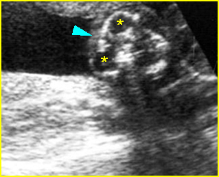
Fig 1: Anencephaly: Coronal scan of the face (12 weeks): no skull (arrowhead) above the orbits (*) with frog face appearance
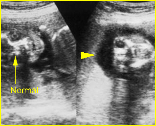
Fig 2: Anencephaly: Coronal scan of the face of twins: normal face of the left twin and no skull (arrowhead) above the orbits (arrowhead) in the right twin
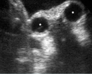
Fig 3: Anencephaly: Oblique coronal scan: no skull above the orbit (*)
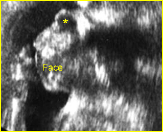
Fig 4: Anencephaly: Facial profile view: no skull above the orbit (*)
Video clips of anencephaly
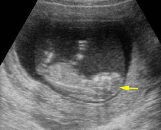
Anencephaly: Sagittal scan of the fetus at 11 weeks; no skull (arrow)
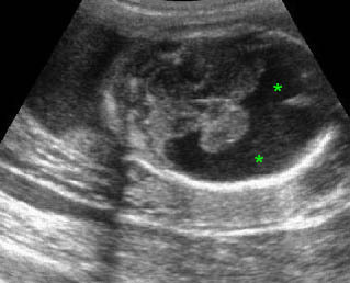
Anencephaly: Coronal scan of the face: no skull above the orbit (arrowhead)
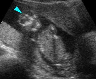
Anencephaly: Coronal scan of the face: no skull above the orbit (arrowhead) with frog face appearance
- Ultrasound is highly accurate for detection of anencephaly during the second trimester, with a detection rate of 100%.
- Lack of bony calvarium above the orbits.
- Absence of the cerebral hemispheres but normal midbrain and posterior fossa.
- An angiomatous mass with multiple cystic spaces replacing the forebrain; its presence must not dissuade the examiner from the correct diagnosis of anencephaly.
- In early diagnosis, spectacle sign, prominent orbits on the coronal view and may look like frog-eye face or Mickey-mouse face; the brain will have an irregular floppy outline due to the absence of the skull or a mobile cranial cyst.
- Polyhydramnios secondary to severe brain dysfunction resulting in ineffective fetal swallowing, found in half of cases and more often in late pregnancy.
- Echogenic amniotic fluid is found in nearly 90% of cases of acrania/anencephaly in the first trimester.
- Associated anomalies such as omphalocele or spina bifida.
- Pitfalls: If the head is deep in the pelvis, the defect may be missed by transabdominal examination and thought to be a technical problem.
- Diagnosable from 10 to 14 weeks of gestation.
Associations: Associated anomalies, found in one-third of cases, such as spina bifida (most common), hydronephrosis, omphalocele, and cardiac defects. About 2% of fetuses with anencephaly have abnormal chromosomes, particularly trisomy 18.
Management: Termination of pregnancy should always be offered.
Prognosis: Uniformly lethal.
Recurrence risk: About 2-4%. Periconceptional folic acid supplementation can significantly decrease the recurrent risk. 50-70% of these defects can be prevented if a woman consumes sufficient folic acid daily before conception and throughout the first trimester of her pregnancy.

