Holoprosencephaly
Holoprosencephaly represents a broad spectrum of malformations due to a lack of separation of the structures of the forebrain, resulting in no midline separation of the cerebral hemispheres and diencephalic structures. The normally bilateral diencephalon and basal ganglia are fused and tend to incorporate into the upper brainstem. Recent discoveries in the fields of genetics and developmental neurobiology have advanced our knowledge of this complex disorder, such as 12 loci on 11 chromosomes, or deletion of distal 7q. It is usually classified as alobar (failure of the brain cleavage, complete fusion), semilobar (partially fused), or lobar (only fused frontal horns). The alobar form, the most common form, has a monoventricular cavity with fused thalami and can be subdivided into three separate configurations: pancake, cup, or ball forms. The pancake type, which is the rarest, occurs when the residual brain is minimal and compressed over the skull base. The ball variation occurs when the cerebral cortex covers the monoventricular cavity. The cup form is intermediate between the two, with the residual brain having a cup-like configuration on a sagittal view. It may also be divided into those with and without a dorsal sac. Thus, alobar holoprosencephaly without a dorsal sac is similar to the ball form, while the cup and pancake forms have a dorsal sac.
Incidence: 1-2 per 10,000 live births and 1 per 250 embryos, male/female, 1:3 for the alobar form and 1:1 for the lobar form.
Fig 1, Fig 2, Fig 3, Fig 4, Fig 5, Fig 6, Fig 7
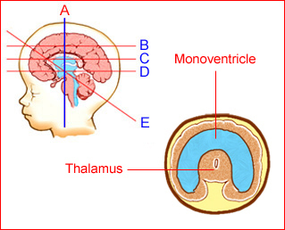
Fig 1: Schematic drawing: Coronal view shows monoventricle with fused thalamus in alobar holoprosencephaly

Fig 2: Schematic drawing: Tranthalamic view shows monoventricle with fused thalamus in alobar holoprosencephaly
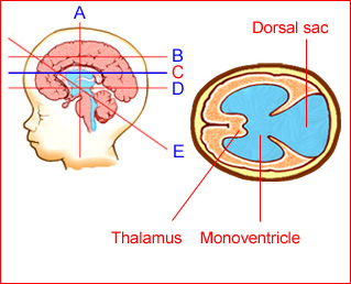
Fig 3: Schematic drawing: Transventricular view shows mono-ventricle with dorsal sac in holoprosencephaly
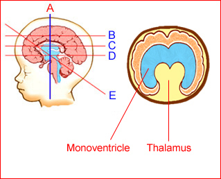
Fig 4: Schematic drawing: Coronal scan shows partial separation of the lateral ventricles in semilobar holoprosencephaly
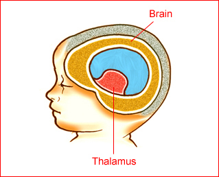
Fig 5: Schematic drawing: Ball type of holoprosencephaly (brain mantle completely covers the mono-ventricle
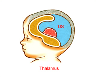
Fig 6: Schematic drawing: Cup type of holoprosencephaly (DS = dorsal sac)
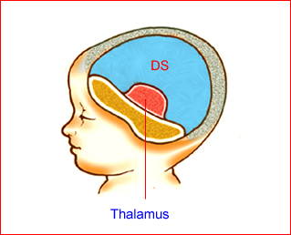
Fig 7: Schematic drawing: Pancake type of holoprosencephaly (DS = dorsal sac)
Sonographic findings:
Alobar holoprosencephaly Fig 8, Fig 9, Fig 10
- fused thalami
- monoventricular cavity, lacking occipital, temporal, and frontal horns
- absent falx cerebri
- absent cavum septum pellucidum
- dorsal sacs (cup and pancake type) communicating widely with monoventricular cavity
- a ridge of cerebral tissue demarcating the boundary between the dorsal sac and the monoventricular cavity is often seen
- often associated with facial abnormalities
- the 3D prenatal US may be helpful, especially in the demonstration of various abnormalities of the face
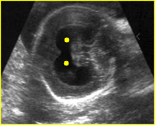
Fig 8: Holoprosencephaly: Oblique coronal scan of the skull: common ventricle (solid circle)
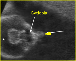
Fig 9: Holoprosencephaly, cyclopia and proboscis Common lateral ventricle (*), fused orbits (arrow), proboscis (arrowhead)
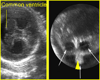
Fig 10: Holoprosencephaly, hypotelorism and proboscis : Left: Fusion of the lateral ventricle Right: hypotelorism (arrow) and proboscis
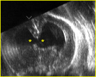
Fig 11: Semilobar holoprosencephaly Oblique coronal scan of the skull: common ventricle (*) with partial separation
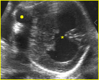
Fig 12: Semilobar holoprosencephaly Oblique coronal scan of the skull: common ventricle (*) with partial separation (solid circle = orbits)
Video clips of holoprosencephaly
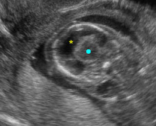
Holoprosencephaly: Transverse coronal scan of the fetal head: fusion of the lateral ventricle (*) (solid circle = fused thalamus)
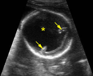
Holoprosencephaly: Dorsal sac (*) with folding of the remaining brain tissue (arrow)
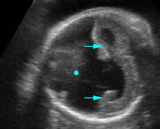
Holoprosencephaly: Dorsal sac (solid circle) with folding of the remaining brain tissue (arrow)
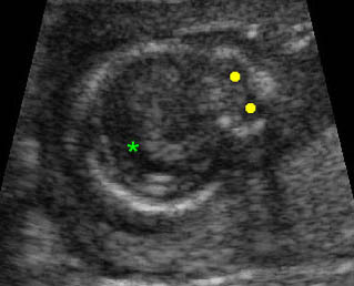
Holoprosencephaly: Transverse scan of the head: fusion of the lateral ventricle (*), no falx cerebri, marked hypotelorism (solid circle)
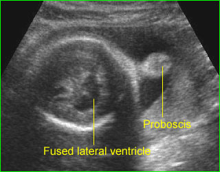
Proboscis (holoprosencephaly) : Midline solid mass at the forehead (proboscis) and fused ventricle
Semilobar holoprosencephaly Fig 11, Fig 12
- incompletely fused thalami
- monoventricular cavity with partial separation
- absent cavum septum pellucidum
- presence of incomplete falx cerebri
- often associated with facial abnormalities
Lobar holoprosencephaly (rarely detected antenatally)
- absent cavum septum pellucidum
- nearly normal intracranial structures
- abnormal course of the anterior cerebral artery on Doppler examination
Common associated anomalies
- face (most common): hypotelorism, cyclopia, proboscis, cleft lip and palate (especially midline cleft)
- abdomen: renal agenesis or cystic dysplasia, esophageal atresia, omphalocele, bladder exstrophy
- chest: pulmonary hypoplasia, cardiac malformations (VSD, ASD, transposition of great vessels)
- others: myelomeningocele, polydactyly, clubfoot
- chromosome abnormalities: trisomy 13 (most common), 13q-, trisomy 18, 8p-, and triploidy
The main differential diagnoses are hydrocephalus and hydranencephaly:
- hydrocephalus can be readily distinguished by visualization of falx cerebri and splayed thalami rather than fused thalami
- hydranencephaly is characterized by complete absence of a cerebral hemisphere, variable presence of the falx cerebri and no facial abnormalities
- a dorsal sac must be differentiated from other cysts such as arachnoid cyst, porencephaly, or Dandy-Walker malformation
The alobar type can be diagnosed as early as 9-11 weeks of gestation.
Associations: Alobar and semilobar holoprosencephaly are often associated with significant facial abnormalities, including cyclopia, ethmoidocephaly, cebocephaly, cleft lip, and trisomy 13. Other associated anomalies include cardiac defect, omphalocele, Dandy-Walker malformation and some cases of trisomy 18.
Management: Termination of pregnancy should be offered and karyotyping and fetal autopsy should be performed.
Prognosis: Fatal in most cases, or resulting in severe handicaps for the survivors.
Recurrence risk: An empirical recurrent risk for sporadic cases is approximately 6%.

