Porencephaly/Schizencephaly
These two entities are often considered together because of their similar appearance. However, they have separate origins. Porencephaly is a locally destructive brain lesion resulting from either a developmental anomaly or intraparenchymal insult. There are two types of porencephaly. Type I is generally due to an antepartum intraparenchymal hemorrhage. Type II lesions are usually developmental anomalies. Various causes of parenchymal damage have been reported, such as trauma or inherited disease. Schizencephaly is a full-thickness cleft of the cerebral mantle considered to be a migrational abnormality rather than a destructive process. The etiology may include encephaloclastic disorder, cytomegalovirus or genetic disorders such as triple X syndrome.
Sonographic findings:
Porencephaly:
Fig 1, Fig 2, Fig 3
- a fluid-filled space within normal brain parenchyma, often communicating with the lateral ventricles
- usually unilateral
- often progressive changes to hydranencephaly (most severe form)
- no pressure effect on the adjacent brain
- the defect lined by white matter (demonstrated by MRI)
Schizencephaly:
Fig 4
- unilateral or bilateral cystic lesion
- may or may not communicate with the lateral ventricle
- typically bilateral clefts in the fetal brain connecting the lateral ventricles with the subarachnoid space
- absence of the cavum septum pellucidum is commonly seen
- MRI is very helpful in confirming the diagnosis
- the defect is lined by gray matter (demonstrated by MRI)
– The main differential diagnoses include arachnoid cyst, interhemispheric cyst in the case of agenesis of the corpus callosum, and dorsal sac in holoprosencephaly.
– Usually diagnosable after 18 weeks.
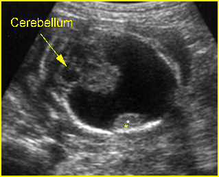
Fig 1: Porencephaly Transthalamic view: only small part of brain tissue left (*)
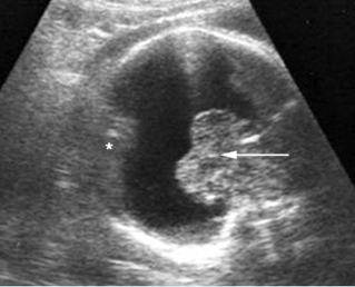
Fig 2: Porencephaly Most of the brain tissue is absent, only small part of brain tissue left (*). (arrow = thalami)
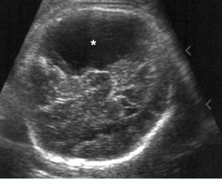
Fig 3: Porencephaly Some part of cerebral tissue is absent (*)
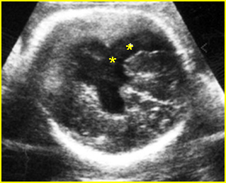
Fig 4: Schizencephaly Cleft of cerebral tissue is absent (*) resulting in cystic brain lesion
Video clips of porencephaly / schizencephaly
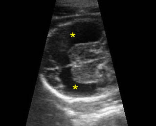
Porencephaly: Transthalamic view: missing large piece of brain (*)
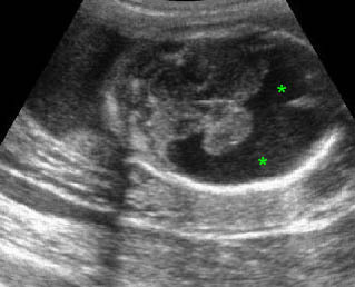
Porencephaly: Transverse scan of the head: fluid collection (*) in the large area of missing brain tissue
Associations: Rare.
Management: In continuing pregnancies, the fetus should be delivered in a tertiary center.
Prognosis: Depends on the size and location of the lesion; very poor for extensive porencephaly, and the bilateral form of schizencephaly.
Recurrence risk: Rare, though hereditary cases have been reported.

