Atrial Septal Defect (ASD)
ASD is an opening of the atrial septum causing communication between the two atria. Embryologically, the first septum (primum) to develop arises from the endocardial cushion and starts at the AV valves, growing toward the free wall of the combined atrium. Later a second septum (secundum) starts at the free wall of the atrium and grows toward the septum primum. The gap between the two is called the ostium primum. When the fusion between the septum primum and endocardial cushion has occurred, the septum primum fenestrates. The resulting communication is called ostium secundum. A second septum then extends on the right side of the septum primum and covers part of the ostium secundum. The remaining orifice is the foramen ovale, and the foramen ovale flap is the lower part of the septum primum. Thus, defects that are close to the endocardial cushion are called the ostium primum, and defects in the area of the foramen ovale are the ostium secundum. The primum ASD is the simplest form of atrioventricular septal defect or partial AV canal.
Fig 1
Incidence: ASD accounts for 8% of structural heart defects; secundum defects are the most common and are found in 0.07 per 1000 infants.
Sonographic findings:
Fig 2, Fig 3
- Secundum ASDs are most commonly isolated.
- Occasionally, ASD is related to other cardiac lesions including mitral, pulmonary, tricuspid or aortic atresia.
- Abnormal oscillatory pattern of the foramen ovale flap motion by M-mode.
- Pitfalls:
- Prenatal diagnosis of secundum ASD is relatively difficult due to the physiologic presence of the foramen ovale. Most likely, only unusually large defects can be recognized with certainty.
- The detection rate was poor for isolated septal defects but higher when associated with other complex lesions or abnormal four-chamber views
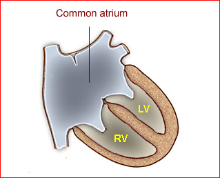
Fig 1: Schematic drawing: Atrial septal defect with common atrium (RV = right ventricle, LV = left ventricle)
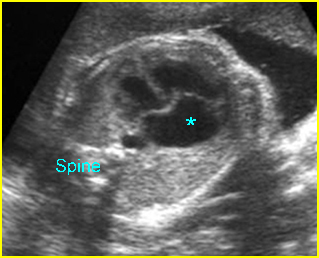
Fig 2: Atrial septal defect (ASD) Four-chamber view: common atrium (*) no atrial septum and flap of foramen ovale
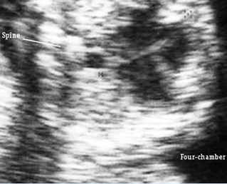
Fig 3: Atrial septal defect (ASD) Four-chamber view: common atrium, no atrial septum and flap of foramen ovale
Video clips of Atrial Septal Defect (ASD)
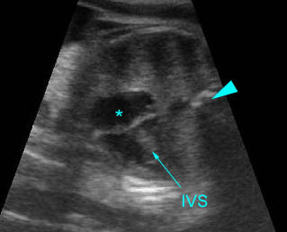
Atrial septal defect: Four-chamber view: abnormal cardiac axis, no atrial septum seen (arrowhead = spine)
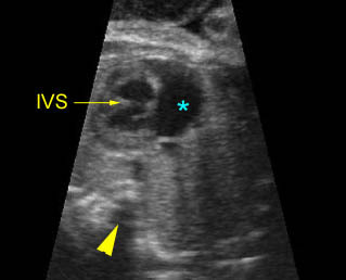
ASD & VSD: Atrial septal and ventricular septal defect
Four-chamber view: abnormal cardiac axis, no atrial septum (*) and incomplete ventricular septum (arrowhead = spine)
ASD occurs in the setting of other defects:
- Ebstein’s anomaly
- Hypoplastic right heart
- Hypoplastic left heart
- Pulmonic stenosis
- Truncus arteriosus
Associations: Most cases of secundum ASD are isolated but may sometimes be found to be part of syndromes such as Holt-Oram syndrome. The primum defects are more associated with other anomalies. (See the section “Atrioventricular canal defect”.)
Management: Careful prenatal and postnatal search for associated anomalies is required; small defects may become smaller or close after birth, however, moderate to large defects often need surgical correction or catheterization closure devices.
Prognosis: ASDs are not a cause of impairment of cardiac function in utero because a large right-to-left shunt at the level of the atria is a physiologic condition in the fetus. Most affected infants are asymptomatic even in the neonatal period.

