Pulmonary Sequestration
Pulmonary sequestration (PS) is a supernumerary lobe of the benign pulmonary tissue, separated from the normal tracheobronchial tree, having its own vascular supply, which usually arises from the thoracic or abdominal aorta. The lesions are pathologically divided into intralobar and extralobar forms (invested by its own pleura). Intralobar sequestrations almost always drain into pulmonary veins whereas most extralobar lesions drain into systemic veins. Approximately 10-15% of extralobar forms are found within or below the diaphragm, usually on the left.
Incidence: 1 in 1000 births.
Fig 1
Sonographic findings:
Fig 2, Fig 3, Fig 4
- Typically homogeneous echogenic mass varying in size in the chest (90% of cases) or abdomen (10% of cases).
- Unilateral in most cases, usually left lower lobe.
- Mediastinal shift, pleural effusion and hydrops fetalis if the mass is large enough.
- Visualization of a feeder vessel arising from the thoracic or abdominal aorta feeding the mass strongly suggestive of extralobar sequestration.
- Regression on the follow-up scans (decreasing in size, echogenic, and mediastinal shift) is often seen.
- Main differential diagnoses are other lung masses, including type III CCAM, CDH, bronchial atresia, and mediastinal teratoma. The extrathoracic form may be differentiated from neuroblastoma, renal tumor, and teratoma. PS usually is echogenic, left-sided, and can be identified in the second trimester whereas neuroblastoma is most often cystic, right-sided, and identified in the third trimester.
- Pitfalls:
- A feeding vessel of pulmonary sequestration in the thorax is not always seen, therefore it is often indistinguishable from Type III CCAM.
- Thoracoabdominal masses including CCAM, CDH, neuroblastoma, lymphangioma, and bronchial atresia, mimicking PS.
- The extrathoracic type is easily overlooked.
- MRI may be helpful in difficult cases.
- Usually detected in the third trimester but some cases are detected early in the second trimester.
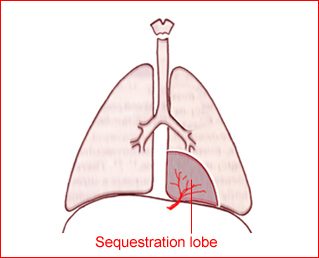
Fig 1: Schematic drawing: Bronchopulmonary extra-lobar seques-tration in the inferior portion of the thorax
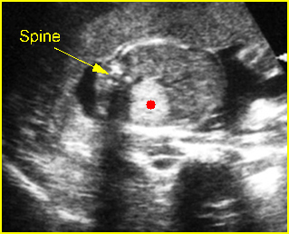
Fig 2: Extralobar pulmonary sequestration Cross-sectional scan of thorax: bright echogenic mass (solid circle) anterior to the spine
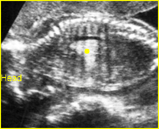
Fig 3: Extralobar pulmonary sequestration Sagittal scan of thorax: intrathoracic bright echogenic mass (solid circle) in the shape of lung lobe
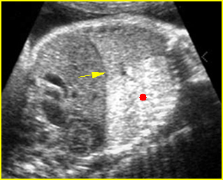
Fig 4: Extralobar pulmonary sequestration Oblique sagittal scan of the fetal trunk: markedly echogenic, lung-shaped mass in the chest (solid circle) (arrow = intact diaphragm)
Video clips of pulmonary sequestration
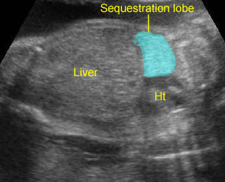
Lung sequestration: Extralobar sequestration: Echogenic lung mass with lobar in shape
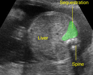
Lung sequestration: Extralobar sequestration in the upper abdomen: Echogenic mass with lobar in shape behind the liver
Associations: Associated abnormalities including cardiac defect, CDH or lung abnormalities are found in 10% of intralobar type cases and 58% of extralobar type cases. Sequestration and CCAM may be seen together, which is suggestive of a common embryogenesis despite diverse morphology.
Management: Conservative and follow-up ultrasound examination is appropriate in most cases. Many extralobar pulmonary sequestrations dramatically decrease in size before birth or after birth. Fetal intervention with a thoracoamniotic shunt may be needed in some hydropic cases. Delivery should be at term in a tertiary center. Cases with residual mass at birth should have surgical resection. An ex utero intrapartum therapy (EXIT) strategy with resection of the mass during the EXIT procedure may be appropriate in selected cases. However, the need for surgery should be based on appropriate postnatal investigations (e.g. CT scans), rather than on antenatal behavior.
Prognosis: Good prognosis in the majority of cases, spontaneous regression occurs in many cases, and the persistent cases are resected safely postnatally; poor if associated with hydrops, though reversible in some cases.

