Congenital Cystic Adenomatoid Malformation (CCAM)
CCAM is a hamartoma of the lung that may involve a whole lung or a single lobe. Multiple cysts replace pulmonary parenchyma. It can also occur in lung sequestration. The cysts are usually unilateral and are classified into three types as follows:
Fig 1
- macrocystic (type I): few in number and large, up to 7 cm, and thick-walled
- mixed (type II): multiple and smaller (about 1 cm)
- microcystic (type III): very small, <0.5 cm, and not visible as distinct cysts by ultrasound; the lesions appear as bulky, intrathoracic, solid masses with increased echogenicity.
Incidence: Unknown.
Sonographic findings:
Fig 2, Fig 3, Fig 4
- Echogenic or sonolucent mass in one side of the chest possibly shifting the heart.
- Unilateral in most cases.
- Hydrops fetalis (<10% of cases), commonly seen with a large mass, in particular a greater CAM volume ratio.
- Polyhydramnios (65% of cases) secondary to esophageal obstruction from the intrathoracic mass, obstructed venous return or decreased absorption of lung fluid.
- Doppler study reveals a normal blood supply, unlike the sequestration.
- Main differential diagnosis:
- If cystic mass: CCAM, CDH, bronchogenic or neuroenteric cyst, esophageal duplication
- If solid mass: microcystic CCAM, pulmonary sequestration, CHD.
- Pitfalls:
- Most often confused with congenital diaphragmatic hernia.
- Acoustic enhancement posterior to the heart mimicking microcystic mass.
- The normal thymus may occasionally simulate hypoechogenic mass.
- First diagnosable between 12 and 18 weeks of gestation
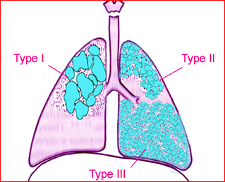
Fig 1: Schematic drawing: The three type of CCAM: type I (large cysts of variable size), type II (small cystic type), and type III (solid appearance)
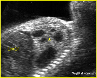
Fig 2: Cystic adenomatoid malformation Oblique sagittal scan of the fetal trunk: solid-cystic mass (*), mostly cystic, in the left chest (CCAM type I)
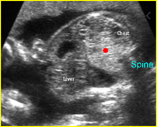
Fig 3: Cystic adenomatoid malformation Oblique sagittal scan of the fetal trunk: echogenic mass in the left chest (solid circle)
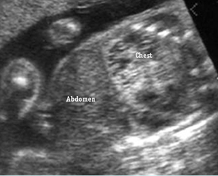
Fig 4: Cystic adenomatoid malformation Oblique sagittal scan of the fetal trunk: partial solid and partial cystic mass in the left chest (CCAM type II)
Video clips of congenital cystic Adenomatoid malformation (CCAM)
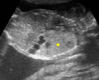
CCAM (Congenital cystic adenomatoid malformation; CCAM) Coronal scan of the chest: large echogenic mass (solid circle) in the left thorax with some cystic changes
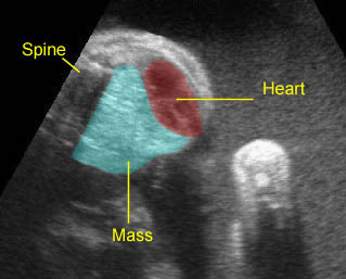
CCAM (Congenital cystic adenomatoid malformation: CCAM) CCAM type III: Large echogenic lung mass with mediastinal shift
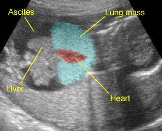
Laryngeal Atresia : Bilateral enlarged echogenic lung caused by high airway obstruction, associated with ascites, simulating bilateral CCAM type III
Associations: Usually not associated with other anomalies.
Prognosis: Good prognosis in the majority of cases especially the cystic type and it can spontaneously regress antenatally, but can not continue after birth; poor prognosis if associated with hydrops, especially microcystic type, lung hypoplasia, prematurity or severe associated malformations.
Management: Follow-up ultrasound examination is required, with no active intervention in the absence of acute polyhydramnios or hydrops. Persistent CCAM needs postnatal surgical removal.
Recurrence risk: Unknown.

