Mediastinal Shift
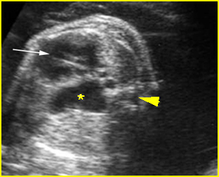
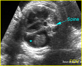
Fig 1, Fig 2
- Unilateral or asymmetric pleural effusion
- Chest masses secondary to any causes
- Unilateral bronchial atresia
- Unilateral agenesis of lung.

Fig 1: Diaphragmatic hernia Cross-sectional scan of thorax at the level of four-chamber view: cystic structure of bowel loop (*) located in the chest with cardiac displacement (arrow) to the right (arrowhead = spine)

Fig 2: Pleural effusion Cross-sectional scan of thorax: pleural effusion (*) in the left thorax with cardiac displacement to the right
Video clips of mediastinal shift

Diaphragmatic hernia: Cross-sectional scan: The stomach is herniated into the chest with cardiac displacement

CCAM (Congenital cystic adenomatoid malformation: CCAM)
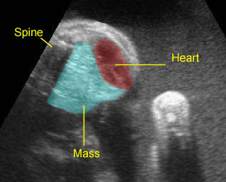
CCAM type III: Large echogenic lung mass with mediastinal shift

