Other Abnormalities
Increased thickness of the ventricular wall
- Normal (bright spot of papillary muscle)
- Right ventricular wall hypertrophy; commonly seen with volume overload or increased afterload
- Cardiomyopathy
- Cardiac tumor
Global cardiomegaly
- Hydrops fetalis
- High-output heart failure (chorioangioma, hemangioma)
- Cardiomyopathy
- Cardiac tumor (rhabdomyoma, teratoma)
Cardiac echogenic mass
- Artifacts: echogenic papillary muscles of the heart
- Rhabdomyoma (most common, accounting for 60%), often related to tuberous sclerosis
- Teratoma (25%)
- Fibroma (12%)
- Myxoma (rare)
- Hemangioma (rare)
- Hamartoma (rare)
Pericardial hypoecho
- Pericardial effusion
- Pleural effusion
- Normal: hypoechoic dropout
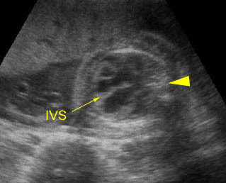
Cardiomegaly / Bradycardia: Four-chamber view: generalized enlargement of the heart with pericardial effusion related to Hb Bart’s disease with bradycardia (arrowhead = spine)
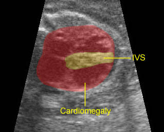
Cardiomyopathy : Marked cardiomegaly with thickening of the interventricular septum (IVS)
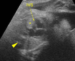
Cardiac rhabdomyoma: Four-chamber view: echogenic mass (*) in the left ventricle (arrowhead = spine)
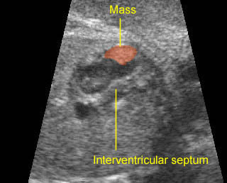
Small rhabdomyoma: Small echogenic mass (5 mm) at the inner surface of the left ventricular wall
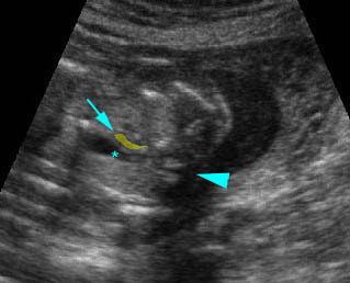
Coarctation of aorta: Arch view at the upper thorax, extremely small aortic arch (arrow) compared to ductal arch (*) (arrowhead = spine)

