Normal Examination
Technique of examination
– Transverse thoracic plane: Fig 1, Fig 2, Fig 3 The ultrasound transducer should be oriented to visualize the upper thorax first, from clavicles, and then move down to the abdomen. A sweep in transverse planes provides important information, including the ribs, thoracic spine, lung echogenicity, cardiac location, size, and axis. The four-chamber view can also be satisfactorily assessed. This view should be examined in all fetuses in the second and third trimesters as part of a routine scan. The thorax is rather round on this plane. The plane at the level of the four-chamber view can also perform thoracic circumference measurements.
– Sagittal thoracic plane: Fig 4 The transducer should be adjusted to obtain the anteroposterior images from one side to the other. The thorax appears dome-shaped on this plane. The diaphragm and thoracic size compared with the abdomen can be assessed.
– Coronal thoracic plane: Fig 5, Fig 6 The transducer is oriented to obtain the coronal images from the spine to the anterior wall. The thorax appears bell-shaped on this plane. The diaphragm and thoracic size compared with the abdomen can be assessed.
– Bones assessment: Fig 7, Fig 8, Fig 9, Fig 10 The fetal thoracic cage is bell-shaped and bordered by the clavicles (which can also be measured) at the apices and the smooth hypoechoic diaphragm inferiorly. The ribs, smoothly marginated and regularly spaced, form the lateral boundaries and extend anteriorly and inferiorly more than halfway around the thorax from their dorsal attachments. The subjective impression of the thoracic size is sufficient in most cases. However, direct measurement of the thoracic circumference may be of value in the diagnosis of lung hypoplasia.
– Lungs: Fig 11 The lungs have homogeneous mid-range echoes, however, the echogenicity may be greater than, less than, or equal to the echogenicity of the liver. The lungs may become progressively more echogenic than the liver as gestation progresses. No abdominal contents should be seen with the lungs.
– Heart: Fig 2, Fig 3 On the transverse plane, the visualization of the normal four-chamber view of the heart with appropriate cardiac position, axis, and size helps to exclude many thoracic abnormalities. The four-chamber view examination can detect 60-70% of cardiac defects and an additional view of the ventricular outflow tracts and great vessels further increases that sensitivity. The heart occupies about one-third of the thoracic volume, and most of the cardiac volume is located in the left anterior quadrant of the chest. The cardiac axis, the angle that the interventricular septum makes with the line drawn from the spine to the anterior chest wall, ranges between 22 and 75 degrees, with a mean of 45 degrees. Echogenic foci caused by a specular reflection from the papillary muscles and chordae tendinae are commonly seen. The bright foci, termed echogenic intracardiac foci, can be associated with chromosome abnormalities.
– Thymus: The thymus, a homogeneous, relatively hypoechoic structure in the anterior fetal mediastinum, could be visualized with some effort sonographically in 74% of 251 normal fetuses.
– Larynx and Trachea: Fig11 The larynx is virtually always visible when the hypopharynx is fluid-filled. The fluid-filled trachea is a relatively easy structure to visualize. The fetus intermittently and frequently breathes amniotic fluid into the trachea. The trachea usually can be traced to its distal end, passing posterior to the aortic arch, but the carina and bronchi are quite difficult to perceive.
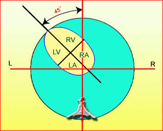
Fig 1: Schematic drawing: Four-chamber view with typical cardiac axis, normal size and appropriate location (LA = left atrium, LV = left ventricle, RA = right atrium, RV = right ventricle)
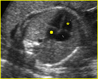
Fig 2: Normal four-chamber view Four-chamber view: normal four chambers; (* = right ventricle, solid circle = left atrium)
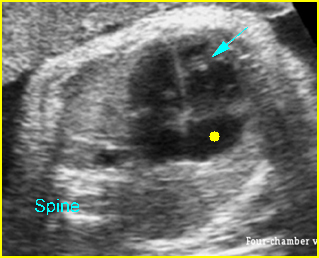
Fig 3: Normal four-chamber view Cross-sectional scan of thorax: normal four-chamber view (arrow = moderator band in right ventricle, solid circle = right atrium)
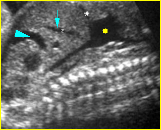
Fig 4: Right atrium Sagittal scan of the thorax: inferior vena cava and superior vena cava empty to the right atrium (solid circle) (* = diaphragm, arrow = umbilical vein, arrowhead = hepatic vein)
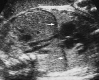
Fig 5: Intact diaphragm Coronal scan of the trunk: normal diaphragm, hypoechoic line (arrow) above the liver
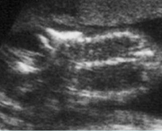
Fig 6: Scapula Coronal view of thorax: normal triangular shape of the scapula
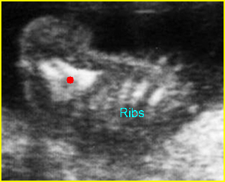
Fig 7: Normal capula Coronal scan of the scapula: normal triangular shape
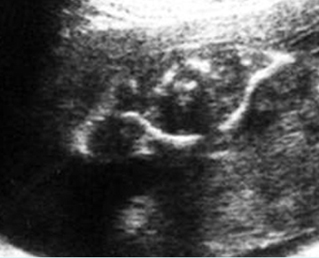
Fig 8: Clavicles Cross-sectional scan of the upper thorax: normal clavicles
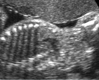
Fig 9: Ribs Coronal scan of thorax: normal cross-section of the ribs with acoustic shadow
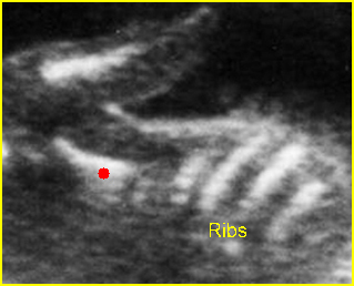
Fig 10: Scapula Coronal view of normal triangular shape scapula (solid circle)

Fig 11: Normal four-chamber view Four-chamber view: normal four chambers; (* = right ventricle, solid circle = left atrium)
Video clips of the normal heart
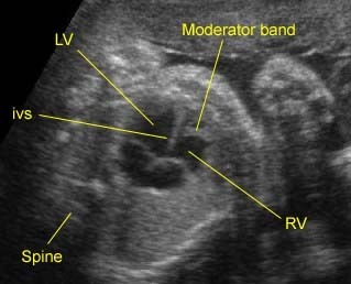
Normal four-chamber view: Typical four-chamber view: note the moderator band in the right ventricle (RV) (LV = left ventricle, IVS = interventricu-lar septum)
Pitfalls
- False diagnosis of a diaphragmatic hernia: Oblique images of the chest and abdomen may include abdominal viscera in the heart. This may be misleading, causing the examiner to misinterpret normal findings as abnormal (i.e. false diagnosis of a diaphragmatic hernia).
- Ribs in the abdomen: The ribs may extend inferiorly into the upper abdomen. Therefore, observation of the ribs and the abdominal structures in the same transverse plane should not be misinterpreted as abnormal.
- Pseudo skin thickening: The thickening of subcutaneous tissue of the middle upper chest, including the scapula, may normally appear somewhat thicker than that of the abdomen or scalp.
Checklists
- Normal heart location, axis and size
- Normal thoracic size (compared with abdominal size)
- No abnormal fluid collection or abnormal mass
- Normal homogeneous echogenicity of lung
- Normal ribs and thoracic spine.

