Sonolucent Bones
Diffuse demineralization of the skull and long bones almost always occurs with fetal skeletal dysplasia syndromes. In the case of severe demineralization of the bony calvarium, the cranium is thin without an acoustic shadow and so poorly ossified that the intracranial structure can easily be seen. This increased visualization of the intracranial structures may be confused with such abnormalities as exencephaly due to acrania or acalvaria. Unlike exencephaly, however, there is an intact but poorly ossified skull. Careful scanning reveals concomitant limb anomalies.
Fig 1, Fig 2, Fig 3, Fig 4
The main differential diagnoses of the sonolucent skull are as follows:
- Osteogenesis imperfecta (most common)
- Hypophosphatasia (rare)
- Achondrogenesis type I (rare).
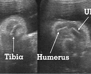
Fig 1: Hypophosphatasia Longitudinal scan of long bones: shortened and poorly ossified bones
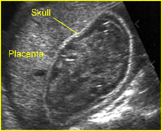
Fig 2: Poorly ossified and compressible skull Cross-sectional scan of skull: deformable skull with sonolucency in osteogenesis imperfecta type IIA
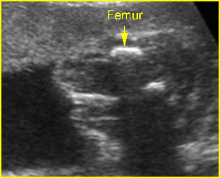
Fig 3: Micromelia in Thanatophoric dysplasia Severe shortenings of long bones (arrow) but normal ossification
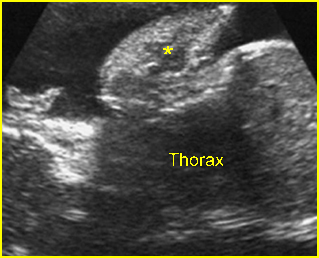
Fig 4: Sonolucent and shortened arm Longitudinal scan of the arm: Sonolucent and shortened humerus (*) in case of achondrogenesis
Video clips of sonolucent bones
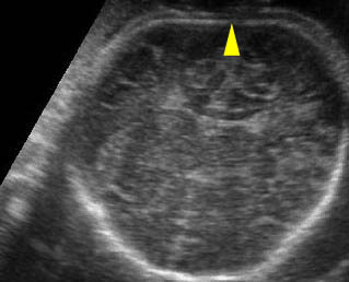
Sonolucent skull : The thin skull is so poorly ossified that cerebral gyri and sulci could be seen easily

Achondrogenesis: Mid-sagittal view of the spine: extremely poor ossification of the spine (arrow)

