Congenital Hypophosphatasia
Congenital hypophosphatasia is a rare autosomal recessive inherited disorder characterized by demineralization of bones with low alkaline phosphatase in serum and other tissues. It is related to the numerous tissue non-specific alkaline phosphatase genes on chromosome 1. Prenatal diagnosis may be made using ultrasound and by assaying alkaline phosphatase in tissue obtained from chorionic villous samplings or in blood from cordocentesis, and can also be made by DNA analysis.
Incidence: 1 per 100,000 births.
Sonographic findings:
Fig 1, Fig 2, Fig 3, Fig 4, Fig 5, Fig 6
- Variable hypomineralization, boneless appearance in some cases.
- Compressible skull with complete absence of acoustic shadow, and probably so severe that the normally-difficult-to-visualize near-field brain is easily seen.
- Ossification centers in both the vertebral bodies and neural arches may be absent, or there may be preservation of some vertebral body ossification centers, unlike achondrogensis in which the vertebral body ossification centers are absent with preserved neural arch ossification centers.
- Severe micromelia.
- Thin or delicate or seemingly absent long bones.
- Spurs along the midshaft of long bones and at the knees and elbows.
- The main differential diagnosis is OI type II, in particular type IIC which involves similarly thin bones. All three forms of OI type II have multiple fractures and wavy or wrinkled bones that should help distinguish them from hypophosphatasia.
- Increased nuchal translucency thickness during late first trimester.
- Pitfalls: It is probably indistinguishable from OI type II and achondrogenesis. However, they have the same lethal prognosis.
- Extremely low levels of alkaline phosphatase in the cord blood can confirm the diagnosis.
- Usually diagnosed in the second half of pregnancy, but possible as early as the late first trimester.
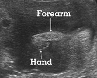
Fig 1: Hypophosphatasia Longitudinal scan of the forearm and hand: poorly ossified radius and ulna as well as hand bones and clubhand
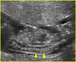
Fig 2: Sonolucent spine Sagittal scan of the fetus at 14 weeks: sonolucent vertebrae of the fetus with hypophosphatasia
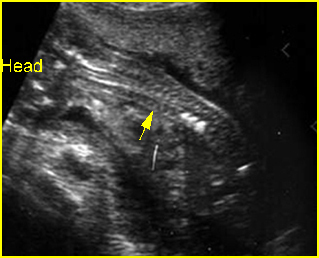
Fig 3: Hypophosphatasia Oblique sagittal scan of the spine: poorly ossified spine (arrow), only 3 vertebral bodies are ossified
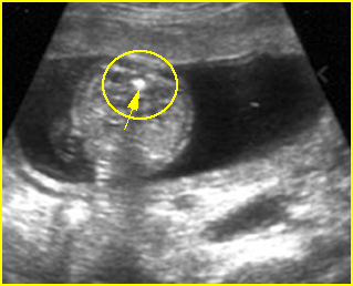
Fig 4: Hypophosphatasia Cross-sectional scan of the spine: poorly ossified posterior ossification centers but normally ossified anterior center (arrow)
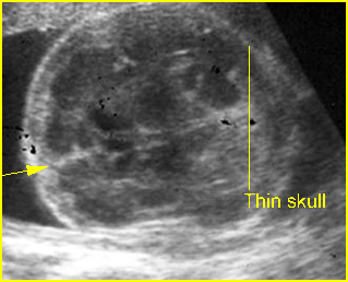
Fig 5: Hypophosphatasia Cross-sectional scan of skull: thin and sonolucent calvarium (arrow = falx cerebri)
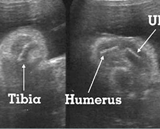
Fig 6: Hypophosphatasia Longitudinal scan of long bones: shortened and poorly ossified bones
Video clips of congenital hypophosphatasia
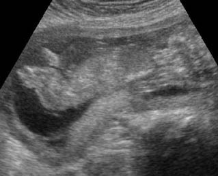
Hypophosphatasia : Sagittal scan of the fetus at 14 weeks; poorly ossified bony structures
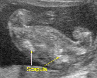
Hypophosphatasia : Transverse section at the upper thorax: shortenings and poorly ossified long bones
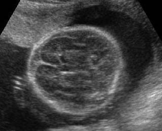
Hypophosphatasia : Transverse scan of the skull: brachycephaly with sonolucent skull
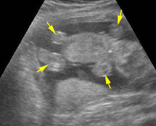
Hypophosphatasia : Early recognition of limb shortenings with poorly ossified bones at 13 weeks of gestation
Associations: Rare.
Management: Termination of pregnancy can be offered.
Prognosis: Lethal, though successful treatment with high-dose pyridoxal phosphate in some non-lethal cases has been reported.
Recurrence risk: 25%, an autosomal recessive disorder.

