Limb Reduction Defects
Limb reduction deformities, presenting alone or as part of a specific syndrome, occur in approximately 0.5 per 10,000 births. Half of them are simple transverse reduction deficiencies of one forearm or hand without associated anomalies and the remaining half have additional anomalies.
Fig 1, Fig 2, Fig 3
Isolated limb reduction:
- isolated limb deficiency more commonly occurs in the upper extremities rather than the leg, which generally occurs within the context of a syndrome, as do bilateral amputations or reduction of all limbs
- sporadic occurrence in most cases with negligible risk of recurrence
Limb reduction associated amniotic band:
- secondary to entanglement with mesodermic bands derived from the chorionic surface of the amnion after the latter has separated from the chorion
- isolated or multiple limb reductions, with or without other defects
Phocomelia:
- typically hands and feet are present, but the intervening arms and legs are absent
- main differential diagnoses include Robert’s syndrome, variants of TAR syndrome, and Grebe’s syndrome
- can be caused by exposure to thalidomide but this is only of historical interest
Radial ray defects: radial clubhand may be part of
- TAR syndrome (thrombocytopenia with absent radius)
- Fanconi’s syndrome: an autosomal recessive disorder consisting of pancytopenia anemia, leukopenia and thrombocytopenia and skeletal anomalies, especially radial clubhand
- Aase’s syndrome: an autosomal recessive disorder characterized by hypoplastic anemia and radial clubhand with bilateral triphalangeal thumb and a hypoplastic radius
- Holt-Oram syndrome: an autosomal dominant disorder characterized by congenital heart defects, radial hypoplasia and triphalangeal or absent thumb
- VATER association: a sporadic disorder resulting from defective mesodermal development during embryogenesis consisting of vertebral segmentation (70%), anal atresia (80%), tracheoesophageal fistula (70%), esophageal atresia, and radial (65%) and renal defects (53%)
- Goldenhar’s syndrome: characterized by hemifacial microsomia, hypoplasia of the malar and maxillary or mandibular region, vertebral anomalies, and radial defects
- Klipple-Feil syndrome
- Chromosomal abnormalities, particularly trisomy 18 and 21.
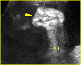
Fig 1: Transverse limb reduction Scan of the foot: absent distal end of foot
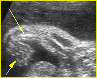
Fig 2: Absent radius syndrome Longitudinal scan of upper limb (15 weeks): short ulna (arrow) without radius with fixed flexion of wrist joint (arrowhead)
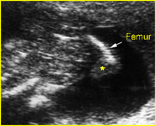
Fig 1: Limb reduction Longitudinal scan of lower limb: absent tibia and fibula, abnormal foot (*) arising from femur (arrow) in the fetus with VATER association
Video clips of limb reduction defects
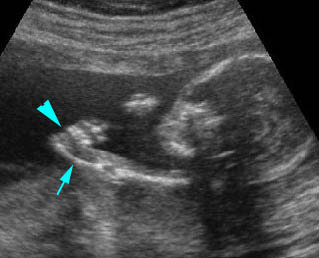
Radial ray defects : Longitudinal scan of the upper extremity: absent radius (arrow) with fixed flexion of the wrist joint (arrowhead)

Radial ray defects : Longitudinal scan of the upper extremity at 13 weeks: absent radius (with fixed flexion of the wrist joint (arrow)
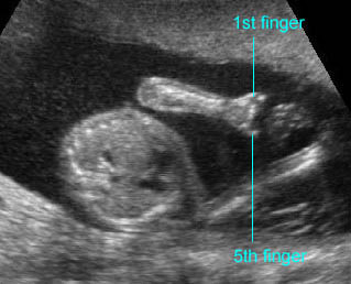
Transverse limb defect : Absent fingers; only the 1st and 5th fingers remained
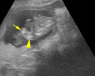
Transverse limb defect : Absent hand with shortenings of all long bones (14 weeks) (arrow = severely shortened forearm, arrow = humerus)

