Achondrogenesis
Achondrogenesis is a lethal chondrodystrophy characterized by severe micromelia, short trunk, absent vertebral bodies ossification, and macrocrania. It has been classified into two types:
– Type I: autosomal recessive type, the more severe form, accounting for 20% of cases, characterized by severe hypoossification and severe micromelia, and thin ribs with fractures (type IA) or not (type IB). Type 1B is associated with mutation in the gene for the DTDST gene on chromosome 5, the same gene identified in the cause of diastrophic dysplasia.
– Type II (Langer-Saldino, autosomal dominant), accounting for 80% of cases, has relatively normal ossification, less severe micromelia, and thicker ribs without fractures. Type II is caused by a new mutation in the COL2A1 gene on chromosome 12, or inherited via germline mosaicism of the healthy parents.
Incidence: About 1 per 4000 births.
Sonographic findings:
Fig 1, Fig 2, Fig 3, Fig 4, Fig 5
- Decreased mineralization of all or many vertebral bodies, sacrum and ischium. The absence of vertebral body ossification may be the unique finding of type II.
- Severe micromelia with bowing, however, it can be found with normally developed extremities in rare cases.
- Enlarged calvarium with poor ossification in type I but relatively normal ossification in type II.
- Thin ribs (type I) with or without fractures.
- Polyhydramnios is common.
- Redundancy and edema of the subcutaneous tissues (pseudohydrops) or nuchal edema even in the first trimester.
- 3D ultrasound is helpful in providing more details such as facial dysmorphism, the relative proportion of the appendicular skeletal elements and the hands and feet.
- The main differential diagnosis includes thanatophoric dysplasia, achondroplasia, and short-rib polydactyly syndrome, all of which may have underdeveloped vertebral bodies, but none of these also have calvarial underossification. Severe hypophosphatasia may result in severe calvarial and spinal underossification, but hypophosphatasia usually has diffuse underossification of the spinal ossification centers or focal loss of the neural arch ossification centers as opposed to focal lack of vertebral body ossification that is seen in achondrogenesis.
- Pitfalls: Normally developed extremities can be seen in type II. Therefore, complete examination of the vertebral body is very important.
- First diagnosable in the late first trimester, but mostly in the second trimester.
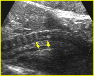
Fig 1: Spinal poor ossification Mid-sagittal scan of the cervical spine: very sonolucent spine (arrow) in association with achondrogenesis
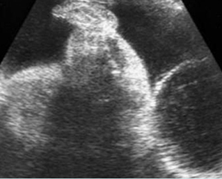
Fig 2: Long bone shortening Longitudinal scan of the thigh and leg: shortening of thigh and leg, compared with foot (achondrogenesis)
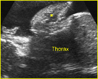
Fig 3: Sonolucent and shortened arm Longitudinal scan of the arm: Sonolucent and shortened humerus (*) in case of achondrogenesis
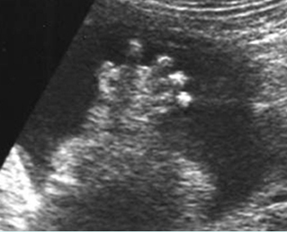
Fig 4: Poor ossification of hand Sonolucency of the hand bones as well as short fingers
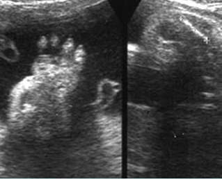
Fig 5: Poor ossification Sonolucency of the hand bones as well as short fingers (left) and short humerus (right)
Video clips of achondrogenesis

Achondrogenesis : Mid-sagittal view of the spine: extremely poor ossification of the spine (arrow)
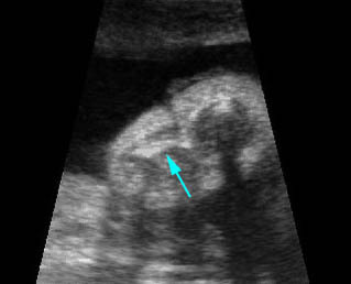
Achondrogenesis : Shortened long bones with poor-ossified bone (arrow) and redundant skin
Associations: Rare, cephalocele, cystic hygroma, and polydactyly in some cases.
Management: Termination of pregnancy can be offered.
Prognosis: Lethal.
Recurrence risk: Type IB is autosomal dominant, and carries a theoretical recurrence risk of 25%. The recurrence risk of type II, new mutations, is rare.

