Abnormal Hands and Feet
Hands and feet should be examined to exclude polydactyly, brachydactyly, abnormal hand posture, sandal gap, clinodactyly, Rocker-bottom foot, clubfoot, etc. These abnormalities are commonly part of several syndromes.
Polydactyly Fig 1
Polydactyly is the presence of an additional digit, which can be postaxial (on the ulnar or fibular side) or preaxial (on the radial or tibial side) and with a central form. This extra digit may range from a fleshy nubbin to a complete digit. Postaxial polydactyly is the most common form and is inherited autosomal dominantly as an isolated finding in most cases. However, it is significantly related to some syndromes, especially trisomy 13 or short-rib polydactyly syndrome. Preaxial polydactyly, especially a triphalangeal thumb, is most likely to be part of a syndrome.
The major differential diagnoses for polydactyly include
- Isolated polydactyly
- Chromosome abnormalities, especially trisomy 13
- Meckel Gruber syndrome
- Asphyxiating thoracic dystrophy
- Short-rib polydactyly syndrome
- Achondroectodermal dysplasia (Ellis-Van Creveld syndrome)
- Smith-Lemli-Opitz syndrome.
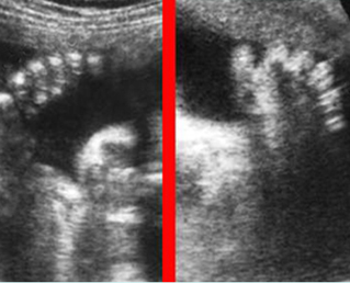
Fig 1: Polydactyly
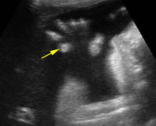
Post-axial polydactyly
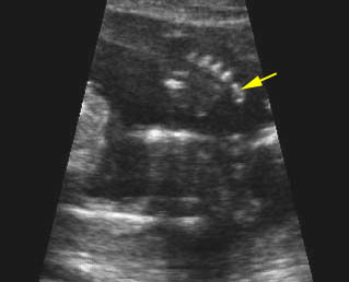
Polydactyly : Post-axial polydactyly (fleshy finger)
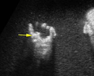
Pre-axial polydactyly
Clubfoot Fig 2
A clubfoot is one of the most common congenital anomalies, occurring in 1:250 to 1 in 1000 births. In 95% of cases, the sole is turned medially (talipes equinovarus). This can be caused by external compression in the case of oligohydramnios or by internal factors including abnormal bone formation, spina bifida, muscular defects or genetic causes (15% have a family history of clubfoot). Most clubfeet are found in otherwise normal infants. However, about 10% of cases are associated with several syndromes including Pena-Shokier phenotype, trisomy 18 and 13, etc.
Rocker bottom foot Fig 3
Vertical talus, or eversion of the planar arch, produces a rocker-bottom (convex outward) appearance of the bottom of the foot. This is most often associated with chromosomal abnormalities, particularly trisomy 13 and 18, although it may also be seen as an isolated anomaly, with caudal dysplasia sequence, neural tube defects, and neuromuscular disorders, and as part of the Potter sequence
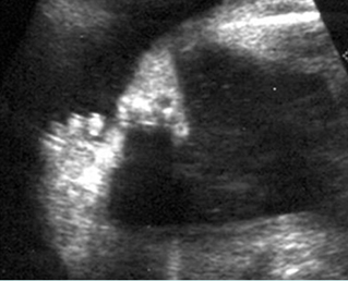
Fig 2: Clubfoot Longitudinal scan of the lower leg: plantar view of the foot seen in the same plane of longitudinal view of tibia and fibula
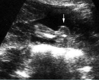
Fig 3: Rocker bottom foot Prominent calcaneous bone (arrow) associated with trisomy 13
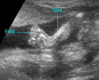
Clubfoot : The plantar view of the foot can be demonstrated on the same plane of the longitudinal view of the tibia
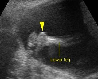
Rocker-bottom foot : Sagittal scan of the fetal foot showing the prominent heel
Abnormal hand posture Fig 4, Fig 5, Fig 6
Fetal hand malformation may be isolated or associated with chromosomal abnormalities, limb reduction defect, a neuromuscular disorder, or skeletal dysplasia. Ultrasound evaluation of the fetal hand requires assessment of the number and configuration of the fingers, the presence and position of the thumb, and the relationship of the thumb and fingers to each other and to the hand and wrist. The phalanges should be evaluated in the long-axis rather than the short-axis view. Short-axis views may represent the metacarpals rather than the phalanges. The normal fetal hand is most often in a resting position with loosely curled fingers, which the fetus periodically opens. Sonographic assessment requires skill on the part of the examiner in following the movement of the hand and documenting the extended fingers and thumb should the hand open during the examination.
Clenched hands Fig 7, Fig 8
Clenched hands or abnormal hand postures are often related to other abnormalities and more than half of cases have aneuploidy, predominantly trisomy 18 (88% of aneuploid fetuses).
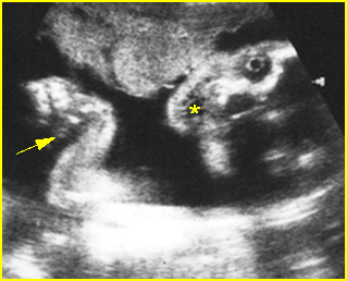
Fig 4: Pena-Shokier phenotype Fixed flexion of both elbow and wrist joint (arrow) as well as persistent mouth opening (*)
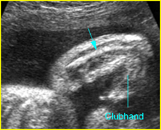
Fig 5: VATER associations Longitudinal scan of forearm: absent radius with abnormal hand posture
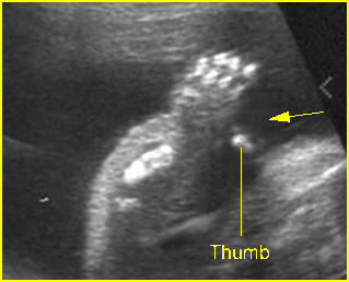
Fig 6: Hitchhiker thumbs Abnormal posture and wide separation of the thumb (arrow) in the fetus with diastrophic dysplasia (25 weeks)
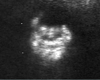
Fig 7: Clenched hand Overlapping fingers of the fetus with trisomy 18
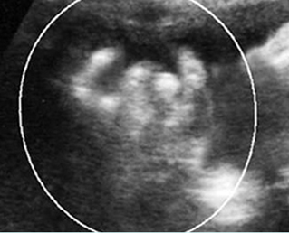
Fig 8: Clenched hand Overlapping fingers of the fetus with trisomy 18
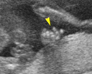
Overlaping fingers : Clenched hand (arrowhead) in trisomy 18
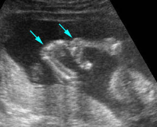
Fixed flexion: Fixed flexion of the wrist joint in association with trisomy 18
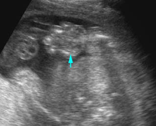
Edematous hand
Clinodactyly Fig 9, Fig 10
Clinodactyly is the permanent curvature or deflection of one or more fingers. It is frequently associated with abnormal chromosomes especially trisomy 21.
Syndactyly
Syndactyly is the fusion of digits and may consist of bony or soft tissue fusion. This may also be difficult to detect sonographically, especially those cases consisting of soft tissue fusion. Syndactyly is associated with a number of syndromes. It may be seen in the SRPD syndromes with the characteristics of polydactyly, and it may be seen in triploidy or Down’s syndrome.

Fig 9: Clinodactyly Curved-in position of the fifth finger (arrow) of the fetus with trisomy 21
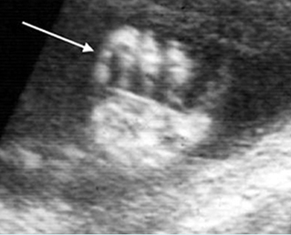
Fig 10: Clinodactyly Scan of the hand: disproportionately small middle phalange of the fifth finger (arrow)
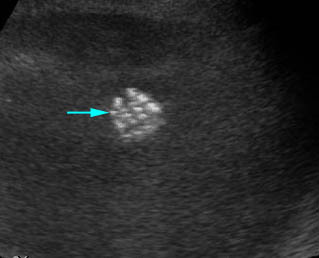
Clinodactyly : Clinodactyly with hypoplasia of the middle phalanx of the fifth finger

