Fracture In Utero
The detection of rib or long bone fractures in association with severe micromelia suggests a diagnosis of osteogenesis imperfecta. Fractures may be subtle or may lead to angulation.
Fig 1, Fig2, Fig 3
The major differential diagnoses for fractures in utero are as follows:
- Osteogenesis imperfecta type II (most common)
- Hypophosphatasia (rare)
- Campomelic dysplasia (not a true fracture but bowing).
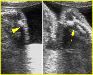
Fig 1: Fracture in utero Longitudinal scan of upper extremity: poorly ossified and fracture in osteogenesis imperfecta type IIA
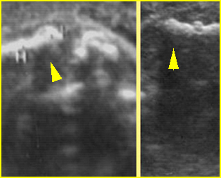
Fig 2: Callus formation of long bone Longitudinal scan of humerus and femur: irregularity due to callus (arrowhead) in osteogenesis imperfecta type IIA
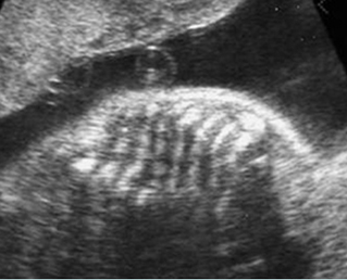
Fig 3: Rib fractures Multiple rib fractures with poor ossification in the fetus with osteogenesis imperfecta type IIA
Video clips of fracture in utero
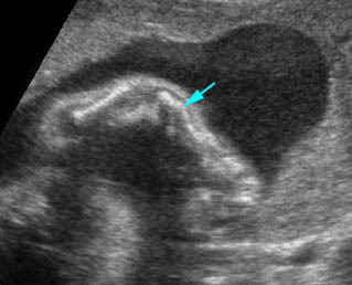
Fracture in utero : Longitudinal scan of upper limb: callus formation (arrow) and irregularity of ulna secondary to previous fracture
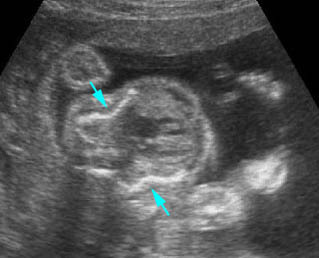
Osteogenesis imperfecta : Cross-sectional scan of the thorax: rib fractures (arrow)

