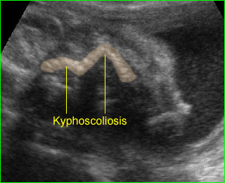
Hypoplastic Thorax

Hypoplastic Thorax

The specific group of autosomal recessive disorders mainly involves ribs and thoracic hypoplasia including asphyxiating thoracic dysplasia, Ellis-van Creveld syndrome, and short-rib polydactyly syndrome. There is phenotypic overlap between these three entities. They may be a spectrum of the same entity.

Sonographic differential diagnoses of the hypoplastic thorax include:

  • Asphyxiating thoracic dysplasia (Jeune syndrome)
  • Ellis-van Creveld syndrome
  • Short-rib polydactyly syndrome
  • Other syndromes with a lesser degree of rib shortenings
    • thanatophoric dysplasia
    • atelosteogenesis
    • fibrochondrogenesis
    • achondrogenesis
    • Jarcho-Levin syndrome.

 Asphyxiating thoracic dysplasia (Jeune syndrome)

Asphyxiating thoracic dysplasia (Jeune syndrome) is an autosomal recessive skeletal dysplasia, an ATD gene located on chromosome 15q13, characterized by a small thorax, a varying degree of short limbs, renal anomaly, and polydactyly.

Incidence: Rare.

Sonographic findings:

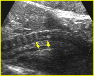
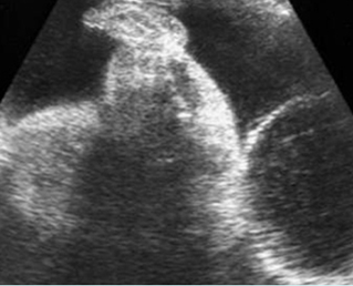
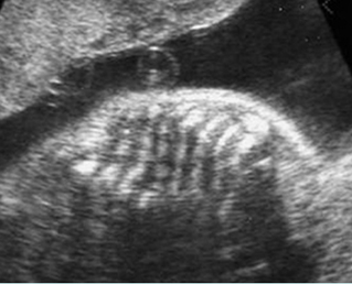
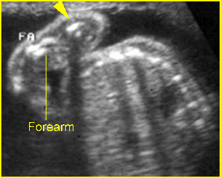
 Fig 1, Fig 2

  • Narrow and bell-shaped thorax, with short, horizontal ribs.
  • Normal or mildly shortened long bones, but not as short as those in short-rib polydactyly syndrome or thanatophoric dysplasia.
  • Polydactyly and cleft lip/palate in many cases.
  • Associated renal abnormalities.
  • Normal bone echogenicity.
  •  Increased nuchal translucency at late first trimester.
  • Associated anomalies: renal cystic dysplasia, pancreatic cyst.
  • Usually diagnosed in the second or third trimester, but diagnosis in the first trimester has been reported.
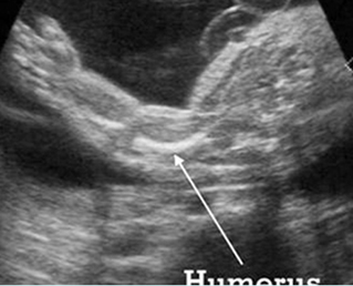
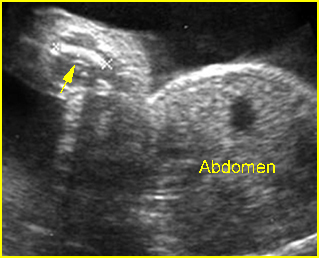
Fig 1:  Short humerus  Longitudinal scan of long bones: shortened but well ossified humerus of the fetus with Juene syndrome
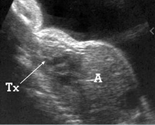
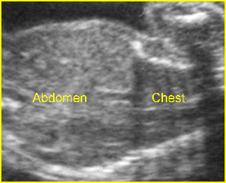
Fig 2:  Small thorax  Sagittal scan of the thorax and abdomen: disproportion in size of the thorax and abdomen of the fetus with Juene syndrome (Tx = chest, A = abdomen)

Management: Termination of pregnancy can be offered when diagnosed before viability.

Prognosis: Poor, but with a wide spectrum of severity from lethal to long-term survival.

Recurrence risk: Theoretically, the recurrent risk of these autosomal recessive disorders is 25%.
Short-rib polydactyly syndrome (SRP)

SRP is an autosomal recessive disorder characterized by short ribs, micromelia and polydactyly. SRP may be associated with a gene defect involving 4q13 or 4p16.

Incidence: Rare.

Sonographic findings:

  • Micromelia, usually severe.
  • Constricted thorax with severe short ribs.
  • Postaxial polydactyly.
  • Normal bone echogenicity.
  • Associated, genitourinary and gastrointestinal anomalies (Saldino-Noonan type).
  • 3D ultrasound may provide additional details.
  • SRP may be divided into three subtypes as follows (these subtypes are probably part of a continuous spectrum with variable expressivity):
    • associated cleft lip and palate (Majewski type)
    • associated renal abnormality (Naumoff type)
    •  associated cleft lip/palate, genitourinary, gastrointestinal anomalies (Beemer-Langer type).
  • Usually diagnosed in the second half of pregnancy but possible as early as 13 weeks.

 Management: Termination of pregnancy can be offered when diagnosed before viability.

Prognosis: Poor, but with a wide spectrum of severity from lethal to long-term survival.

Recurrence risk: Theoretically, the recurrent risk of these autosomal recessive disorders is 25%.

 Chondroectodermal Dysplasia (Ellis-van Creveld syndrome; EVC)

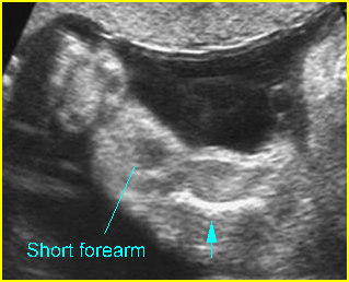
EVC is an autosomal recessive disorder characterized by short ribs, short limbs, polydactyly, dysplastic nails and teeth. EVC may be associated with a gene defect involving 4p16. DNA analysis for the first trimester diagnosis has been reported.

Incidence: Rare.

Sonographic findings:

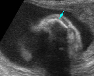
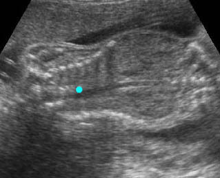
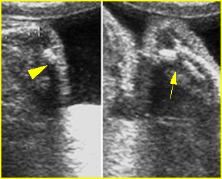
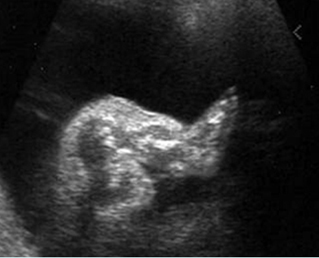
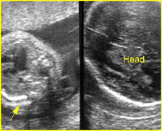
Fig 3, Fig 4, Fig 5

  • Acromesomelia with normal spine and skull.
  • Long and narrow thorax with short ribs.
  • Postaxial polydactyly.
  • Congenital heart defects in 60% of cases.
  • Normal bone echogenicity.
  • Usually diagnosed in the second half of pregnancy but possible to diagnose as early as the late first trimester.

 Management: Termination of pregnancy may be considered when diagnosed before viability. For the continuing pregnancy, ECV should not alter the standard obstetric management.

Prognosis: Depends on the severity, which varies from lethal to long-term survival. Overall the prognosis is rather good, but there is a significant mortality rate, due primarily to cardiorespiratory failure.

Recurrence risk: Theoretically, the recurrent risk of these autosomal recessive disorders is 25%.

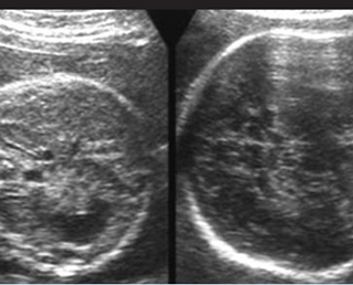
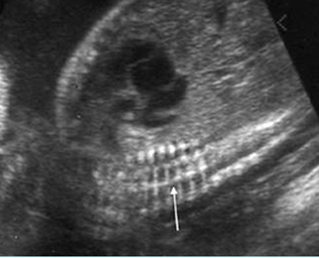
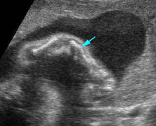
Fig 3:  Small thorax   Cross-sectional scan of the thorax and skull: disproportion in size of the thorax and head of the fetus with Ellis-van Creveld syndrome (arrow = short rib)
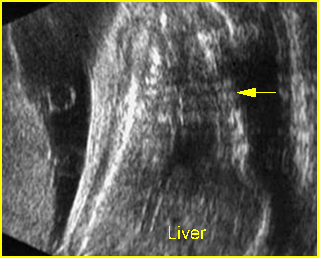
Fig 4:  Small thorax  Coronal scan: showing disproportion in size between thorax and abdomen of the fetus with Ellis-van Creveld syndrome
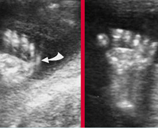
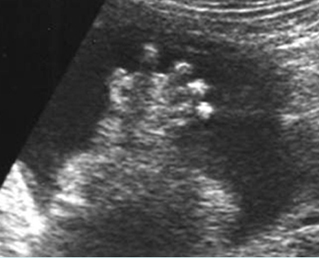
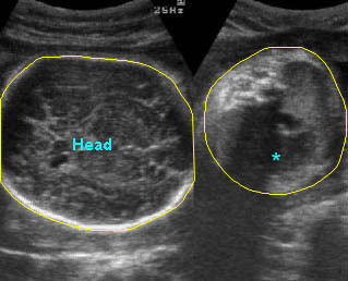
Fig 3:  Polydactyly

Video clips of hypoplastic thorax
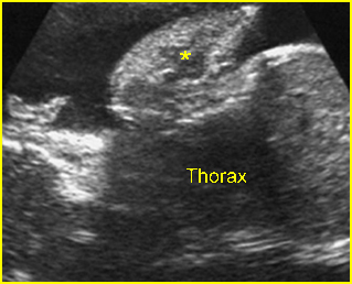
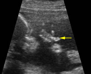
Lung hypoplasia / SRP Syndrome :  Small thorax (*) with short ribs compared to the head size in case of fetal short-rib polydactyly syndrome
Post-axial polydactyly
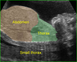
Short-rib polydactyly syndrome :  Markedly small thorax compared to the abdomen in case of short-rib polydactyly syndrome

Read More

Diastrophic Dysplasia

Diastrophic Dysplasia

Diastrophic dysplasia, an autosomal recessive disorder related to a mutation in the DTDST gene, is characterized by micromelia, clubfoot hand deformities, multiple joint flexion contractures, and scoliosis. DNA analysis for prenatal diagnosis is possible.

Incidence: Rare.

Sonographic findings:

Fig 1, Fig 2, Fig 3, Fig 4

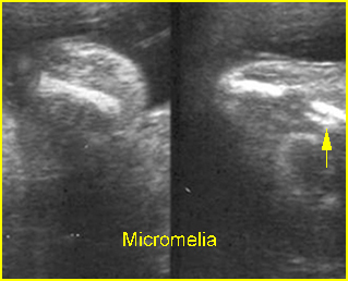
  • Micromelia, typically severe rhizomelic-type limb shortening and bowing of all long bones but a wide spectrum of severity.
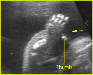
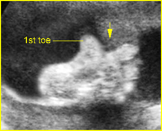
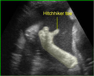
  • Hand deformities with abducted positions of the thumbs or toes called hitchhiker thumbs and toes which are relatively unique for diastrophic dysplasia; 3D ultrasound may provide better details.
  • Long bones are not bowed, unlike most other lethal syndromes.
  • Micrognathia, facial clefts and cardiac defects are occasionally seen.
  • Differential diagnoses include thanatophoric dysplasia, achondroplasia, arthrogryposis multiple congenita, atelosteosgenesis type II and pseudodiastrophic dysplasia.
  • Usually diagnosable in the second trimester, but diagnosis in the late first trimester has been reported.

Fig 1:  Hitchhiker thumbs  Abnormal posture and wide separation of the thumb (arrow) in the fetus with diastrophic dysplasia (25 weeks)

Fig 2:  Shortenings of long bones   Short humerus, ulna and radius but normal bone density in the fetus with diastrophic dysplasia

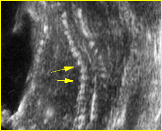
Fig 3:  Scoliosis  Coronal scan of the spine: scoliosis of thoracic spine in the fetus with diastrophic dysplasia

Fig 4:  Hitchhiker toe  Abnormal posture and wide separation of the toe (arrow) in the fetus with diastrophic dysplasia (26 weeks)

Video clips of diastrophic dysplasia

Hitchhiker toe  :  Persistent wide separation of the thumb and the second toe

Associations: Rare.

Management: Termination of pregnancy may be an option when diagnosed before viability. For the continuing pregnancy, diastrophic dysplasia should not alter the standard obstetric management.

Recurrence risk: Because it is an autosomal recessive disorder, the theoretical risk of recurrence is 25%.

Prognosis: Non-lethal, normal intellectual development, but a wide spectrum of morbidity.

Read More

Campomelic Dysplasia

Campomelic Dysplasia

Campomelic dysplasia is a rare lethal disorder characterized by bowing of long bones of the lower extremities, an enlarged and elongated skull with a peculiar small facies, and hypoplastic scapulae. This disorder is related to a single mutation in the SOX-9 gene located at 17q24, an SRY-related gene. An apparent preponderance of females with this condition is due to 46,XY male with sex reversal.

Incidence: Approximately 1 in 110,000 births.

Sonographic findings:

Fig 1, Fig 2, Fig 3

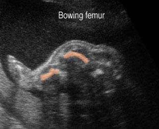
  • Bowing, sometimes angulation, of the femur and tibia.
  • Hypoplastic or absent fibulae is commonly seen.
  • Other tubular bones are normal in length.
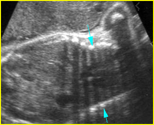
  • Bell-shaped narrow thorax.
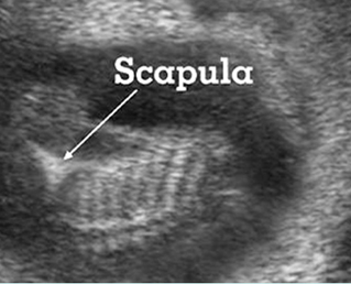
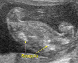
  • Hypoplastic scapulae.
  • Normal bone echogenicity.
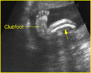
  • Occasional associated deformities: clubfeet, micrognathia, pyelectasis, and ventriculomegaly.
  • 3D ultrasound may provide information supplementary to that provided by 2D ultrasound for early diagnosis.
  • The main differential diagnoses include any cause with bowing long bones such as thanatophoric dysplasia, and variants of osteogenesis imperfecta.
  • Pitfalls: A variant of the same entity disorder may show no bowed limbs, a so-called acampomelic campomelic dysplasia.
  • Usually diagnosed in the second half of pregnancy.

Fig 1:  Campomelic dysplasia   Longitudinal scan of tibia: anterior bowing of well ossified tibia

Fig 2:  Hypoplastic scapula  Coronal scan of the scapula: scapula hypoplasia of the fetus with campomelic syndrome
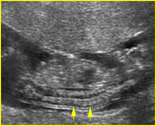
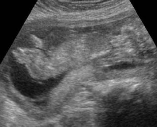
Fig 3:  Small thorax  Coronal scan of fetal trunk: disproportion of thorax (arrow) and abdomen size of the fetus with campomelic dysplasia

Video clips of campomelic dysplasia

Campomelia :  Anterior bowing of the tibia o (arrow)

Campomelia :  Coronal scan of the trunk:: markedly small thorax (solid circle)

Associations: Rare, several non-specific anomalies such as micrognathia, hydronephrosis and cardiac defects.

Management: Termination of pregnancy can be offered.

Prognosis: Lethal in most cases.

Recurrence risk: The majority of cases are caused by new mutations, therefore, the recurrence risk is rare.

Read More

Thanatophoric Dysplasia (TD)

Thanatophoric Dysplasia (TD)

TD is the most common lethal skeletal dysplasia in fetuses and neonates. It is caused by generalized disruption of the growth plate resulting from new autosomal dominant mutations in FGFR3. It is characterized by extreme rhizomelia, a normal trunk length with a narrow thorax, and a large head with a prominent forehead. Two subtypes have been identified:

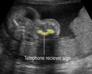
–     Type 1: severe rhizomelia, typical bowed “telephone receiver” femurs, narrow thorax, a relatively large head and absent cloverleaf skull.

–     Type 2: severe cloverleaf skull and short and straight long bones.

Prenatal diagnosis with DNA analysis is currently possible.

Incidence: 1 in 4000-15,000 births, relatively increased with paternal age.

Sonographic findings:

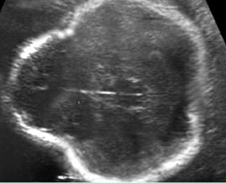
Fig 1, Fig 2, Fig 3, Fig 4, Fig 5, Fig 6

  • Normal bone echodensity.
  • Severe micromelia with typical telephone receiver femurs (type I).
  • Long bones may grow slowly and steadily at first and stop in late pregnancy.
  • Short and straight long bones (type II).
  • Short and broad tubular bones in hands and feet.
  • Relatively large calvarium with a prominent forehead.
  • Cloverleaf skull in about 14% of cases (all cases of type II).
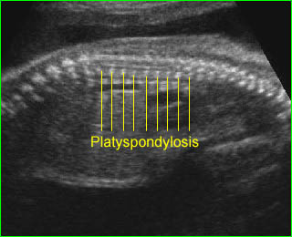
  • Platyspondylosis may be documented in some cases.
  • Polyhydramnios is nearly always present, especially in the third trimester.
  • Ventriculomegaly in some cases.
  • Increased NT in the late first trimester in some cases.
  • The main differential diagnoses include all short-limbed dwarfism with normal echodensity such as short rib polydactyly syndrome, homozygous achondroplasia, and asphyxiating thoracic dysplasia (slight shortening of long bones and normal vertebrae).
  • 3D ultrasound is helpful in providing more details such as the relative proportion of the appendicular skeletal elements and the hands and feet.
  • Pitfalls: A normal sonographic appearance in the late first trimester can not exclude the possibility of TD.
  • Usually diagnosed after the first trimester.

Fig 1:  Micromelia in Thanatophoric dysplasia  Severe shortenings of humerus (arrow) but normal ossification

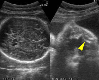
Fig 2:  Thanatophoric dysplasia   Cross-sectional scan of the skull: Cloverleaf skull; prominent parietal bone

Fig 3:  Large head   Cross-sectional scan of the abdomen and skull: disproportion in size of the trunk and head of the fetus with Thanatophoric dysplasia

Fig 4:  Micromelia in Thanatophoric dysplasia   Severe shortenings of humerus (arrow) but normal ossification

Fig 5:  Platyspondylosis   Oblique sagittal scan of the spine: relatively flattened vertebra (arrow) in case of Thanatophoric dysplasia

Fig 6:  Small thorax in Thanatophoric dysplasia   Coronal scan of fetal trunk: disproportionately small thorax, compared to the abdomen

Video clips of thanatophoric dysplasia (TD)

Long bone shortening :  Severe long bone shortening with bowing and normal echogenicity in case of thanatophoric dysplasia

Thanatophoric dysplasia :  Severe shortening of the long bone (arrowhead) compared to the skull

Thanatophoric dysplasia :  Shortened femur with bowing resulting in telephone receiver appearance

Thanatophoric dysplasia :  Platyspondylosis: flattened vertebral bodies

Associations: Hydrocephalus in some cases.

Management: Termination of pregnancy can be offered.

Prognosis: Lethal; in the report of one survivor, mental and bone maturation were severely retarded.

Recurrence risk: The majority of cases are caused by new mutations, therefore, the recurrence risk is rare.

Read More

Achondrogenesis

Achondrogenesis

Achondrogenesis is a lethal chondrodystrophy characterized by severe micromelia, short trunk, absent vertebral bodies ossification, and macrocrania. It has been classified into two types:

–     Type I: autosomal recessive type, the more severe form, accounting for 20% of cases, characterized by severe hypoossification and severe micromelia, and thin ribs with fractures (type IA) or not (type IB). Type 1B is associated with mutation in the gene for the DTDST gene on chromosome 5, the same gene identified in the cause of diastrophic dysplasia.

–     Type II (Langer-Saldino, autosomal dominant), accounting for 80% of cases, has relatively normal ossification, less severe micromelia, and thicker ribs without fractures. Type II is caused by a new mutation in the COL2A1 gene on chromosome 12, or inherited via germline mosaicism of the healthy parents.

Incidence: About 1 per 4000 births.

Sonographic findings:

Fig 1, Fig 2, Fig 3, Fig 4, Fig 5

  • Decreased mineralization of all or many vertebral bodies, sacrum and ischium. The absence of vertebral body ossification may be the unique finding of type II.
  • Severe micromelia with bowing, however, it can be found with normally developed extremities in rare cases.
  • Enlarged calvarium with poor ossification in type I but relatively normal ossification in type II.
  • Thin ribs (type I) with or without fractures.
  • Polyhydramnios is common.
  • Redundancy and edema of the subcutaneous tissues (pseudohydrops) or nuchal edema even in the first trimester.
  • 3D ultrasound is helpful in providing more details such as facial dysmorphism, the relative proportion of the appendicular skeletal elements and the hands and feet.
  • The main differential diagnosis includes thanatophoric dysplasia, achondroplasia, and short-rib polydactyly syndrome, all of which may have underdeveloped vertebral bodies, but none of these also have calvarial underossification. Severe hypophosphatasia may result in severe calvarial and spinal underossification, but hypophosphatasia usually has diffuse underossification of the spinal ossification centers or focal loss of the neural arch ossification centers as opposed to focal lack of vertebral body ossification that is seen in achondrogenesis.
  • Pitfalls: Normally developed extremities can be seen in type II. Therefore, complete examination of the vertebral body is very important.
  • First diagnosable in the late first trimester, but mostly in the second trimester.

Fig 1:  Spinal poor ossification   Mid-sagittal scan of the cervical spine: very sonolucent spine (arrow) in association with achondrogenesis

Fig 2:  Long bone shortening   Longitudinal scan of the thigh and leg: shortening of thigh and leg, compared with foot (achondrogenesis)

Fig 3:  Sonolucent and shortened arm   Longitudinal scan of the arm: Sonolucent and shortened humerus (*) in case of achondrogenesis

Fig 4:  Poor ossification of hand   Sonolucency of the hand bones as well as short fingers

Fig 5:  Poor ossification   Sonolucency of the hand bones as well as short fingers (left) and short humerus (right)

Video clips of achondrogenesis

Achondrogenesis :  Mid-sagittal view of the spine: extremely poor ossification of the spine (arrow)

Achondrogenesis :  Shortened long bones with poor-ossified bone (arrow) and redundant skin

Associations: Rare, cephalocele, cystic hygroma, and polydactyly in some cases.

Management: Termination of pregnancy can be offered.

Prognosis: Lethal.

Recurrence risk: Type IB is autosomal dominant, and carries a theoretical recurrence risk of 25%. The recurrence risk of type II, new mutations, is rare.

Read More

Congenital Hypophosphatasia

Congenital Hypophosphatasia

Congenital hypophosphatasia is a rare autosomal recessive inherited disorder characterized by demineralization of bones with low alkaline phosphatase in serum and other tissues. It is related to the numerous tissue non-specific alkaline phosphatase genes on chromosome 1. Prenatal diagnosis may be made using ultrasound and by assaying alkaline phosphatase in tissue obtained from chorionic villous samplings or in blood from cordocentesis, and can also be made by DNA analysis.

Incidence: 1 per 100,000 births.

Sonographic findings:

Fig 1, Fig 2, Fig 3, Fig 4, Fig 5, Fig 6

  • Variable hypomineralization, boneless appearance in some cases.
  • Compressible skull with complete absence of acoustic shadow, and probably so severe that the normally-difficult-to-visualize near-field brain is easily seen.
  • Ossification centers in both the vertebral bodies and neural arches may be absent, or there may be preservation of some vertebral body ossification centers, unlike achondrogensis in which the vertebral body ossification centers are absent with preserved neural arch ossification centers.
  • Severe micromelia.
  • Thin or delicate or seemingly absent long bones.
  • Spurs along the midshaft of long bones and at the knees and elbows.
  • The main differential diagnosis is OI type II, in particular type IIC which involves similarly thin bones. All three forms of OI type II have multiple fractures and wavy or wrinkled bones that should help distinguish them from hypophosphatasia.
  • Increased nuchal translucency thickness during late first trimester.
  • Pitfalls: It is probably indistinguishable from OI type II and achondrogenesis. However, they have the same lethal prognosis.
  • Extremely low levels of alkaline phosphatase in the cord blood can confirm the diagnosis.
  • Usually diagnosed in the second half of pregnancy, but possible as early as the late first trimester.

Fig 1:  Hypophosphatasia    Longitudinal scan of the forearm and hand: poorly ossified radius and ulna as well as hand bones and clubhand

Fig 2:  Sonolucent spine   Sagittal scan of the fetus at 14 weeks: sonolucent vertebrae of the fetus with hypophosphatasia

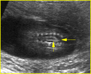
Fig 3:  Hypophosphatasia  Oblique sagittal scan of the spine: poorly ossified spine (arrow), only 3 vertebral bodies are ossified

Fig 4:  Hypophosphatasia   Cross-sectional scan of the spine: poorly ossified posterior ossification centers but normally ossified anterior center (arrow)

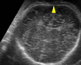
Fig 5:  Hypophosphatasia   Cross-sectional scan of skull: thin and sonolucent calvarium (arrow = falx cerebri)

Fig 6:  Hypophosphatasia   Longitudinal scan of long bones: shortened and poorly ossified bones

Video clips of congenital hypophosphatasia

Hypophosphatasia :  Sagittal scan of the fetus at 14 weeks; poorly ossified bony structures

Hypophosphatasia :  Transverse section at the upper thorax: shortenings and poorly ossified long bones

Hypophosphatasia :  Transverse scan of the skull: brachycephaly with sonolucent skull

Hypophosphatasia :  Early recognition of limb shortenings with poorly ossified bones at 13 weeks of gestation

Associations: Rare.

Management: Termination of pregnancy can be offered.

Prognosis: Lethal, though successful treatment with high-dose pyridoxal phosphate in some non-lethal cases has been reported.

Recurrence risk: 25%, an autosomal recessive disorder.

Read More

Osteogenesis Imperfecta (OI)

Osteogenesis Imperfecta (OI)

OI is genetically a heterogeneous disorder consisting of both autosomal dominant and autosomal recessive entities caused by mutations in one or two structural genes for type I procollagen. The clinical heterogeneity is due to the different mutations in the genes: COL1A1 and COL1A2. OI is divided into four types as follows:

  • Type I (autosomal dominant) is characterized by bone fragility, blue sclera, hearing loss, and normal calvarium, with fractures ranging from none to multiple. DNA-based analysis is possible for early prenatal diagnosis.
  • Type II (new dominant mutations and autosomal recessive in <5%) is a perinatal lethal variety characterized by almost no ossification of the skull, beaded ribs, shortened crumpled long bones and multiple fractures in utero.
  • Type III (autosomal recessive, rare) is a non-lethal variety characterized by blue sclera (becoming white with time) and multiple fractures present at birth.
  • Type IV (autosomal dominant) is the mildest form (mild to moderate osseous fragility). Long bones and sclera are normal.

Incidence: 0.4 per 10,000 births for OI, and 0.2 per 10,000 births for OI type II.

Sonographic findings (OI type II) :

Fig 1, Fig 2, Fig 3, Fig 4, Fig 5, Fig 6

  • Variable hypomineralization, which may result in complete absence of acoustic shadow and may be so severe that the normally-difficult-to-visualize near-field brain is easily seen.
  • Micromelia (type IIB and IIC may be less severe).
  • Fractures of long bones.
  • Thickened long bones due to frequent fractures and secondary callus formation.
  • Small bell-shaped thorax with multiple rib fractures or beaded ribs.
  • Increased nuchal translucency thickness in the first trimester in some cases.
  • MRI complemented sonography may be helpful in further differentiating clinical and sonographic findings.
  • Sonographic features are variable among the three subtype as follows:
    • Type IIA: thick bones, multiple fractures, hypomineralization, beaded ribs
    • Type IIB: thick bones, multiple fractures, less beading of ribs; lower extremities are affected more than upper extremities
    • Type IIC: thin bones, multiple fractures, thin beaded ribs.
  • Pitfalls: Variable short limb lengths without fractures may be the only clue in the mild form.
  • Usually diagnosed in the second and third trimesters but possible in the late first trimester.

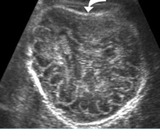
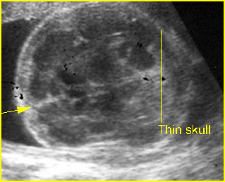
Fig 1:  Sonolucent and compressible skull   Poorly ossified and compressible cranium (arrow), cerebral sulci and gyri could easily be seen

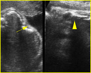
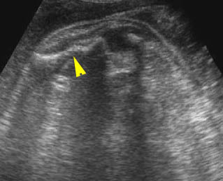
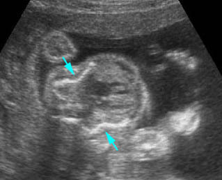
Fig 2:  Rib fractures   Multiple rib fractures with poor ossification in the fetus with osteogenesis imperfecta type IIA

Fig 3:  Micromelia   Irregularity and severe shortening of long bone (arrowhead)

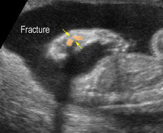
Fig 4:  Fracture in utero   Longitudinal scan of upper extremity: poorly ossified and fracture in osteogenesis imperfecta type IIA

Fig 5:  Osteogenesis imperfecta type IIA   Longitudinal scan of lower extremity: shortened and irregular ossified long bones

Fig 6:  Fracture of long bones   Longitudinal scan of rib and humerus: fracture rib (arrow) and humerus (arrowhead) with moderate ossification

Video clips of osteogenesis imperfecta (OI)

Osteogenesis imperfecta (IIA) :  Irregular long bone (arrowhead) with poorly ossified

Osteogenesis imperfecta (IIA) :  Cross-sectional scan of the thorax: rib fractures (arrow)

Osteogenesis imperfecta:  Long bone fracture of the forearm

Fracture in utero :  Longitudinal scan of upper limb: callus formation (arrow) and irregularity of ulna secondary to previous fracture

Sonolucent skull :  The thin skull is so poorly ossified that cerebral gyri and sulci could be seen easily

Associations: Rare, though concurrent anencephaly has been reported.

Management: Termination of pregnancy can be offered. For term pregnancy, cesarean delivery does not decrease fracture rates at birth in infants with non-lethal forms of OI.

Prognosis: OI type II is lethal.

Recurrence risk: Unlikely because OI type II is due to new mutations in most cases and there are only a few reports of autosomal recessive inheritance.

Read More

Spinal Dysraphism

Spinal Dysraphism

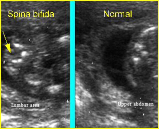
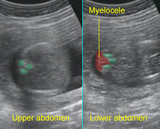
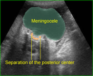
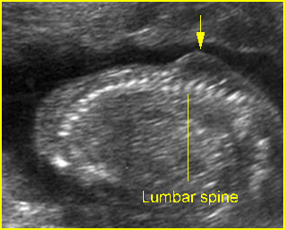
Spinal dysraphism, splaying of the posterior ossification centers of the spine, usually indicates spina bifida resulting from failure of closure of the posterior neuropore. However, spinal dysraphism can be due to other abnormalities as follows:

Fig 1, Fig 2, Fig 3

Major differential diagnoses

  • Spina bifida
    • meningocele
    • meningomyelocele
    • myeolochisis
  • Arnold-Chiari malformation type II
  • Pseudodysraphism: technical pitfalls (described previously)

Minor differential diagnoses

  • Diastematomyelia: a splitting of the spinal cord
  • Lipomyelomeningocele: asymmetric lesion of lipoma originating in the spinal cord or cauda equina
  • etc.

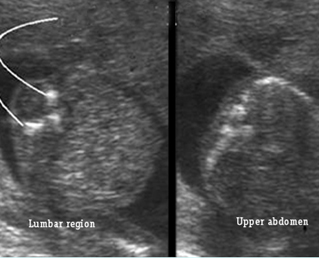
Fig 1:  Spina bifida   Cross-sectional scan of the lumbar and thoracic spine: separation of posterior ossification centers at the lumbar region with small protrusion of the overlying tissue

Fig 2:  Spina bifida   Coronal scan of the lumbosacral spine: separation of the ossification centers of the spine (arrow)

Fig 3:  Spina bifida  Cross-sectional scan of the lumbar and thoracic spine: separation of posterior ossification centers at the lumbar region with small protrusion of the overlying tissue (arrow)

Video clips of spinal dysraphism

Spina bifida :  Cross-sectional scan of the normal spine at the level of upper abdomen, but wide separation of the posterior ossification centers at the lumbar spine with the protrusion of the spinal cord

Large meningocele :  Cross-sectional scan of the normal spine at the level of upper abdomen, but wide separation of the posterior ossification centers at the lumbar spine with the protrusion of the meningeal sac

Read More

Masses on the Back

Masses on the Back

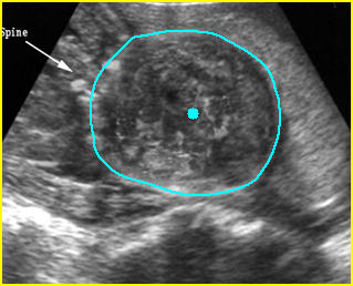
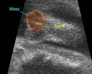
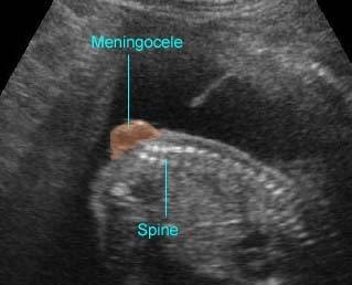
Lumbosacral masses can be caused by a number of specific anomalies, especially meningomyelocele, which is the most common cause. The differential diagnosis for lumbosacral mass can be summarized as follows:

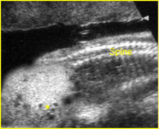
Fig 1, Fig 2, Fig 3

Major differential diagnoses

Meningomyelocele (most common)

  • spinal dysraphism
  • solid-cystic mass located at the lumbosacral spine in most cases
  • associated cranial signs of spina bifida, such as lemon sign, banana sign, and ventriculomegaly

Sacrococcygeal teratoma (SCT) (uncommon)

  • solid-cystic, solid predominantly in most cases but entirely cystic in 15% of cases
  • high vascularization
  • intra-abdominal components in most cases with a displacement effect on internal structures
  •  usually located in the sacrococcygeal area

Limb-body wall complex (uncommon)

  • solid-cystic asymmetric mass
  • abnormal spinal curvature
  • no specific location
  • severe abdominal wall defects
  •  limb defects
  • no or very short umbilical cord

Amniotic band syndrome (uncommon)
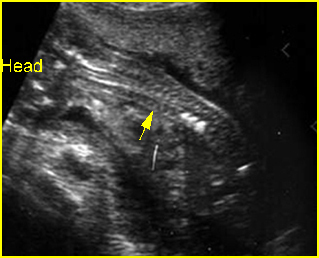
  • solid-cystic asymmetric mass
  • no specific location
  • associated limb reduction/constriction defects
  • amniotic band in the amniotic cavity.

Minor differential diagnoses

  • Artifacts: extrafetal mass, such as chorioangioma attached to the fetal back
  • Rare tumors: lipomas, lipomyelomeningocele, and large hemangioma.

Fig 1:  Sacrococcygeal teratoma   Sagittal scan of the spine: abnormal complex solid mass (*) beneath the sacral spine

Fig 2:  Large meningomyelocele   Scan of lower spine: large solid mass with heterogeneous echodensity (solid circle) (arrow = sacrum)

Fig 3:  Lumbar meningocele   Sagittal scan of the spine: small solid mass protruding from lumbar region

Video clips of masses on the back

Sacrococcygeal Teratoma :  Mass on the back: the complex cyst at the caudal end of the spine

Spina bifida :  Sagittal scan of the fetal spine showing sacral meningocele

Read More

Abnormal Spinal Curvature

Abnormal Spinal Curvature

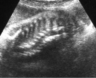
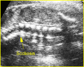
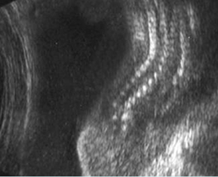
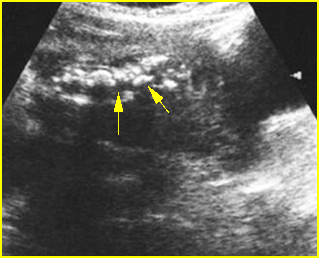
These abnormalities include kyphoscoliosis, hemivertebrae, angulations or disorganized vertebral structures. Scoliosis, an abnormal lateral deflection of the spinal column, is the most common in this category.

Fig 1, Fig 2, Fig 3, Fig 4

Sonographic differential diagnosis:

  • Normal
  • Meningomyelocele (common)
  • LBWC (uncommon)
  • Amniotic band syndrome (uncommon)
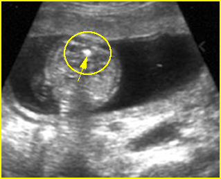
  • Skeletal dysplasia
  • VATER or VACTERL associations (rare)
  • Isolated hemivertebrae
  • Iniencephaly (very rare)
  • Caudal regression syndrome
Note:

  • Hemivertebrae are an example of congenital scoliosis resulting from failure of segmentation, which is mostly associated with other anomalies.
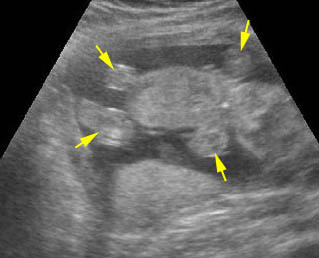
  • The normal flexible skeleton can be subject to strong deformation forces in utero resulting in transient scoliosis.
  • Gross skeletal defects, including absent ribs or hemivertebrae, associated with lateral spinal deflection probably represent truly pathologic scoliosis.

Fig 1:  Scoliosis   Coronal scan of the spine: scoliosis of spine at the thoracic region (arrow)

Fig 2:  Angulation of spine   Coronal scan of the spine: angulation of spine at the thoraco-lumbar region (arrow)

Fig 3:  VATER associations   Coronal scan of the spine: scoliosis

Fig 4:  Disorganized spine :   Coronal scan of the spine: scoliosis and disorganized

Video clips of abnormal spinal curvature

Limb-body wall complex:  Coronal scan of the fetal spine: scoliosis associated with limb body wall complex

Severe kyphoscoliosis :  Severe kyphoscoliosis in case of iniencephaly

Read More