Masses on the Back
Lumbosacral masses can be caused by a number of specific anomalies, especially meningomyelocele, which is the most common cause. The differential diagnosis for lumbosacral mass can be summarized as follows:
Fig 1, Fig 2, Fig 3
Major differential diagnoses
Meningomyelocele (most common)
- spinal dysraphism
- solid-cystic mass located at the lumbosacral spine in most cases
- associated cranial signs of spina bifida, such as lemon sign, banana sign, and ventriculomegaly
Sacrococcygeal teratoma (SCT) (uncommon)
- solid-cystic, solid predominantly in most cases but entirely cystic in 15% of cases
- high vascularization
- intra-abdominal components in most cases with a displacement effect on internal structures
- usually located in the sacrococcygeal area
Limb-body wall complex (uncommon)
- solid-cystic asymmetric mass
- abnormal spinal curvature
- no specific location
- severe abdominal wall defects
- limb defects
- no or very short umbilical cord
Amniotic band syndrome (uncommon)
- solid-cystic asymmetric mass
- no specific location
- associated limb reduction/constriction defects
- amniotic band in the amniotic cavity.
Minor differential diagnoses
- Artifacts: extrafetal mass, such as chorioangioma attached to the fetal back
- Rare tumors: lipomas, lipomyelomeningocele, and large hemangioma.
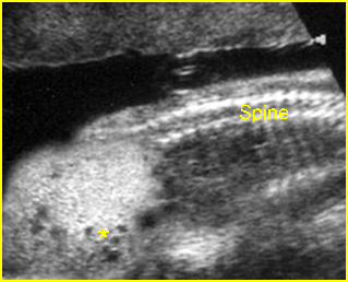
Fig 1: Sacrococcygeal teratoma Sagittal scan of the spine: abnormal complex solid mass (*) beneath the sacral spine
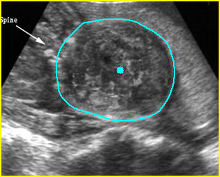
Fig 2: Large meningomyelocele Scan of lower spine: large solid mass with heterogeneous echodensity (solid circle) (arrow = sacrum)
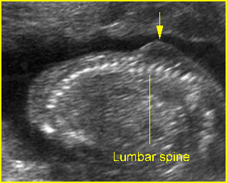
Fig 3: Lumbar meningocele Sagittal scan of the spine: small solid mass protruding from lumbar region
Video clips of masses on the back
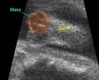
Sacrococcygeal Teratoma : Mass on the back: the complex cyst at the caudal end of the spine
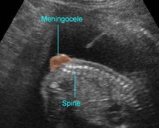
Spina bifida : Sagittal scan of the fetal spine showing sacral meningocele

