Abnormal Hands and Feet

Abnormal Hands and Feet

Hands and feet should be examined to exclude polydactyly, brachydactyly, abnormal hand posture, sandal gap, clinodactyly, Rocker-bottom foot, clubfoot, etc. These abnormalities are commonly part of several syndromes.

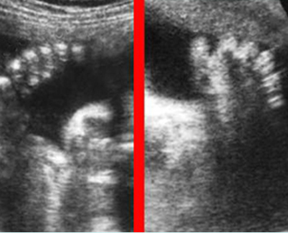
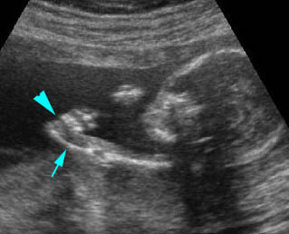
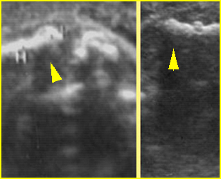
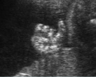
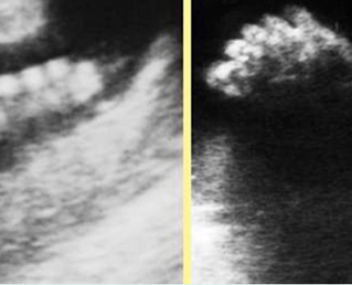
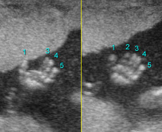
Polydactyly Fig 1

Polydactyly is the presence of an additional digit, which can be postaxial (on the ulnar or fibular side) or preaxial (on the radial or tibial side) and with a central form. This extra digit may range from a fleshy nubbin to a complete digit. Postaxial polydactyly is the most common form and is inherited autosomal dominantly as an isolated finding in most cases. However, it is significantly related to some syndromes, especially trisomy 13 or short-rib polydactyly syndrome. Preaxial polydactyly, especially a triphalangeal thumb, is most likely to be part of a syndrome.

The major differential diagnoses for polydactyly include

  • Isolated polydactyly
  • Chromosome abnormalities, especially trisomy 13
  • Meckel Gruber syndrome
  • Asphyxiating thoracic dystrophy
  • Short-rib polydactyly syndrome
  • Achondroectodermal dysplasia (Ellis-Van Creveld syndrome)
  • Smith-Lemli-Opitz syndrome.

Fig 1:  Polydactyly
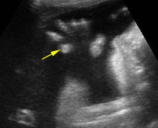
Post-axial polydactyly
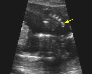
Polydactyly :  Post-axial polydactyly (fleshy finger)
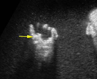
Pre-axial polydactyly

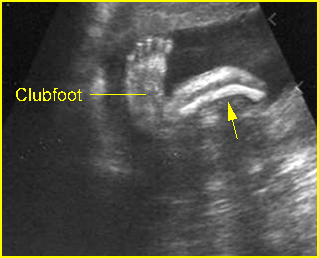
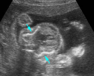
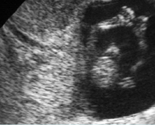
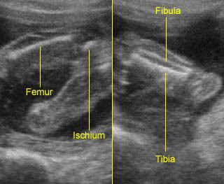
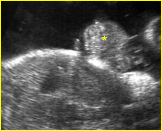
Clubfoot Fig 2

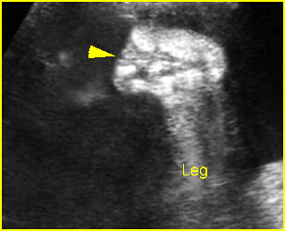
A clubfoot is one of the most common congenital anomalies, occurring in 1:250 to 1 in 1000 births. In 95% of cases, the sole is turned medially (talipes equinovarus). This can be caused by external compression in the case of oligohydramnios or by internal factors including abnormal bone formation, spina bifida, muscular defects or genetic causes (15% have a family history of clubfoot). Most clubfeet are found in otherwise normal infants. However, about 10% of cases are associated with several syndromes including Pena-Shokier phenotype, trisomy 18 and 13, etc.

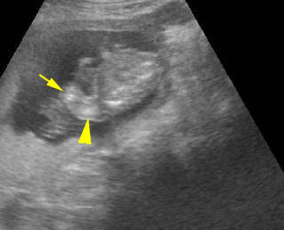
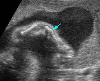
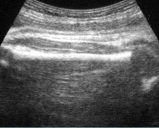
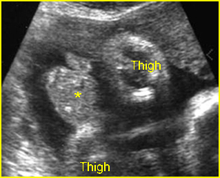
Rocker bottom foot Fig 3

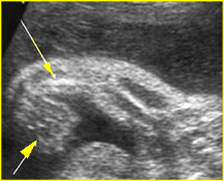
Vertical talus, or eversion of the planar arch, produces a rocker-bottom (convex outward) appearance of the bottom of the foot. This is most often associated with chromosomal abnormalities, particularly trisomy 13 and 18, although it may also be seen as an isolated anomaly, with caudal dysplasia sequence, neural tube defects, and neuromuscular disorders, and as part of the Potter sequence
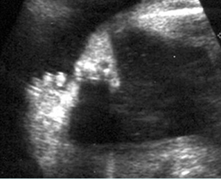
Fig 2:  Clubfoot  Longitudinal scan of the lower leg: plantar view of the foot seen in the same plane of longitudinal view of tibia and fibula
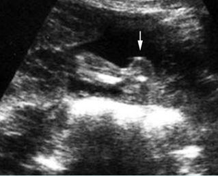
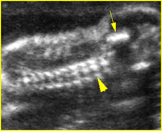
Fig 3:  Rocker bottom foot   Prominent calcaneous bone (arrow) associated with trisomy 13
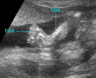
Clubfoot :  The plantar view of the foot can be demonstrated on the same plane of the longitudinal view of the tibia
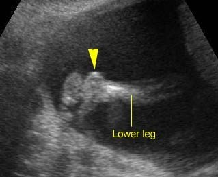
Rocker-bottom foot :  Sagittal scan of the fetal foot showing the prominent heel

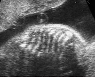
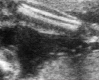
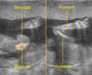
Abnormal hand posture Fig 4, Fig 5, Fig 6

Fetal hand malformation may be isolated or associated with chromosomal abnormalities, limb reduction defect, a neuromuscular disorder, or skeletal dysplasia. Ultrasound evaluation of the fetal hand requires assessment of the number and configuration of the fingers, the presence and position of the thumb, and the relationship of the thumb and fingers to each other and to the hand and wrist. The phalanges should be evaluated in the long-axis rather than the short-axis view. Short-axis views may represent the metacarpals rather than the phalanges. The normal fetal hand is most often in a resting position with loosely curled fingers, which the fetus periodically opens. Sonographic assessment requires skill on the part of the examiner in following the movement of the hand and documenting the extended fingers and thumb should the hand open during the examination.

Clenched hands Fig 7, Fig 8

Clenched hands or abnormal hand postures are often related to other abnormalities and more than half of cases have aneuploidy, predominantly trisomy 18 (88% of aneuploid fetuses).
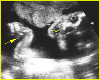
Fig 4:  Pena-Shokier phenotype  Fixed flexion of both elbow and wrist joint (arrow) as well as persistent mouth opening (*)
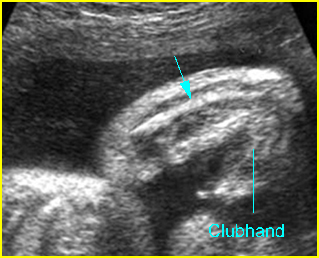
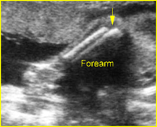
Fig 5:  VATER associations  Longitudinal scan of forearm: absent radius with abnormal hand posture
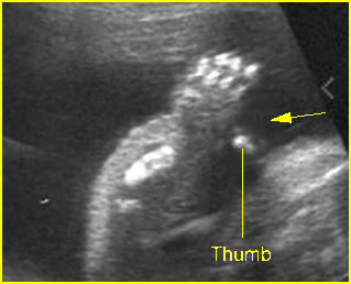
Fig 6:  Hitchhiker thumbs   Abnormal posture and wide separation of the thumb (arrow) in the fetus with diastrophic dysplasia (25 weeks)
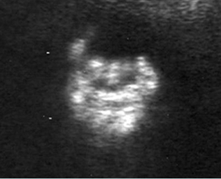
Fig 7:  Clenched hand   Overlapping fingers of the fetus with trisomy 18
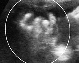
Fig 8: Clenched hand   Overlapping fingers of the fetus with trisomy 18
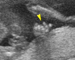
Overlaping fingers :  Clenched hand (arrowhead) in trisomy 18
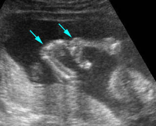
Fixed flexion:  Fixed flexion of the wrist joint in association with trisomy 18
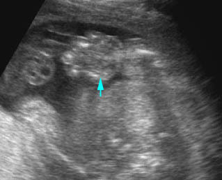
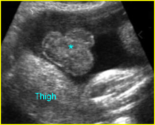
Edematous hand

Clinodactyly Fig 9, Fig 10

Clinodactyly is the permanent curvature or deflection of one or more fingers. It is frequently associated with abnormal chromosomes especially trisomy 21.

Syndactyly

Syndactyly is the fusion of digits and may consist of bony or soft tissue fusion. This may also be difficult to detect sonographically, especially those cases consisting of soft tissue fusion. Syndactyly is associated with a number of syndromes. It may be seen in the SRPD syndromes with the characteristics of polydactyly, and it may be seen in triploidy or Down’s syndrome.

Fig 9: Clinodactyly   Curved-in position of the fifth finger (arrow) of the fetus with trisomy 21
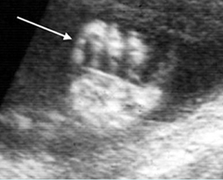
Fig 10:  Clinodactyly  Scan of the hand: disproportionately small middle phalange of the fifth finger (arrow)
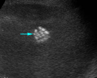
Clinodactyly :  Clinodactyly with hypoplasia of the middle phalanx of the fifth finger

Read More

Limb Reduction Defects

Limb Reduction Defects

Limb reduction deformities, presenting alone or as part of a specific syndrome, occur in approximately 0.5 per 10,000 births. Half of them are simple transverse reduction deficiencies of one forearm or hand without associated anomalies and the remaining half have additional anomalies.

Fig 1, Fig 2, Fig 3

Isolated limb reduction:

  • isolated limb deficiency more commonly occurs in the upper extremities rather than the leg, which generally occurs within the context of a syndrome, as do bilateral amputations or reduction of all limbs
  • sporadic occurrence in most cases with negligible risk of recurrence

Limb reduction associated amniotic band:

  • secondary to entanglement with mesodermic bands derived from the chorionic surface of the amnion after the latter has separated from the chorion
  • isolated or multiple limb reductions, with or without other defects

Phocomelia:

  • typically hands and feet are present, but the intervening arms and legs are absent
  • main differential diagnoses include Robert’s syndrome, variants of TAR syndrome, and Grebe’s syndrome
  • can be caused by exposure to thalidomide but this is only of historical interest

Radial ray defects: radial clubhand may be part of

  • TAR syndrome (thrombocytopenia with absent radius)
  • Fanconi’s syndrome: an autosomal recessive disorder consisting of pancytopenia anemia, leukopenia and thrombocytopenia and skeletal anomalies, especially radial clubhand
  • Aase’s syndrome: an autosomal recessive disorder characterized by hypoplastic anemia and radial clubhand with bilateral triphalangeal thumb and a hypoplastic radius
  • Holt-Oram syndrome: an autosomal dominant disorder characterized by congenital heart defects, radial hypoplasia and triphalangeal or absent thumb
  • VATER association: a sporadic disorder resulting from defective mesodermal development during embryogenesis consisting of vertebral segmentation (70%), anal atresia (80%), tracheoesophageal fistula (70%), esophageal atresia, and radial (65%) and renal defects (53%)
  • Goldenhar’s syndrome: characterized by hemifacial microsomia, hypoplasia of the malar and maxillary or mandibular region, vertebral anomalies, and radial defects
  • Klipple-Feil syndrome
  • Chromosomal abnormalities, particularly trisomy 18 and 21.

Fig 1:  Transverse limb reduction  Scan of the foot: absent distal end of foot

Fig 2:  Absent radius syndrome  Longitudinal scan of upper limb (15 weeks): short ulna (arrow) without radius with fixed flexion of wrist joint (arrowhead)

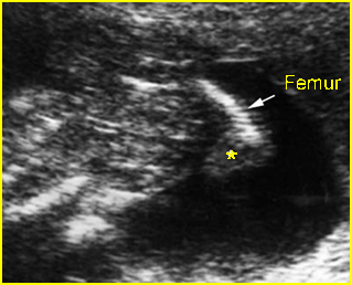
Fig 1:  Limb reduction   Longitudinal scan of lower limb: absent tibia and fibula, abnormal foot (*) arising from femur (arrow) in the fetus with VATER association

Video clips of limb reduction defects

Radial ray defects :  Longitudinal scan of the upper extremity: absent radius (arrow) with fixed flexion of the wrist joint (arrowhead)

Radial ray defects :  Longitudinal scan of the upper extremity at 13 weeks: absent radius (with fixed flexion of the wrist joint (arrow)

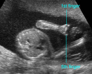
Transverse limb defect :  Absent fingers; only the 1st and 5th fingers remained

Transverse limb defect :  Absent hand with shortenings of all long bones (14 weeks) (arrow = severely shortened forearm, arrow = humerus)

Read More

Long Bone Bowing

Long Bone Bowing

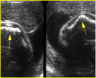
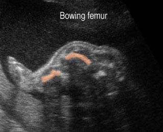
The degree of long-bone curvature should be examined. Campomelic (bent-bow) dysplasia is characterized by ventral bowing of the long bones. Thanatophoric dysplasia and osteogenesis imperfecta also have bowed extremities.

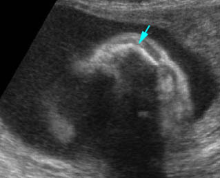
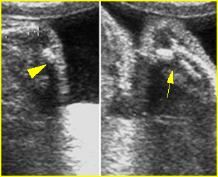
Fig 1, Fig  2

The major differential diagnoses are as follows:

  • Campomelic dysplasia (bowing but no shortening)
  • Osteogenesis imperfecta type II (related to fracture and callus formation)
  • Thanatophoric dysplasia (severe shortened long bones)
  • Achondrogenesis
  • Hypophosphatasia

Fig 1:  Campomelic dysplasia   Longitudinal scan of tibia: anterior bowing of well ossified tibia

Fig 2:  Osteogenesis imperfecta type IIB  Irregular and angular fracture of long bones (arrow) but rather well-ossified

Video clips of fracture in utero

Long bone shortening :  Severe long bone shortening with bowing and normal echogenicity in case of thanatophoric dysplasia

Campomelia :  Anterior bowing of the tibia (arrow)

Read More

Fracture In Utero

Fracture In Utero

The detection of rib or long bone fractures in association with severe micromelia suggests a diagnosis of osteogenesis imperfecta. Fractures may be subtle or may lead to angulation.

Fig 1, Fig2, Fig 3

The major differential diagnoses for fractures in utero are as follows:

  • Osteogenesis imperfecta type II (most common)
  • Hypophosphatasia (rare)
  • Campomelic dysplasia (not a true fracture but bowing).

Fig 1:  Fracture in utero  Longitudinal scan of upper extremity: poorly ossified and fracture in osteogenesis imperfecta type IIA

Fig 2:  Callus formation of long bone  Longitudinal scan of humerus and femur: irregularity due to callus (arrowhead) in osteogenesis imperfecta type IIA

Fig 3:  Rib fractures  Multiple rib fractures with poor ossification in the fetus with osteogenesis imperfecta type IIA

Video clips of fracture in utero

Fracture in utero :  Longitudinal scan of upper limb: callus formation (arrow) and irregularity of ulna secondary to previous fracture

Osteogenesis imperfecta :  Cross-sectional scan of the thorax: rib fractures (arrow)

Read More

Sonolucent Bones

Sonolucent Bones

Diffuse demineralization of the skull and long bones almost always occurs with fetal skeletal dysplasia syndromes. In the case of severe demineralization of the bony calvarium, the cranium is thin without an acoustic shadow and so poorly ossified that the intracranial structure can easily be seen. This increased visualization of the intracranial structures may be confused with such abnormalities as exencephaly due to acrania or acalvaria. Unlike exencephaly, however, there is an intact but poorly ossified skull. Careful scanning reveals concomitant limb anomalies.

Fig 1, Fig 2, Fig 3, Fig 4

The main differential diagnoses of the sonolucent skull are as follows:

  • Osteogenesis imperfecta (most common)
  • Hypophosphatasia (rare)
  • Achondrogenesis type I (rare).

Fig 1:  Hypophosphatasia  Longitudinal scan of long bones: shortened and poorly ossified bones

Fig 2:  Poorly ossified and compressible skull   Cross-sectional scan of skull: deformable skull with sonolucency in osteogenesis imperfecta type IIA

Fig 3:  Micromelia in Thanatophoric dysplasia   Severe shortenings of long bones (arrow) but normal ossification

Fig 4:  Sonolucent and shortened arm   Longitudinal scan of the arm: Sonolucent and shortened humerus (*) in case of achondrogenesis

Video clips of sonolucent bones

Sonolucent skull : The thin skull is so poorly ossified that cerebral gyri and sulci could be seen easily

Achondrogenesis:  Mid-sagittal view of the spine: extremely poor ossification of the spine (arrow)

Read More

Bone Shortening

Bone Shortening

All long bones should be measured in each extremity, although subjective evaluation may be sufficient in most cases. The severity of bone shortening varies from extreme to mild.

Fig 1, Fig 2, Fig 3

The major differential diagnoses of bone shortenings are as follows:

Severe shortening

  • thanatophoric dysplasia (micromelia) (common)
  • osteogenesis imperfecta type II (micromelia) (common)
  • achondrogenesis (micromelia) (common)
  • short-rib polydactyly syndrome (uncommon)
  • diastrophic dysplasia (uncommon)
  • homozygous achondroplasia (uncommon)
  • mesomelic dysplasia (uncommon)
  • chondrodysplasia punctata, rhizomelic type

Mild-to-moderate shortening

  • osteogenesis imperfecta type II (common)
  • heterozygous achondroplasia (common)
  • asphyxiating thoracic dysplasia (common)
  • campomelic dysplasia (uncommon)
  • hypophosphatasia (uncommon)
  • chondroectodermal dysplasia (uncommon).

Fig 1:  Long bone shortening   Longitudinal scan of the thigh and leg: shortening of thigh and leg, compared with foot (achondrogenesis)

Fig 2:  Short humerus  Longitudinal scan of long bones: shortened but well ossified humerus of the fetus with Juene syndrome

Fig 3:  Micromelia in Thanatophoric dysplasia   Severe shortenings of long bones (arrow) but normal ossification

Video clips of bone shortening

Thanatophoric displasia:  Severe shortenings of the long bone (arrowhead) compared to the skull

Long bone shortening : Severe long bone shortening with bowing and normal echogenicity in case of thanatophoric dysplasia

Read More

Normal Examination Spine

Normal Examination

Normal Spine

Fig 1, Fig 2, Fig 3, Fig 4, Fig 5, Fig 6

–     Mineralization of the spine begins at 8 weeks of gestation. The three ossification centers of individual vertebrae include a single ventral center for the vertebral body (centrum) and two paired dorsal centers that will become the lateral masses and the posterior arch. They are well visualized from 15 to 16 weeks onwards. However, spina bifida can be detected much earlier. The posterior ossification centers begin at the base of the transverse processes. As ossification progresses, the laminae become visible, usually after 19 weeks. The inward angulation of the normal laminae is the opposite of the outward splaying of the laminae seen in spina bifida, an optimal situation for detecting this anomaly. The arch of the upper sacral region is not consistently recognizable until after 25 weeks.

  • Technique of examination: The spine can be systematically examined during the second and third trimesters as follows:
    • Adjust the transducer on the maternal abdomen to achieve a cross-section image of the fetal abdomen or thorax.
    • Identify the location of the fetal spine, noting the acoustic shadow behind the spine, and carefully adjust the transducer to demonstrate all three ossification centers.
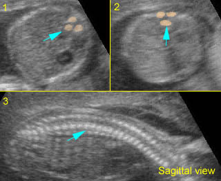
    • To obtain the sagittal view, from the previous image, make an attempt to bring both posterior centers to the middle of the image, lying horizontally or perpendicular to the ultrasound beam, and then rotate the transducer 90 degrees and meticulously adjust it to obtain the best image of the coronal view.
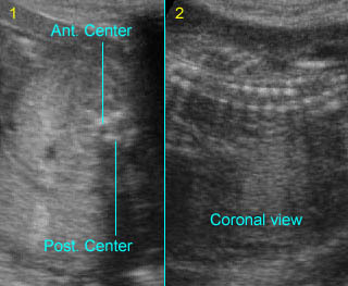
    • To obtain the coronal view, from the cross-section image, make an attempt to bring both posterior centers to the middle of the image, lying vertically or parallel to the ultrasound beam, and then rotate the transducer 90 degrees and meticulously adjust it to obtain the best image of the coronal view.
  • Evaluation of the three views of the spine:
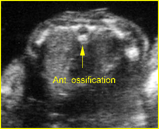
    • On the transverse (axial) view, the anterior ossification center (vertebral body) and posterior ossification centers (pedicles, transverse processes, laminae, and spinous processes) may all be identified as echogenic structures. This view may be superior to longitudinal views in demonstrating small spinal defects, since all three ossification centers can be imaged simultaneously.
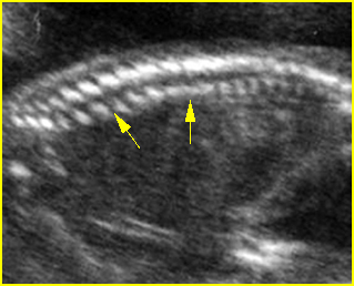
    • On the sagittal view, many vertebrae can be visualized on a single image. The curvature can be best evaluated<?is this sentence complete enough?>. The normal spine appears as two parallel lines formed by the vertebral bodies anteriorly and the ossification centers of the lateral processes converging in the sacrum. The lines correspond to the posterior elements of the vertebrae and the vertebral body. This view can optimally demonstrate interruption of the overlying integument when myelomeningocele is present.
    • On the coronal view, many vertebrae can be visualized on a single image. Hence, scoliosis, hemivertebrae, and disorganized vertebrae are optimally visualized on this view. <?can you check the following sentence>The images oriented through the dorsal ossification centers can demonstrate the two parallel rows of echogenicity in these centers and also the extent of a dysraphic defect compared to adjacent vertebrae in the case of spina bifida.

Fig 1:  Normal spine   Cross-sectional scan of the abdomen: posterior ossification centers of spine align vertically

Fig 2:  Normal thoracic spine  Coronal scan of the thoracic spine: normal echodensity and alignment

Fig 3:  Normal spine :   Cross-sectional scan of the abdomen posterior ossification centers of spine align horizontally (arrow = anterior ossification center)

Fig 4:  Normal spinal curvature  Sagittal view of the spine: normal curvature (arrow) with complete overlying skin

Fig 5:  Normal lumbar spine  Coronal scan of the lumbar spine (arrowhead) (arrow = iliac bone)

Video clips of normal examination

Normal spine (sagittal scan) :  Rotating the transducer: from cross-sectional view (post. ceters in the horizontal plane) to sagittal view of the spine

Normal spine (coronal scan):  Rotating the transducer: from cross-sectional view (post. ceters in the vertical plane) to coronal view of the spine

Normal spine (sagittal scan):  Rotating the transducer: from cross-sectional view (post. centers in the horizontal plane) to sagittal view of the spine
(arrow = anterior ossification center)

Normal spine (coronal view) :  Rotating the transducer: from cross-sectional view (post. centers in the vertical plane) to coronal view of the spine

Potential pitfalls:

    • Incomplete ossification: Scanning in early pregnancy reveals incomplete ossification of the lateral centers. Hence, on the transverse view, the posterior centers may appear to be parallel to one another rather than converging to the midline. This may be misinterpreted as spinal dysraphism.
    • Pseudodyspharism: On the coronal plane of the cervical and lumbar spine, the two parallel lines of posterior centers normally diverge. This divergence should not be mistaken for a spinal dysraphism.
    • Pseudodyspharism: <?please check the following sentence>On the transverse scan of the lumbosacral spine, if the ultrasound beam angled obliquely across the vertebral body of one vertebra but missed the posterior element or the posterior centers of another, this could simulate dyspharism. A normal appearance is restored when the ultrasound beam is reoriented perpendicular to the spinal axis.
  • High-quality sonograms can also show the spinal cord within the spinal canal. The central canal is readily detectable within the cord. The more echogenic cauda equina (nerve roots) can be seen distal to the conus medullaris (distal spinal cord). With growth of the spine, the position of the conus medullaris ascends with gestational age.
  • The spinal cord neural tissue, like that of most brain tissue, is echopenic. The conus medullaris and the craniocervical junction can be seen, albeit inconsistently, in nearly all fetuses by 18-20 weeks. The tissues surrounding the cord (leptomeninges) are brightly echogenic, as are those that surround the brain, and the dura is usually also seen discretely as a linear bright reflector. In fetuses with myelomeningoceles, determination of the most cephalic spinal level lesion is an important factor in prognosis. This level can be determined by counting up from the last ossified vertebral segment.
Read More

Normal Examination Limbs

Normal Examination

Normal limbs

      Fig 1, Fig 2, Fig 3, Fig 4, Fig 5, Fig 6, Fig 7, Fig 8

  1. Development: During the 8th week from the last menstrual period (LMP), a hyaline cartilage outline of the future appendicular skeleton begins to appear. The primary ossification centers of a long bone are located in the center of the shaft (diaphysis) and appear between weeks 7 and 12. Transabdominal and transvaginal sonography can clearly define the fetal skeleton even in the first trimester. The secondary ossification centers located in the epiphyses are at the distal ends of the long bone. During bone growth, the epiphyseal plate intervenes between the epiphysis and the end of the diaphysis (i.e. metaphysis). When the epiphyseal plate is replaced by bone, growth of the bone ceases. By 15-16 gestational weeks both the appendicular and axial skeleton can be consistently well imaged by sonography, although phalanges may be difficult to perceive in some instances. In the second trimester, the scapula, clavicle, humerus, radius, ulna metacarpals, femur, tibia, fibula, metatarsals and phalanges can be appreciated well sonographically in most cases.
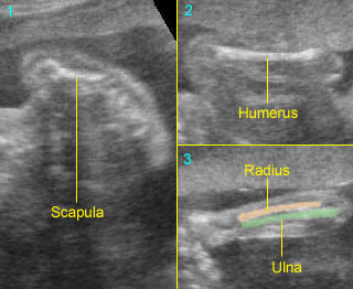
  2. Systematic approach: All long bones should be visualized with a systematic and routine sonographic approach to minimize errors in detecting limb defects. For example, in a survey of the upper limbs, the scapula should first be located and then the probe is laterally moved to identify the humerus, which at first may be in the cross-section or longitudinal view of the long bones. After completion of the upper arm assessment, the transducer is moved to visualize the distal end of the humerus or the elbow joint and then the probe is meticulously adjusted to locate the proximal end of the radius and ulna. Similarly, after thorough examination of the forearm the wrist joints and hands will be demonstrated respectively. The lower survey must be approached in the same way from the iliac wing through the foot.
  3. Identification of long bones: The simple technique to identify the types of long bones is to obtain planes of sections that traverse the short axis of the limb. Such a plane through the forearm and calf will demonstrate two bones. In the lower leg, the more lateral bone is the fibula, and the medial bone is the tibia. The tibia and fibula and radius and ulna end at the same level distally, however, proximally the ulna is longer than the radius. This allows ready differentiation of these two bony sets and of the radius from the ulna in the upper extremity set.
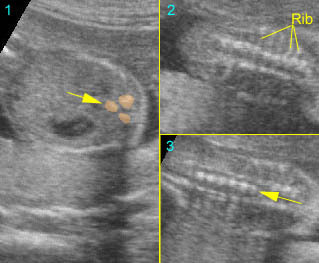
  4. Femur length measurement: Femur length measurement is usually used as a means of predicting gestational age. The measurement is technically the easiest of the most common biometric measurements. The transducer need only be aligned to the long axis of the bone to obtain a proper plane of the section. Only the ossified portions of the diaphysis and metaphysis are measured. The cartilaginous ends of the femur are excluded.
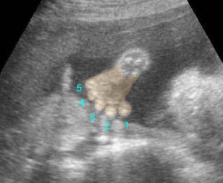
  5. Identification of hands: With patience one can usually visualize all four fingers and the thumb. The hand is frequently clenched in a fist-like fashion, which can complicate the counting of fingers. The toes, although smaller than the fingers, can be seen relatively well. If difficulty arises, it is usually the functionally less important fourth and fifth toes that are not seen.
  6. If a skeletal dysplasia is suspected early in the second trimester, a follow-up study to assess the interval long bone growth should be performed. The growth rate of the long bones decreases before the absolute length falls below the cut-off point.
  7. Fetal parts ratios: The proportions between specific fetal body parts may be helpful in the diagnosis of a skeletal dysplasia. For example, foot length is not affected by most skeletal dysplasias whereas most other long bones are affected. In a normal fetus, the femur length/foot ratio (age-independent) is approximately equal to 1, and can effectively distinguish skeletal dysplasias from fetal growth restriction. Furthermore, the femur/head circumference and abdominal circumference/thoracic circumference ratios are also reliable for the detection of severe skeletal dysplasia.
  8. Degree of skeletal ossification: This should be routinely assessed, although it is usually evaluated by subjective impression. Examination of the acoustic shadow and the echogenicity of the bone itself are often helpful. However, the degree of skeletal ossification cannot be well judged. Only in the most extremely osteopenic bone can one appreciate diminished ossification on sonograms.

Fig 1:  Normal scapula  Coronal plane of the scapula: triangular shape

Fig 2:  Normal upper limb

Fig 3:  Normal radius and ulna   Note: proximal end not in the same level (arrow)

Fig 4:  Normal hand

Fig 5:  Normal hand in late first trimester

Fig 6:  Normal femur   Longitudinal scan of femur: standard plane for femur measurement

Fig 7:  Normal tibia and fibula  Note: proximal and distal end of the both bones are in the same level

Fig 8:  Normal toes

Video clips of normal examination

Normal upper limbs:  Scapula, humerus, radius and ulna; Note: ulna longer than radius at the proximal end, unlike tibia and fibula
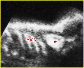
Normal lower limbs:  Femur, tibia and fibula
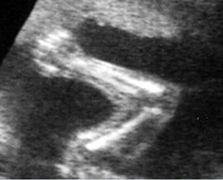
Normal upper limb
Normal foot & toes

Normal hand

Pitfalls

  1. Fetal position: The fetal position is very important for a complete examination. All limbs are best visualized when floating freely in the amniotic fluid and imaging is difficult when they are tucked under the fetal body. Usually the limbs are readily imaged when the fetus is in the supine position whereas the posterior elements of the spine may be easily imaged in prone or decubitus positions.
  2. FL measurement error: A non-osseous tissue at the end of the femur can give equal brightness. Reflection is returned from tissues distal to the epiphyseal plate but in immediate contiguity with the distal femoral metaphysis.
  3. Short long bones: A fetal long bone length greater than 2 standard deviations below the mean for the gestational age does not necessarily indicate the presence of a skeletal dysplasia. Other possibilities for short fetal long bone include a normal physiologic variation in bone length, intrauterine growth restriction and chromosome abnormalities and other syndromes that may have a skeletal abnormality as part of their presentation.
Read More

Bladder and Cloacal Exstrophy

Bladder and Cloacal Exstrophy

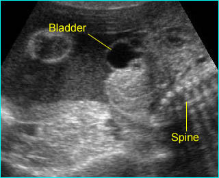
This is a rare congenital anomaly characterized by the exteriorization of the viscera on the abdominal surface, low insertion of the umbilical cord, divergent pubic rami and abnormal exterior genitalia. Bladder and cloacal exstrophy share a common embryologic origin in abnormal cloacal development, but are different in terms of severity and extent of involvement. Bladder exstrophy is a lower abdominal wall defect with herniation of the bladder whereas cloacal exstrophy is much more complex, including omphalocele, bladder exstrophy, imperforate anus, spine malformation (also known as OEIS complex), spina bifida and intersex.

Incidence: Sporadic occurrence with a prevalence of 1 in 30,000 births with a male to female ratio of 2:1 for bladder exstrophy, 1 in 50,000 births with a higher frequency in twins for cloacal exstrophy, and 1 in 200,000-300,000 for OEIS complex.

Sonographic findings:

  • Soft tissue mass at the lower abdominal wall
    Fig 1, Fig 2, Fig 3, Fig 4
  • Persistently absent fetal bladder with a large midline infraumbilical cystic or solid mass.
  • A large cystic mass in the pelvis representing a persistent cloaca can be seen in some cases.
  • Omphalocele is usually seen.
  • Lumbosacral abnormalities.
  • Associated anomalies such as renal agenesis, myelomeningocele, horseshoe kidney, and clubfeet.
  • Umbilical arteries running alongside the mass suggestive of bladder exstrophy.

Fig 1:  Bladder extrosphy  Sagittal view of the fetal abdomen: complex extra-abdominal mass (*) below the umbilicus, finally proven to be bladder extrosphy

Fig 2:  Cloacal extrosphy  Free floating complex mass (*) connecting with fetal perineum

Fig 3:  Cloacal extrosphy  Free floating complex mass (*) connecting with fetal perineum

Fig 4:  Cloacal extrosphy  Free floating complex mass (solid circle) connecting with fetal perineum, abnormal fetal limb (arrow) (* = femur)

Video clips of bladder and cloacal exstrophy

Bladder extrosphy : Bladder is located outside the abdomen, presented as a cystic mass below the cord insertion

Associations: Genital defects in both conditions; renal, skeletal, neural tube, intestinal, cardiovascular and omphaloceles defects in cloacal exstrophy.

Management: Termination of pregnancy can be offered when diagnosed before viability. Isolated bladder exstrophy requires postnatal surgery, either with initial repair using the staged approach or a complete primary repair technique with continent diversion. Neonatal assignment of genetic males to the female sex is often necessary in cloacal exstrophy because of severe phallic inadequacy resulting in unpredictable sexual identification.

Prognosis: Recently good in isolated bladder exstrophy, but poor in cloacal exstrophy, with a high rate of urinary tract infection in adult women with sexual problems in several cases.

Recurrence risk: Sporadic (rare recurrence) but there is a significant genetic predisposition in some familial cases.

Read More

Limb-Body Wall Complex (LBWC)

Limb-Body Wall Complex (LBWC)

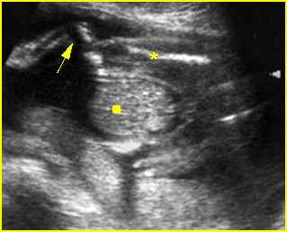
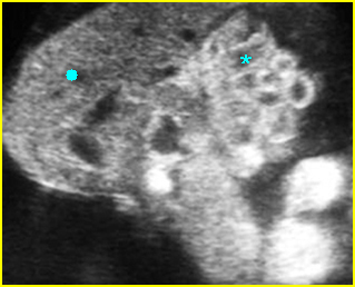
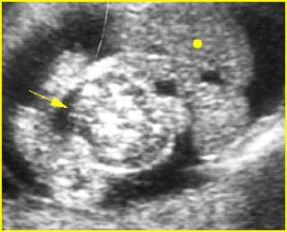
LBWC or body-stalk anomaly or cyllosomas is a complex set of disruptive abnormalities consisting of failure of the anterior abdominal wall to close, short umbilical cord, disruption of the lateral body wall, distinctive scoliosis of the spine, limb defects, non-fusion of the amnion and chorion. Therefore, the amnion does not cover the cord but extends as a sheet from the margin of the cord to be continuous with the body wall and placenta.

Incidence: Sporadic, 1 in 15,000 (1:3000-42,000) births; a higher incidence was observed among mothers with a history of cigarette, alcohol and marijuana use.

Sonographic findings:

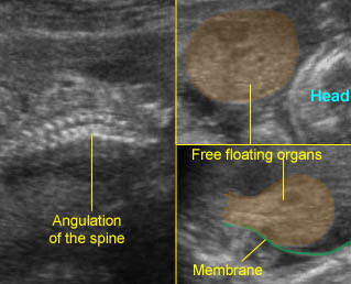
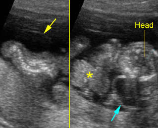
  • Large body wall defect allowing abdominal contents to herniate. Fig1, Fig2
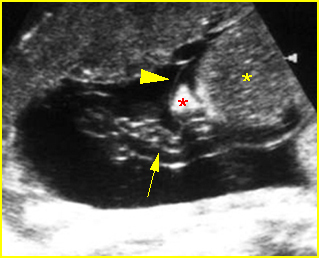
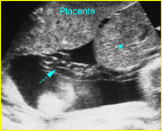
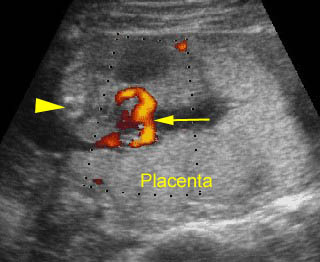
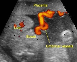
  • The umbilical cord is absent or is a very short segment with a single umbilical artery. Fig3, Fig4
  • The fetal abdomen is connected directly to the placenta.
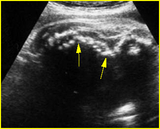
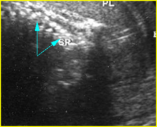
  • Scoliosis is evident in most cases. Fig5, Fig6
  • Limb anomalies in most cases, including clubfeet, oligodactyly, arthrogryposis, absence of limbs, single forearms, split hand and feet.
  • Craniofacial defects frequently seen.
  • An extremely elevated level of maternal serum-alpha-fetoprotein (MSAFP) is also indicative of LBWC.
  • Other associated anomalies.
  • Three-dimensional ultrasound may be helpful.
  • For first trimester diagnosis, the upper part of the fetal body is in the amniotic cavity, whereas the lower part is in the coelomic cavity.
  • Differential diagnoses include other types of abdominal wall defects and amniotic band syndrome which may have very similar features.
  • Pitfalls: Some cases have been initially misdiagnosed as omphalocele or gastroschisis, for which the prognosis is much better.

Fig 1:  Limb-body wall complex  Free floating complex mass containing abnormal shaped liver (solid circle) and bowel (*)

Fig 2:  Limb-body wall complex  Free floating complex bowel (arrow) and liver (solid circle) in the amniotic fluid

Fig 3:  Limb-body wall complex  Free floating complex mass containing liver (*) with covering membrane (arrowhead), short umbilical cord (arrow) originating from the placenta to the mass

Fig 4:  Limb-body wall complex  Free floating complex mass containing liver (*) with short umbilical cord (arrow) originating from the placenta to the mass

Fig 5:  Kyphoscoliosis   Sagittal scan of the fetal trunk: disorganized spine in association with limb-body wall complex

Fig 6:  Disorganized spine  Coronal scan of the fetal spine: disorganized spine (arrow) in association with limb-body wall complex

Video clips of limb-body wall complex (LBWC)

LBWC :  Sagittal to cross-sectional scan of the abdomen: Extensive abdominal wall defect with free floating visceral structures Note: covering membrane and spinal angulation

Limb-body wall complex :  Cross-sectional scan of the abdomen: the total length of the cord from the fetal insertion to placental insertion (arrow) (arrow = spine)

Limb-body wall complex :  Coronal scan of the fetal spine: scoliosis associated with limb body wall complex

LBWC :  Limb-body-wall complex: the umbilical vessels running directly from the placenta to the free floating visceral structures without true umbilical cord

Associations: Other than typical defects, other anomalies are also often seen, especially craniofacial or neural tube defects.

Management: Termination of pregnancy can be offered.

Prognosis: Uniformly fatal.

Recurrence risk: Sporadic (rare recurrence).

Read More