Head-Sign: Skull Defects

Skull Defects

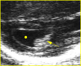
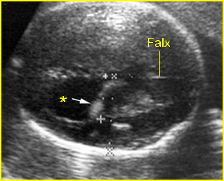
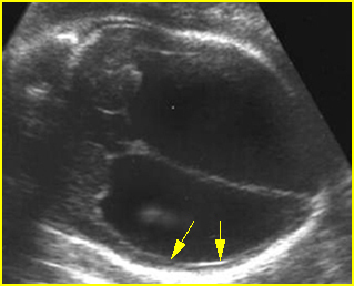
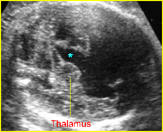
Cranial defects vary from a small meningocele, which is very difficult to visualize, to complete absence of the cranium such as with anencephaly or exencephaly. In practice, there is no problem in the diagnosis of complete absence of the skull or a large defect, however, severe bone demineralization, such as osteogenesis imperfecta, may give the appearance of a large skull defect. Partial defects, or small cephaloceles are occasionally difficult to visualize.

Fig 1, Fig 2, Fig 3
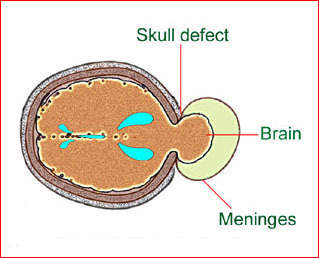
Fig1: Schematic drawing: Occipital meningocele, protrusion of the brain tissue and meninges through the skull defect
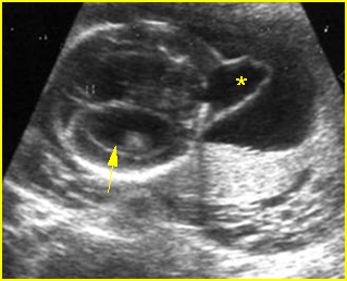
Fig 2: Occipital cephalocele  Transverse scan of the skull: small defect of occipital bone with meningocele (*) (arrow = ventriculomegaly)
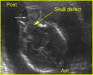
Fig 3: Occipital cephalocele  Transverse scan of the skull: defect of occipital bone with meningoencephalocele (arrow)

Video clips of intracranial massive fluid collection
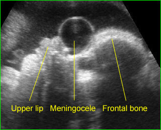
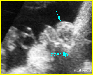
Anterior meningocele:  Sagittal scan of the fetal face: frontal bone defect with midline cystic mass (meningocele)
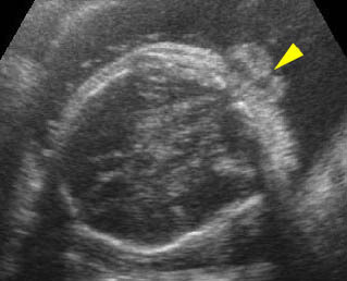
Occipital cephalocele:  Transverse scan the level of cerebellum: small defect of occipital bone with encephalocele

Differential diagnosis

The differential diagnoses and some sonographic features of skull defects are as follows:

Anencephaly:

  • absent bony calvarium above the orbits
  • orbits well visualized
  • absence of supratentorial brain
  • residual brain
  • possibly spina bifida
  • polyhydramnios

Excencephaly

  • calvarium absent
  • disorganized supratentorial brain tissue

Amniotic band syndrome (ABS)

  • asymmetric or bizarre cephalocele
  • other deformities, including limb amputation

Limb-body wall complex

  • asymmetric or bizarre encephaloceles
  • similar to ABS except fetal body adherent to placenta
  • usually more severe than ABS
  • associated bizarre defects of fetal body

Cephalocele

  • midline defect
  • extracranial cyst or brain tissue
  • possibly ventriculomegaly
  • lemon sign may be present

Open neural tube defects

  • lemon sign
  • banana sign
  •  mild ventriculomegaly
  • spinal defect

Fetal demise

  • overlapping sutures (Spalding’s sign)
  • poor visualization of intracranial structures
  • associated findings of fetal demise

Microcephaly

  • calvarium present
  • decreased brain tissue
  • head circumference 2-3 standard deviations below that expected for the menstrual age

Craniosynostosis

  • complete or partial
  • deformed skull
  • possibly microcephaly
  • abnormal cephalic index.

In addition, considerable overlap may be found in these features among abnormalities. For instance, the lemon sign was originally described with opened NTD, however, it may not be present in an opened NTD identified in the third trimester of pregnancy, and a mild lemon sign may be identified in normal fetuses.

    Read More

    Head-Sign: Cystic Brain Lesions

    Cystic Brain Lesions

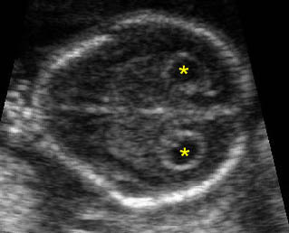
    Cystic lesions of the brain have a number of causes but ventriculomegaly and choroid plexus cyst are the most common. Usually, the location and associated abnormalities are very helpful in the differential diagnosis. For example, interhemispheric cyst is commonly related to agenesis of the corpus callosum. Likewise, ventriculomegaly and dorsal sac in the case of holoprosencephaly are often associated with other abnormalities, whereas others are mostly isolated findings. The choroid plexus cyst is always a lateral cyst located in the choroid plexus and is often isolated and not difficult to diagnose. Cystic lesions of Dandy-Walker malformations are always located in the posterior fossa.

    Fig 1, Fig 2, Fig 3, Fig 4

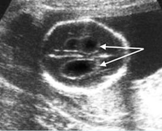
    Fig1: Choroid plexus cysts  Transverse scan of the skull: bilateral large choroid plexus cyst (arrow)

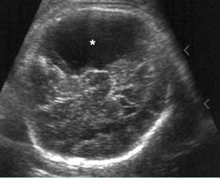
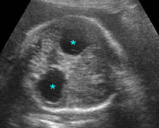
    Fig 2: Porencephaly Some part of cerebral tissue is absent (*)

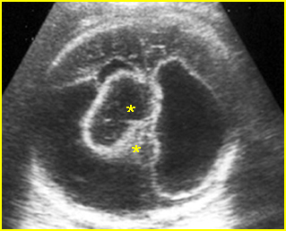
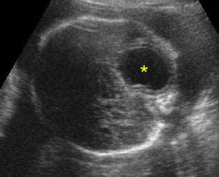
    Fig 3: Ependymoma  Complex cystic mass (*) in the skull combined with fluid collection and partial absence of brain tissue

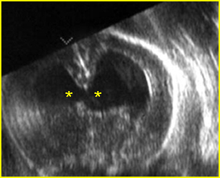
    Fig 4: Semilobar holoprosencephaly  Oblique coronal scan of the skull: common ventricle (*) with partial separation

    Video clips of cystic brain lesions

    Choroid plexus cyst: Transvere scan at the level of lateral ventricles: bilateral choroid plexus cyst (*) with lemon sign

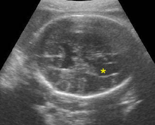
    Arachnoid cyst: Transverse scan of the head: isolated cystic, circular, thin-walled brain lesion (*)

    Colpocephaly:  Transverse scan of the head: bilateral marked dilatation of occipital horn (*) of lateral ventricles

    Differential diagnosis

    The differential diagnoses of intracranial cystic lesion are as follows:

    Midline cyst

    • prominent cavum septum pellucidi (very common)
    • dorsal sac of holoprosencephaly (common)
    • interhemispheric cyst of agenesis of corpus callosum (uncommon)
    • vein of Galen aneurysm (rare)
    • arachnoid cyst (rare)
    • enlarged cavum vergae interhemispheric cystic lesions

    Lateral cyst

    • choroid plexus cyst (very common)
    • unilateral hydrocephalus (uncommon)
    • porencephaly/schizencephaly (uncommon)
    • arachnoid cyst (rare)
    • neoplastic cyst (very rare)
    • periventricular cyst formation secondary to CMV infection

    Posterior fossa cyst

    • enlarged cisterna magna (>10 mm (very common)
    • Dandy-Walker malformation (common)
    • arachnoid cyst (very rare)
    • median retrocerebellar fluid collections remain the most difficult to prognosticate, retrocerebellar cysts often being difficult to differentiate from enlarged cisterna magna and Dandy-Walker complex.
    Read More

    Head-Sign: Massive Intracranial Fluid Collections

    Massive Intracranial Fluid Collections

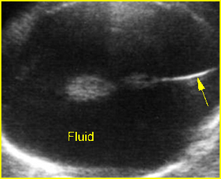
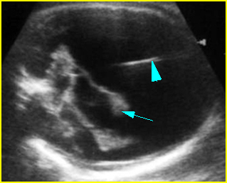
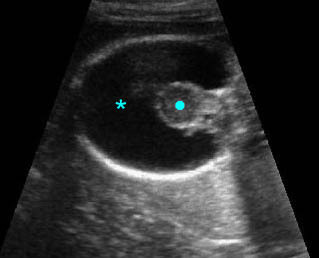
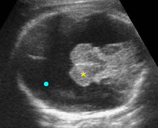
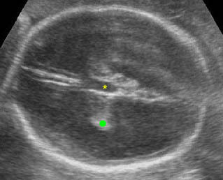
    The three common causes of massive intracranial fluid collections with an intact cranial vault are hydrocephalus, which is usually secondary to aqueductal obstruction, holoprosencephaly, and hydranencephaly. The unique features of each condition can distinguish these three entities from each other.

    Fig 1, Fig 2

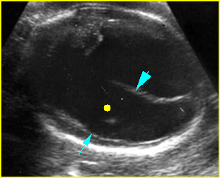
    Fig1: Hydranencephaly  Transverse scan of the skull: absent brain tissue, present falx cerebri (arrow)

    Fig 2: Severe hydrocephalus Markedly dilated ventricles with extremely thin brain, choroid plexus dangling (arrow) (arrowhead = falx cerebri)

    Video clips of intracranial massive fluid collection

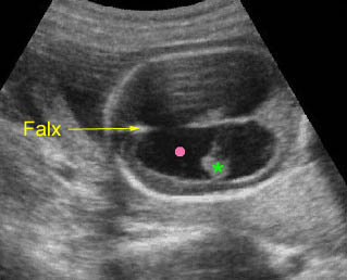
    Holoprosencephaly: Absent brain tissue (*), without falx cerebri, and fused thalamus (solid circle)

    Hydranencephaly: Transthalamic view: totally destructive brain lesion (solid circle) but preserving thalamus (*) and posterior fossa

    Severe hydrocephalus: Transverse scan of the head: markedly dilated ventricles with extremely thin cerebral tissue (solid circle = dangling choroid plexus, * = dilated third ventricle)

    Differential diagnosis

    • Hydrocephalus secondary to aqueductal stenosis (about 1 in 2000 births):
      • thin cerebral mantle with falx cerebri
      • separated thalami and dilated third ventricle
      • obvious choroid dangling sign
      • usually no midline face defects
    • Hydranencephaly (uncommon; 1.0-2.5 in 10,000 births):
      •  no cerebral mantle with falx cerebri
      • normal thalami (not separated or fused)
      • minimal choroid dangling sign
      • no midline face defects
    • Holoprosencephaly (rare; 1 in 16,000 neonates):
      • cerebral mantle with or without dorsal sac, no falx cerebri
      • fused thalami
      • minimal choroid dangling sign
      • associated midline face defects (very common).
    Read More

    Head-Sign: Ventriculomegaly

    Ventriculomegaly

    Ventriculomegaly is the enlargement of the cerebral ventricles with the cause unknown. The term hydrocephalus is usually reserved for more severe cases of ventriculomegaly, and this usually implies obstruction of flow of the CSF. Obstructive hydrocephalus within the fetus is most commonly non-communicating (blockage within ventricles) rather than communicating (extraventricular blockage). Moreover, underdevelopment of the brain and destructive brain lesions can also cause ventriculomegaly.

    Brain destruction is another cause of ventriculomegaly and may include in utero damage due to a CNS infection. CNS infections may be associated with brain atrophy, with resultant microcephaly and ventriculomegaly.

    The main causes of fetal ventriculomegaly are aqueductal stenosis, Chiari II malformation, Dandy-Walker complex and agenesis of the corpus callosum.

    Fig1, Fig2, Fig3, Fig4

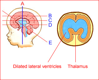
    Fig1: Schematic drawing: Coronal view: Ventriculomegaly with wide separation of the thalami

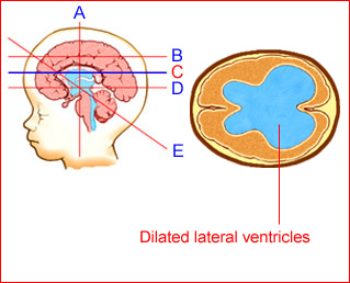
    Fig 2: Schematic drawing: Transventricular view: Ventriculomegaly secondary to aqueductal stenosis

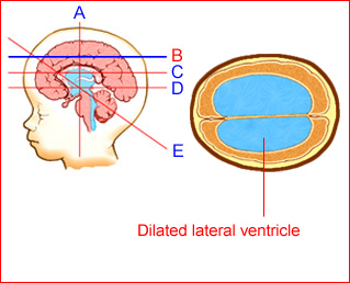
    Fig 3: Schematic drawing: Transventricular view: Ventriculomegaly secondary to aqueductal stenosis

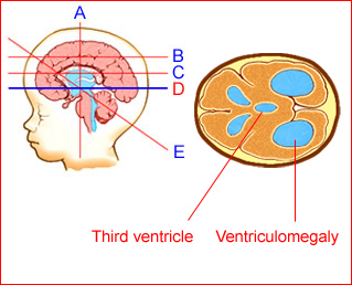
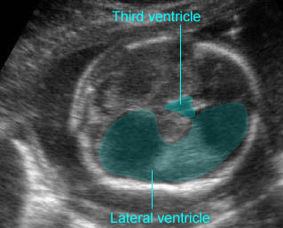
    Fig 4: Schematic drawing: Transthalamic view: Ventriculomegaly with dilated third ventricle secondary to aqueductal stenosis

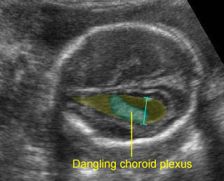
    Fig 5: Mild ventriculomegaly Lateral ventricle (solid circle), width of 11 mm, with choroid plexus dangling sign (arrow)

    Fig 6: Ventriculomegaly  Lateral ventricle (*), width of 15 mm, with choroid plexus dangling sign (arrow)

    Fig 7: Severe hydrocephalus Markedly dilated ventricles with extremely thin brain (arrow)

    Fig 8: Hydrocephalus Transthalamic view: wide separation the third ventricle (*) and dilated lateral ventricles

    Fig 9: Severe hydrocephalus Markedly dilated ventricles (solid circle) with extremely thin brain (arrow), (arrowhead = falx cerebri)

    Fig 10: Bossing forehead secondary to hydrocephaly Facial profile view: sunken nose related to forehead bossing in case of hydrocephaly

    Mild (borderline) ventriculomegaly [Fig5, Fig6] is used when the atrial width is 10-15 mm. Some consider early ventricular dilatation as separation (>3 mm) of the dependent choroid from the medial ventricular wall. This occurs bilaterally in 0.15-0.7% of fetuses and unilaterally in 0.07% of pregnancies. This can be related to an increased risk of CNS and non-CNS anomalies. Isolated borderline ventriculomegaly is normal variant in most cases, however, it may represent the earliest manifestation of cerebral anomalies (4%), chromosomal abnormalities (4%) (mostly trisomy 21) and overall abnormal outcomes (20%).

    However, early and unexplained mild ventriculomegaly appears to have a good prognosis but a long postnatal clinical follow-up is required.

    Overt (or moderate to severe) ventriculomegaly [Fig7, Fig8, Fig9, Fig10] is usually defined for an atrial width of more than 15 mm in the second and third trimesters(8). This may involve isolated cases (30-60%) or cases associated with other CNS anomalies, most frequently neural tube defects (NTDs) and midline defects. The degree of antenatal ventriculomegaly is related to pediatric neurological morbidity and, when it is >15 mm, it is associated with an increase in abnormal neurological development.

    Unilateral ventriculomegaly is a rare finding usually due to focal obstruction of the CSF pathway at the level of Monro’s foramen. Examination of the lateral ventricles using the angled technique helps to distinguish between unilateral and bilateral hydrocephalus. Unilateral ventriculomegaly is usually an isolated finding and when isolated has little measurable effect on developmental outcome.

    Hydrocephalus: Transverse scan of the head: markedly dilated ventricles (solid circle) with very thin cerebral tissue (* = dangling choroid plexus)

    Mild ventriculomegaly: Transverse scan of the head: lateral ventricle (*) width of 10 mm diameter

    Hydrocephaly: Overt ventriculomegaly with dilatation of the third ventricle

    Mild ventriculomegaly: Transventricular view: mild dilatation of the lateral ventricle (10 mm.) with dangling choroid plexus

    Severe hydrocephalus: Transverse scan of the head: markedly dilated ventricles with extremely thin cerebral tissue (solid circle = dangling choroid plexus, * = dilated third ventricle)

    Sonographic findings:

    • Enlargement of the lateral ventricle atrium >10 mm.
    • Minimal separation of the choroid plexus in either proximal or distal cerebral ventricle.
    • Dangling choroid plexus with an abnormal choroid angle (more than 29 degrees)(14). Dangling can be easily recognized without measuring it in most cases.
    • When ventriculomegaly is detected, a careful anatomic survey of the fetus is necessary to determine any associated anomalies. Furthermore, hydrocephalus is associated with a significant risk of chromosomal abnormalities. When objective difficulties preclude detailed sonographic examination, MRI is helpful(15).
    • Sonographic differential diagnoses of ventriculomegaly include
      • Arnold-Chiari malformation type II:
        • deformed cranium (lemon sign), which usually disappears in the third trimester
        • obliteration of cisterna magna (banana sign)
        • associated open spinal defect
        • minimal hydrocephalus
        • pointed frontal horns
      • Dandy-Walker malformation:
        • midline posterior fossa cyst
        • cyst communicates with the fourth ventricle
        • vermian agenesis
        • hypoplastic cerebellar hemispheres
        • elevated tentorium
        • mild to moderate hydrocephalus
      • Agenesis of the corpus callosum:
        • third ventricle elevated
        • separation of frontal horns
        • associated colpocephaly
        • mild ventriculomegaly
      • Aqueduct stances:
        • moderate to massive hydrocephalus
        • normal fourth ventricle
        • normal posterior fossa
        • normal cranial configuration
      • Cephalocele:
        • open defect, usually occipital skull base
        • obliteration of the cisterna magna
        • occasional lemon sign
        • mild to moderate hydrocephalus
        • polyhydramnios.
    Read More

    About MFM

    เกี่ยวกับอะไร?

    • การบริบาลครรภ์เสี่ยงสูง (High risk pregnancy management)
    • การประเมินสุขภาพทารกในครรภ์ (Fetal surveillance)
    • การคัดกรองและการวินิจฉัยก่อนคลอด (Prenatal screening and diagnosis)
    • การตรวจคลื่นเสียงความถี่สูงทางสูติศาสตร์ (Obstetric ultrasound) ในระดับสูง
    • การรักษาทารกในครรภ์ (Fetal therapy)

    ความคาดหวัง

    • สุขภาพอนามัยแม่และเด็ก ตลอดจนครอบครัวดีขึ้น
    • อัตราภาวะทุพพลภาพและการตายปริกำเนิดลดลง
    • อัตราภาวะทุพพลภาพและการตายของมารดาลดลง
    • มีความก้าวหน้าทางวิทยาการด้านเวชศาสตร์มารดาและทารกมากขึ้นอย่างสอดคล้องกับสังคมไทย
    • มีการวิจัยค้นคว้าและเผยแพร่สู่ระดับนานาชาติเพิ่มมากขึ้น
    • ยกระดับมาตรฐานวิชาชีพสูตินรีแพทย์ของประเทศไทยสู่สากล
    • เพิ่มแพทย์ประกอบวิชาชีพที่ทรงคุณธรรม อันเป็นตัวอย่างของคนในสังคมต่อไป

    พันธกิจ

    รับผิดชอบในการบริบาลมารดาและทารกในครรภ์ที่มีปัญหาซับซ้อน อันประกอบด้วยการสร้างเสริมสุขภาพ ป้องกัน รักษา และฟื้นฟูสภาพ รวมทั้งบริหารทรัพยากรอย่างมีประสิทธิภาพและประสิทธิผล ผลิตแพทย์ผู้เชี่ยวชาญด้านเวชศาสตร์มารดาและทารก ศึกษาค้นคว้าอย่างต่อเนื่อง ก่อให้เกิดองค์ความรู้ใหม่ ๆ และเผยแพร่ไปสู่สาธารณะ อันจะนำมาซึ่งความเจริญรุ่งเรืองทางวิทยาการด้านเวชศาสตร์มารดาและทารกในครรภ์ สู่สังคมไทย และนานาชาติ โดยได้รับการส่งเสริมและรับประกันคุณภาพจากราชวิทยาลัยสูตินรีแพทย์แห่งประเทศไทย

    History MFM

    เมื่อปี 2520 ร.ศ.พ.ญ.สุรีย์ สิมารักษ์ เป็นผู้บุกเบิกในการบริบาลผู้ป่วยครรภ์เสี่ยงสูงเป็นพิเศษ และได้แยกหอผู้ป่วยโรคพิษแห่งครรภ์ออกมา พร้อมกับเริ่มต้นด้วยการเปิดคลินิกครรภ์เสี่ยงสูง นับเป็นจุดต้นการก่อตัวของเวชศาสตร์มารดาและทารกท่ามกลางขีดจำกัดของทรัพยากรในการดูแล และท่านได้พัฒนาหน่วยนี้อย่างต่อเนื่องจนสามารถให้บริการการดูแลครรภ์เสี่ยงสูงได้มากขึ้นและดีขึ้น เป็นลำดับ เช่น มีเครื่อง fetal monitor เครื่องแรกเมื่อปี 2525 ในระยะนั้นมีจำนวนการคลอดประมาณ 7000-8000 รายต่อปี ได้มีการแยกหน่วยบริบาลครรภ์เสี่ยงสูงอย่างเป็นรูปธรรม ปี 2532: มีการแยก หน่วยอนุสาขาเวชศาสตร์มารดาและทารกอย่างเป็นทางการ โดยมีหัวหน้าหน่วยคือ ร.ศ.พ.ญ.สุรีย์ สิมารักษ์ และทีมงานได้แก่ น.พ.ชัยรัตน์ คุณาวิกติกุล (หัวหน้าหน่วยในปี 2536-2540, พ.ญ.พรรณี ศิริวรรธนาภา (หัวหน้าหน่ายจากปี 2540 จนถึงปัจจุบัน), น.พ. ธีระ ทองสง, น.พ. ชเนนทร์ วนาภิรักษ์

      • เปิดบริการตรวจคลื่นเสียงความถี่สูงเป็นครั้งแรกโดยร.ศ.แพทย์หญิงพรรณี ศิริวรรธนาภา ในปี 2531
      •  สามารถให้บริการเจาะน้ำครำเพื่อวินิจฉัยโครโมโซมผิดปกติ ตั้งแต่ปี 2528
      • สามารถให้บริการเจาะเลือดสายสะดือทารกโดยร.ศ.น.พ. ชัยรัตน์ คุณาวิกติกุล เป็นครั้งแรกในปี 2531
      • เปิดบริการอัลตราซาวด์อย่างเต็มรูปแบบ และพัฒนามาเป็น referral center พร้อมทั้งฝึกอบรม แพทย์ประจำบ้านด้านอัลตราซาวด์ทางสูติ-นรีเวช ตั้งแต่ปี 2532 เป็นต้นมา
      • เปิดบริการคัดกรองและวินิจฉัยก่อนคลอดธาลัสซีเมียชนิดรุนแรง โดย ร.ศ.น.พ.ชเนนทร์ วนาภิรักษ์ และศ.น.พ. ต่อพงษ์ สงวนเสริมศรี เป็นผู้เริ่มต้นบุกเบิกตั้งแต่ปี 2537
      • เปิดฝึกอบรมต่อยอดสาขาเวชศาสตร์มารดาและทารก ต่อเนื่องมาจากปี 2546 จนกระทั่งถึงปัจจุบันนับว่าหน่วยงานเล็ก ๆ นี้ยังคงเติบโตด้านบริการและวิชาการอย่างต่อเนื่องตามสติกำลัง และมีสมาชิกทีมงานที่เข้มแข็งมาร่วมทีมอีกหลายท่าน คือ พ.ญ.สุพัตรา ศิริโชติยะกุล, น.พ.วีรวิทย์ ปิยะมงคล, น.พ.พฤหัส จันทร์ประภาพ (ย้ายไปอยู่ MFM ศิริราช ปี 2548) พ.ญ. เฟื่องลดา ทองประเสริฐ (2548) พ.ญ. เกษมศรี ศรีสุพรรณดิฐ (2549) พ.ญ. สุชยา ลือวรรณ (2551) และได้ยังมีบุคลากรที่ไม่อาจเอ่ยนามได้หมด (โดยเฉพาะพี่ต๋อย นาย ออม อุบล)

    อาจารย์พิเศษ / อาจารย์ที่เกษียณอายุราชการไปแล้ว

    1. ร.ศ. พ.ญ. สุรีย์ สิมารักษ์, พ.บ.; Associate Professor Suri Simarak, M.D. (อดีตหัวหน้าหน่วย)
    2. ร.ศ.น.พ.ชัยรัตน์ คุณาวิกติกุล, พ.บ.; Associate Professor Chairat Kunaviktikul, M.D. (อดีตหัวหน้าหน่วย)
    3. ร.ศ.พ.ญ.พรรณี ศิริวรรธนาภา, พ.บ.; Associate Professor Pannee Sirivatanpa, M.D. (อดีหัวหน้าหน่วย)

    อาจารย์ประจำหน่วย

    1. ศ.นพ. ธีระ ทองสง, พ.บ.;Professor Theera Tongsong, M.D.
    2. รศ.นพ. ชเนนทร์ วนาภิรักษ์, พ.บ.; Associate Professor Chanane Wanapirak, M.D. (หัวหน้าหน่วย)
    3. รศ. ดร. นพ. วีรวิทย์ ปิยะมงคล, พ.บ.; Associate Professor Dr. Wirawit Piyamongkol, M.D., PhD.
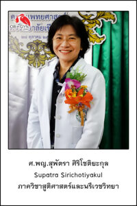
    4. ศ.พญ. สุพัตรา ศิริโชติยะกุล, พ.บ.; Professor Supatra Sirichotiyakul, M.D.
    5. รศ. พญ. เฟื่องลดา ทองประเสริฐ, พ.บ.; Associate Professor Professor Fuanglada Tongprasert, M.D.
    6. รศ.พญ. เกษมศรี ศรีสุพรรณดิฐ, พ.บ.; Associate Professor Kasemsri Srisupundit, M.D.
    7. รศ.พญ. สุชยา ลือวรรณ, พ.บ.; Associate Professor Suchaya Luewan, M.D.
    8. รศ.พญ. กุณฑรี ไตรศรีศิลป์, พ.บ.: Associate Professor Kuntharee Traisrisilp, M.D.
    9. ผศ.นพ. ภูดิศ เจต๊ะวรรณ, พ.บ.; Assistant Professor Phudit Jatavan, M.D.
    10. อ. พญ. ศิรินาถ ศิริเลิศ, พ.บ.; Instructor Sirinart Sirilert, M.D.

    ทีมงานสายสนับสนุน

    1. รัตนาภรณ์ เศขรฤทธิ์, วท.บ.; Ratanaporn Sekararithi BSc.
    2. อภิรดี ตรรกไพจิตร, วท.บ.; Apiradee Taggapichitti, BSc.
    3. มัทนาพร วงศ์ใหญ่, Muttanaporn Wongyai
    4. สุกัญญา จันตา; Sukunya Junta
    Read More

    อ. ปุย ได้รับการแต่งตั้งเป็น รศ.

    อ. ปุย ได้รับการแต่งตั้งเป็น รศ.
    ภาควิชาฯ ขอแสดงความยินดีกับ อ.ปุย (กุณฑรี ไตรศรีศิลป์ หมื่นพินิจ) ที่ได้รับการแต่งตั้งให้ดำรงตำแหน่งทางวิชาการเป็นตำแหน่งรองศาสตราจารย์ (รศ.) ตั้งแต่วันที่ 9 ธันวาคม 2562 (เป็น รศ. มาแล้วเกือบสองปี)

    Read More

    รศ.พญ.ประภาพร สู่ประเสริฐ  : รองคณบดีด้านวิชาการ

    รศ.พญ.ประภาพร สู่ประเสริฐ  : รองคณบดีด้านวิชาการ

    ภาควิชาขอแสดงความยินดีกับ อ.ต่าย ที่ได้รับการแต่งตั้งเป็นรองคณบดีด้านวิชาการ นับเป็นอีกก้าวโชคดีของคณะที่ได้เปิดโอกาสให้ผู้ที่มุ่งมั่น จริงจัง กับการพัฒนาการเรียนการสอนของคณะ และสร้างแพทย์รุ่นต่อไปในนามของแพทย์มช. แพทย์คุณภาพ

    Prapaporn
    Read More