Massive Intracranial Fluid Collections
The three common causes of massive intracranial fluid collections with an intact cranial vault are hydrocephalus, which is usually secondary to aqueductal obstruction, holoprosencephaly, and hydranencephaly. The unique features of each condition can distinguish these three entities from each other.
Fig 1, Fig 2
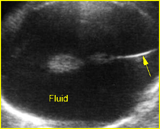
Fig1: Hydranencephaly Transverse scan of the skull: absent brain tissue, present falx cerebri (arrow)
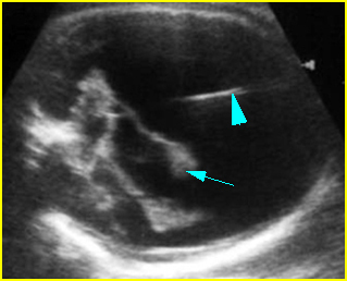
Fig 2: Severe hydrocephalus Markedly dilated ventricles with extremely thin brain, choroid plexus dangling (arrow) (arrowhead = falx cerebri)
Video clips of intracranial massive fluid collection
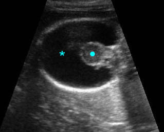
Holoprosencephaly: Absent brain tissue (*), without falx cerebri, and fused thalamus (solid circle)
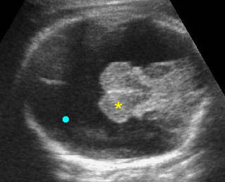
Hydranencephaly: Transthalamic view: totally destructive brain lesion (solid circle) but preserving thalamus (*) and posterior fossa
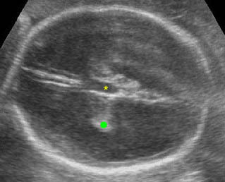
Severe hydrocephalus: Transverse scan of the head: markedly dilated ventricles with extremely thin cerebral tissue (solid circle = dangling choroid plexus, * = dilated third ventricle)
Differential diagnosis
- Hydrocephalus secondary to aqueductal stenosis (about 1 in 2000 births):
- thin cerebral mantle with falx cerebri
- separated thalami and dilated third ventricle
- obvious choroid dangling sign
- usually no midline face defects
- Hydranencephaly (uncommon; 1.0-2.5 in 10,000 births):
- no cerebral mantle with falx cerebri
- normal thalami (not separated or fused)
- minimal choroid dangling sign
- no midline face defects
- Holoprosencephaly (rare; 1 in 16,000 neonates):
- cerebral mantle with or without dorsal sac, no falx cerebri
- fused thalami
- minimal choroid dangling sign
- associated midline face defects (very common).

