Neck Masses
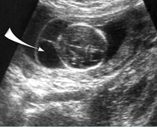
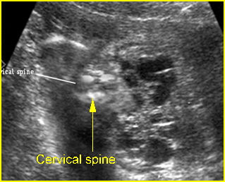
Fig 1, Fig 2
Differential diagnosis of common neck masses:
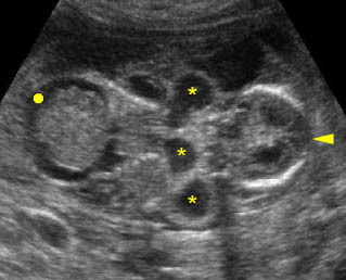
- Cystic hygroma (most common; multiseptate, no solid part; posterolateral; bilateral, hydrops)
- Cervical meningomyelocele (cystic, complex or solid; posterior midline, separation of cervical spine)
- Occipital cephalocele (cystic, complex or solid; posterior midline, skull defect)
- Goiter (generalized hypoechoic solid; anterior, bilateral)
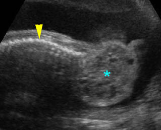
- Cervical teratoma (solid-cystic; anterolateral, unilateral)
- Hemangioma (echogenic, variable in location, positive Doppler signals).

Fig 1: Cystic hygroma Oblique cross-sectional scan at the level of cerebellum: anechoic cyst (arrow) with central septum

Fig 2: Cervical hemagioma Cross-sectional scan at the neck: enlarged complex mass lateral to the cervical spine (positive Doppler signal)
Video clips of neck masses

Cervical teratoma: Sagittal scan of the thorax and neck: solid-cystic mass (*) lateral to the neck (arrowhead = spine)

Cystic hygroma: Coronal scan of the fetal trunk: multiple cystic areas (*) around the neck as well as ascites (solid circle)

