Normal Examination
Practically, most practitioners evaluate the face qualitatively, using three views: the coronal view, looking at bone and soft tissue from the surface well into the palate and orbits; the transverse view, looking at the mandible, maxilla, tooth buds, and orbits; and finally the sagittal view, evaluating the nasal bridge, the frontal bone, and the mandible. The measurements of various aspects of the fetal face have been studied in an attempt to quantify facial anomalies. Normative data of the fetal eye length, nose width and nostril distance, fetal upper lip and chin, binocular distance, and biparietal distance (BPD)/mandibular body length (MBL) ratio for diagnosis of micrognathia have been established. These data are expected to serve as a basis for the objective assessment of the fetal face in high-risk conditions.
Technique:
Fig 1, Fig 2, Fig 3, Fig 4, Fig 5, Fig 6, Fig 7
Axial view
- From the BPD plane, move the transducer down to the face slowly to obtain the biorbital view (axial orbit view). Orbital sizes and distances could be assessed. The biorbital distance can be used in estimating the gestational age and allows detection of hypo-/hypertelorism.
- Move the transducer down directly to the lower level of the face. Maxilla (smooth C-shaped curve of the alveolar ridge). This is important for exclusion of cleft lip/palate. Furthermore, tooth sockets can also be assessed and the exact number after 19 weeks can be determined.
- Normal scan: symmetry of the orbits, normal inter and intra-orbital diameter.
- Abnormalities identified with the axial orbit view included hypotelorism, hypertelorism, microphthalmia, intra-ocular abnormalities, and periorbital mass, and with the axial maxilla view they included anterior cleft palate associated with cleft lip.
Coronal view
- At the biorbital view, slide and rock the transducer so that the biorbital diameter becomes parallel to the vertical line, and move the biorbital diameter line to the middle of the image.
- Rotate the transducer 90 degrees with fine adjustment to get the coronal view; the variations of movement will reveal various images of the coronal view of upper lip, lower lip, nose, nostrils, chin, or cheeks.
- Normal scan: normal lips, nose (nostrils), chin, eyelids, or lens should be seen. Fetal blinking is very common and is increased in frequency with vibroacoustic stimulation.
- Abnormalities identified with this view included cleft lip and oropharyngeal mass.
- The coronal and sagittal planes obtained from the submandibular triangle can be used well for displaying the soft and hard palates in particular.
Facial profile view (sagittal midline view)
- At the biorbital view, slide and rock the transducer so that the biorbital diameter becomes perpendicular to the ultrasound beam, and move the biorbital diameter line to the middle of the image.
- Rotate the transducer 90 degrees with fine adjustment to get the sagittal view; the variations of movement will reveal various images of the facial profile view, especially the relationship of the forehead, nose, lip, and chin.
- Normal scan: normal forehead, nose, lips and chin and no abnormal masses.
- Evaluation of the jaw is usually subjective, however mandibular measurement may be necessary in some cases.
- Abnormalities identified with this view included micrognathia, premaxillary protrusion, proboscis, frontal bossing, flattened nasal bridge, and mass (neck, oral).
Ear
- From the facial area, move the transducer laterally to identify the ear and make fine adjustments to reveal various views of the ear. It is easily identified as a complex soft tissue protrusion externally to the skull.
- The location in relation to the skull and the shape of the ear should be demonstrated.
- Abnormalities identified with this examination included ear malformation, low set ear.
Neck
- Move the transducer downward from the axial plane of the face to the neck or just below the occipital region from the transcerebellar view; fine manipulation of the transducer is needed to visualize the axial plane of the neck.
- Nuchal skin thickness should be routinely evaluated in the transcerebellar plane; the critical landmarks include the cavum septum pellucidi, cerebral peduncles and cerebellar hemispheres. If the transducer is not angled in the correct plane but is too steep this will produce incorrect measurements for nuchal skin thickness.
- The cross-section of the neck appears round, containing hyperechoic spine and soft tissue structures anteriorly including esophagus, trachea, larynx and great vessels.
- Examination of the posterior neck and the base of the skull is important to detect abnormalities such as cystic hygroma, occipital cephalocele, and cervical meningomyelocele.
- Examination of the anterior and anterolateral neck is important to detect such abnormalities as goiter, hemangioma and teratoma. In the anterior neck, thyroid and the fluid-filled trachea and hypopharynx can normally be seen.
4D-US may be superior over 2D and 3D-US in the evaluation of complex facial activity and expression. Facial surface analysis and expressions such as eyelid and mouthing movements, smiling and scowling can be precisely observed using 4D-US.
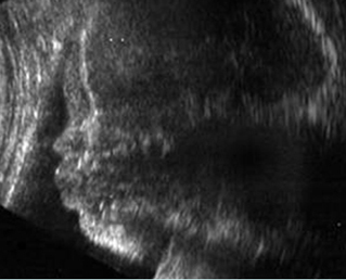
Fig 1: Normal facial profile view
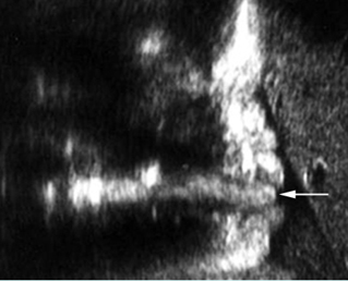
Fig 2: Tongue Facial profile view: normal tongue (arrow)
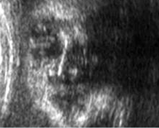
Fig 3: Normal face : Coronal scan of the normal face
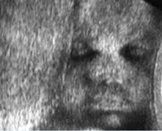
Fig 4: Normal face Coronal scan of the face: normal face
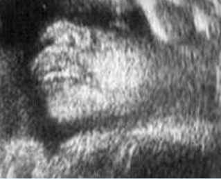
Fig 5: Normal face : Coronal scan of the face: nose with nostrils, lips and cheek
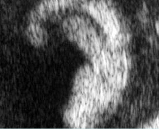
Fig 6: Normal ear
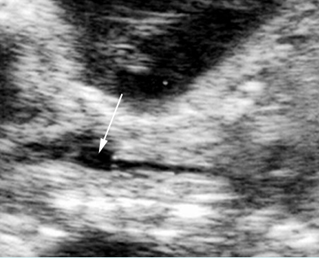
Fig 7: Hypolarynx Coronal scan of the neck: anechoic loculated fluid representing hypolarynx (arrow), continuing from the trachea (arrow)
Video clips of normal face
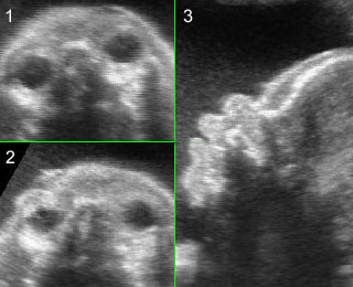
Normal facial profile: Rotation from biorbital view to the facial profile view (mid-sagittal view)
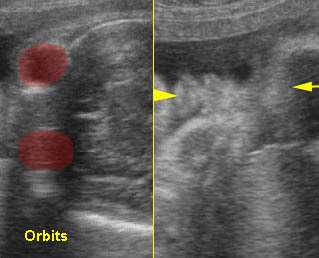
Normal face: Rotation from biorbital view to coronal view of the face: normal lip (arrowhead) (arrow = forehead)
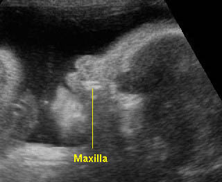
Normal facial profile : Speaking
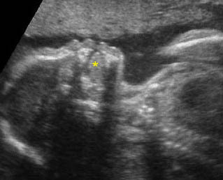
Normal facial profile : Normal facial profile (* = tongue)
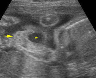
Yawn: Coronal scan: fetal yawning (* = opening mouth, arrow = nose)
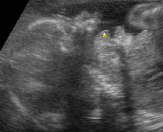
Yawn: Facial profile: scan: fetal yawning (* = opening mouth)
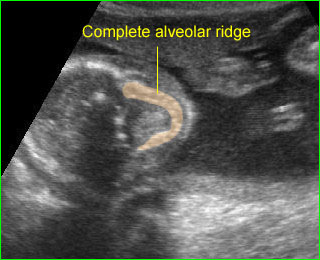
Alveolar ridge: Transverse scan of the fetal face at the level of alveolar ridge; note complete C-shape in case of no cleft palate
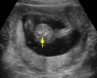
Normal neck: Cross-sectional scan of the fetal neck (arrow = cervical spine)
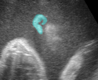
Normal ear
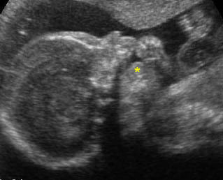
Tongue: Facial profile view: sucking the finger, (* = tongue)
Checklists
Axial view:
- normal for both orbits
- normal biorbital distance
- normal maxilla
Facial profile view:
- normal forehead (no bossing or flat)
- normal nose
- normal lips
- normal chin (no micrognathia)
Coronal view:
- normal upper lips
- normal nose and nostrils
- normal tongue (no persistent protrusion)
Ear:
- normal shape
- normal location
Neck:
- no abnormal mass
- no nuchal thickenings.
Pitfalls
- Fetal positioning, maternal abdominal wall thickness and oligohydramnios can interfere with obtaining the optimal images of the fetal face.
- Midline groove of the upper lip: be careful not to mistake this for cleft lip.
- Nuchal cord: the umbilical cord wraps around the neck, and the nuchal cord is sometimes mistaken for increased thickening of the skin fold. This could be simply overcome by performing either pulse Doppler or color Doppler.
- Non-fused amnion versus cystic neck mass: this can give the artificial appearance of a cystic mass around the occiput, therefore, one should check elsewhere within the uterus for incomplete fusion of the amnion or check the fetus when it has moved into a different anatomic position in which the neck is separate from the amnion.
- Pseudonuchal thickening: too steep angulation and more coronal plane will artificially give the appearance of increased thickness of the skin over the posterior occiput.

