Dandy-Walker Malformation

Dandy-Walker Malformation (DWM)

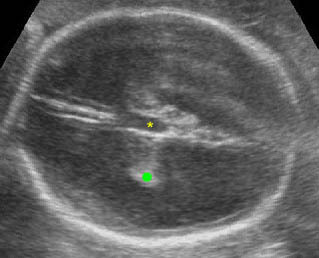
DWM is characterized by a posterior fossa cyst, a defect of the cerebellar vermis, resulting in a communication between the cyst and the fourth ventricle, and variable hydrocephalus. It is one of the most common midline cysts located within the posterior fossa. DWM represents marked genetic and etiologic heterogeneity. It can be subdivided into two subgroups: classic DWM (dilated fourth ventricle with absence of the cerebellar vermis) and DWM variant (partial absence of the cerebellar vermis with a small posterior fossa cyst).

Incidence: 1 per 30,000 births, occurring in 3.5-12% of cases of hydrocephalus.

Sonographic findings:

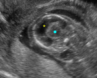
Fig 1, Fig 2, Fig 3
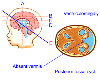
Fig 1: Schematic drawing: Posterior fossa cyst with absent cerebellar vermis in Dandy-Walker malformation
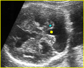
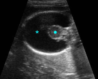
Fig 2: Dandy-Walker malformation  Transcerebellar view: posterior fossa cyst, separation of cerebellar hemisphere (*), absent vermis (solid circle)
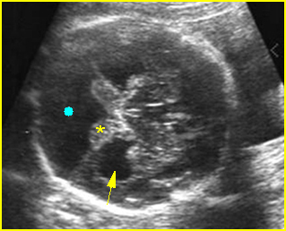
Fig 3: Dandy-Walker malformation  Transcerebellar view: posterior fossa cyst (solid circle), separation of cerebellar hemisphere (*), absent vermis (arrow = associated ventriculomegaly)

  • Partial or complete absence of the cerebellar vermis (the direct continuity of the posterior fossa cyst with the posterior aspect of the midbrain can be visualized without an intervening vermis).
  • Midline posterior fossa cyst caused by an enlargement of the fourth ventricle, communicating with cisterna magna.
  • Splaying of the cerebellar hemispheres.
  • Measurement of cisterna magna of more than 10 mm in classic DWM but normal in DWM variant.
  • Classic DWM is associated with ventriculomegaly (30-80% of cases)  but DWM variant is not.
  • The main differential diagnosis is arachonoid cyst, usually not located in the midline and not associated with absent vermis.
  • Pitfalls:
    • An enlarged cisterna magna of >10 mm, but with a normally formed cerebellum is often mistaken for DWM variant.
    • Isolated enlargement of the fourth ventricle at 14-16 weeks in the absence of vermian changes may be a normal variant.
  • A small cyst inferior to the cerebellum is often a normal variant.
  • Cerebellar hemorrhage followed by a porencephalic cyst of the cerebellum may mimic the classic form of Dandy-Walker malformation.
    • Usually diagnosed in the second half of pregnancy, although it has been detected as early as the first trimester.
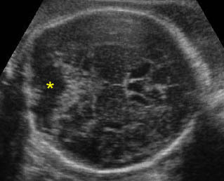
Dilated cisterna magna:  Transcerebellar view: dilated cisterna magna (*)
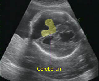
Dandy-Walker malformation:  Cystic dilatation of the posterior fossa with separation of the cerebellar hemisphere and the absence of the vermis
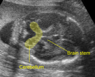
Dandy-Walker malformation:  Separation of the cerebellar hemisphere and the absence of the vermis

Associations: Associated intracranial anomalies are found in about 50% of cases and associated extracranial anomalies in about 35%. Chromosome abnormalities are seen in about 15-30%, especially trisomy 18. DWM is also associated with several other chromosome abnormalities and several syndromes and is X-link recessive in some cases.

Management: Termination of pregnancy may be offered when diagnosed for classic DWM before viability. In continuing pregnancies, a careful search for associated anomalies and postnatal follow-up is indicated. Early delivery may be considered when there is progressive and severe hydrocephalus.

Prognosis: High morbidity and mortality rate, mostly related with other anomalies, but good for DWM variant without associated anomalies.

Recurrence risk: About 1-5% for an isolated case.

Read More

Porencephaly / Schizencephaly

Porencephaly/Schizencephaly

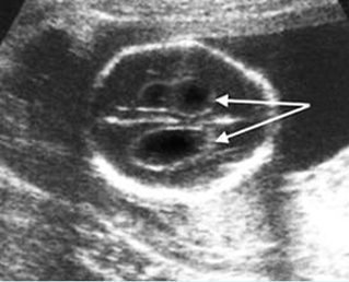
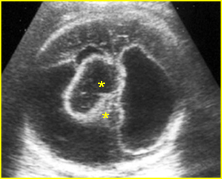
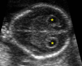
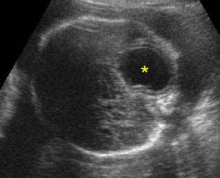
These two entities are often considered together because of their similar appearance. However, they have separate origins. Porencephaly is a locally destructive brain lesion resulting from either a developmental anomaly or intraparenchymal insult. There are two types of porencephaly. Type I is generally due to an antepartum intraparenchymal hemorrhage. Type II lesions are usually developmental anomalies. Various causes of parenchymal damage have been reported, such as trauma or inherited disease. Schizencephaly is a full-thickness cleft of the cerebral mantle considered to be a migrational abnormality rather than a destructive process. The etiology may include encephaloclastic disorder, cytomegalovirus or genetic disorders such as triple X syndrome.
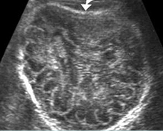
Sonographic findings:

Porencephaly:

Fig 1, Fig 2, Fig 3

  • a fluid-filled space within normal brain parenchyma, often communicating with the lateral ventricles
  • usually unilateral
  • often progressive changes to hydranencephaly (most severe form)
  • no pressure effect on the adjacent brain
  • the defect lined by white matter (demonstrated by MRI)

Schizencephaly:

Fig 4

  • unilateral or bilateral cystic lesion
  • may or may not communicate with the lateral ventricle
  •  typically bilateral clefts in the fetal brain connecting the lateral ventricles with the subarachnoid space
  • absence of the cavum septum pellucidum is commonly seen
  • MRI is very helpful in confirming the diagnosis
  •  the defect is lined by gray matter (demonstrated by MRI)

–     The main differential diagnoses include arachnoid cyst, interhemispheric cyst in the case of agenesis of the corpus callosum, and dorsal sac in holoprosencephaly.

–     Usually diagnosable after 18 weeks.

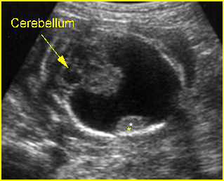
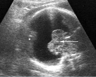
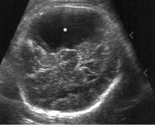
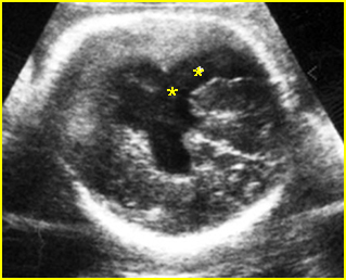
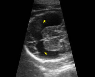
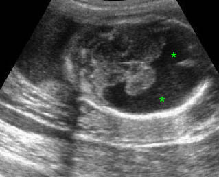
Fig 1: Porencephaly  Transthalamic view: only small part of brain tissue left (*)

Fig 2: Porencephaly  Most of the brain tissue is absent, only small part of brain tissue left (*). (arrow = thalami)

Fig 3: Porencephaly  Some part of cerebral tissue is absent (*)

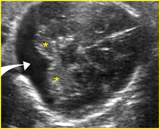
Fig 4: Schizencephaly  Cleft of cerebral tissue is absent (*) resulting in cystic brain lesion

Video clips of porencephaly / schizencephaly

Porencephaly: Transthalamic view: missing large piece of brain (*)

Porencephaly: Transverse scan of the head: fluid collection (*) in the large area of missing brain tissue

Associations: Rare.

Management: In continuing pregnancies, the fetus should be delivered in a tertiary center.

Prognosis: Depends on the size and location of the lesion; very poor for extensive porencephaly, and the bilateral form of schizencephaly.

Recurrence risk: Rare, though hereditary cases have been reported.

Read More

Hydranencephaly

Hydranencephaly

Hydranencephaly is the most severe form of destructive brain lesion, probably due to bilateral internal carotid artery occlusion as well as primary vascular malformations, and possibly intrauterine infection. It is characterized by a complete lack of cerebral tissue and there is covering calvarium, skin dura and leptomeninges.

Incidence: Sporadic occurrence with an incidence of 1-2.5 per 10,000 births.

Sonographic findings:

Fig 1, Fig 2

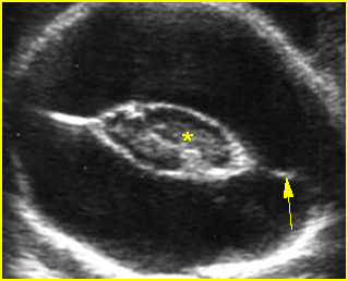
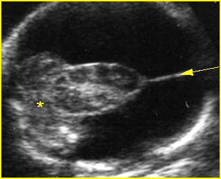
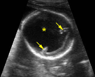
    Hydranencephaly:  Transverse scan of the skull: absent brain tissue, present falx cerebri (arrow), abnormal fused thalamus (*)

    Hydranencephaly:  Transverse scan of the skull: absent cerebral tissue, present falx cerebri (arrow), abnormal fused thalamus, and normal posterior fossa (*)

    Hydranencephaly:  Transthalamic view: totally destructive brain lesion (solid circle) but preserving thalamus (*) and posterior fossa

    • A fluid-filled cranium with a complete lack of cerebral tissue.
    • Presence of falx cerebri.
    • Macrocephaly.
    • Normal midbrain, brain stem, basal ganglia, and posterior fossa.
    • Polyhydramnios in most cases.
    • The main differential diagnosis is severe hydrocephalus, hydrolethalis and alobar holoprosencephaly, especially the pancake type (see holoprosencephaly).
    • Pitfalls:
      • The preserved dura and arachnoid can be confused with very severe hydrocephalus. Color Doppler proves useful for the differentiation. The presence of frontal cerebral cortex is indicative of severe hydrocephalus instead of hydranencephaly.
      • The initial diagnosis may be difficult when the infarction and hemorrhage is an evolving process. Recent hemorrhage is echogenic whereas an organizing clot has a more translucent texture. Layering of the debris may masquerade as cortical tissue.
    • Usually diagnosed between 20 and 30 weeks, although it may be detected as early as 11-13 weeks of gestation.

    Associations: Rare.

    Management: Termination of pregnancy should be offered.

    Prognosis: Uniformly fatal, though a case of survival of 20 years with a minimally conscious state was reported.

    Recurrence risk: Sporadic.

    Read More

    Holoprosencephaly

    Holoprosencephaly

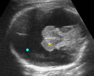
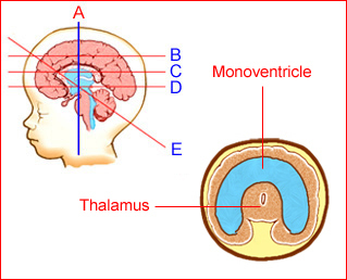
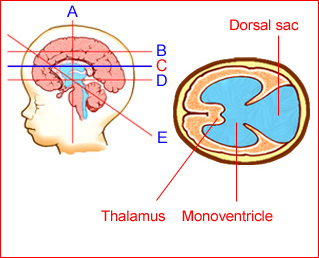
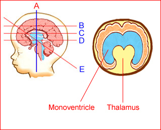
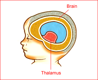
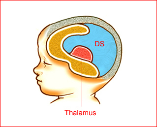
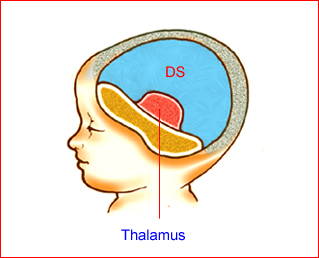
    Holoprosencephaly represents a broad spectrum of malformations due to a lack of separation of the structures of the forebrain, resulting in no midline separation of the cerebral hemispheres and diencephalic structures. The normally bilateral diencephalon and basal ganglia are fused and tend to incorporate into the upper brainstem. Recent discoveries in the fields of genetics and developmental neurobiology have advanced our knowledge of this complex disorder, such as 12 loci on 11 chromosomes, or deletion of distal 7q. It is usually classified as alobar (failure of the brain cleavage, complete fusion), semilobar (partially fused), or lobar (only fused frontal horns). The alobar form, the most common form, has a monoventricular cavity with fused thalami and can be subdivided into three separate configurations: pancake, cup, or ball forms. The pancake type, which is the rarest, occurs when the residual brain is minimal and compressed over the skull base. The ball variation occurs when the cerebral cortex covers the monoventricular cavity. The cup form is intermediate between the two, with the residual brain having a cup-like configuration on a sagittal view. It may also be divided into those with and without a dorsal sac. Thus, alobar holoprosencephaly without a dorsal sac is similar to the ball form, while the cup and pancake forms have a dorsal sac.
    Incidence: 1-2 per 10,000 live births and 1 per 250 embryos, male/female, 1:3 for the alobar form and 1:1 for the lobar form.

    Fig 1, Fig 2, Fig 3, Fig 4, Fig 5, Fig 6, Fig 7

    Fig 1: Schematic drawing: Coronal view shows monoventricle with fused thalamus in alobar holoprosencephaly

    Fig 2: Schematic drawing: Tranthalamic view shows monoventricle with fused thalamus in alobar holoprosencephaly

    Fig 3: Schematic drawing: Transventricular view shows mono-ventricle with dorsal sac in holoprosencephaly

    Fig 4: Schematic drawing: Coronal scan shows partial separation of the lateral ventricles in semilobar holoprosencephaly

    Fig 5: Schematic drawing: Ball type of holoprosencephaly (brain mantle completely covers the mono-ventricle

    Fig 6: Schematic drawing: Cup type of holoprosencephaly (DS = dorsal sac)

    Fig 7: Schematic drawing: Pancake type of holoprosencephaly (DS = dorsal sac)

    Sonographic findings:

    Alobar holoprosencephaly Fig 8, Fig 9, Fig 10

    • fused thalami
    • monoventricular cavity, lacking occipital, temporal, and frontal horns
    • absent falx cerebri
    • absent cavum septum pellucidum
    • dorsal sacs (cup and pancake type) communicating widely with monoventricular cavity
    • a ridge of cerebral tissue demarcating the boundary between the dorsal sac and the monoventricular cavity is often seen
    • often associated with facial abnormalities
    • the 3D prenatal US may be helpful, especially in the demonstration of various abnormalities of the face

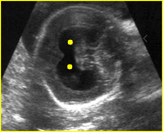
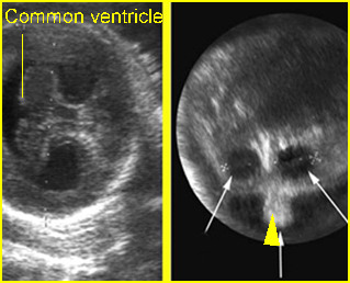
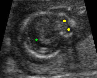
    Fig 8: Holoprosencephaly:  Oblique coronal scan of the skull: common ventricle (solid circle)

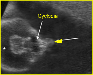
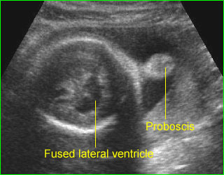
    Fig 9: Holoprosencephaly, cyclopia and proboscis  Common lateral ventricle (*), fused orbits (arrow), proboscis (arrowhead)

    Fig 10: Holoprosencephaly, hypotelorism and proboscis : Left: Fusion of the lateral ventricle Right: hypotelorism (arrow) and proboscis

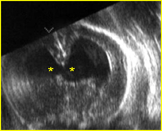
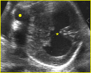
    Fig 11: Semilobar holoprosencephaly  Oblique coronal scan of the skull: common ventricle (*) with partial separation

    Fig 12: Semilobar holoprosencephaly  Oblique coronal scan of the skull: common ventricle (*) with partial separation (solid circle = orbits)

    Video clips of holoprosencephaly

    Holoprosencephaly: Transverse coronal scan of the fetal head: fusion of the lateral ventricle (*) (solid circle = fused thalamus)

    Holoprosencephaly:  Dorsal sac (*) with folding of the remaining brain tissue (arrow)

    Holoprosencephaly:  Dorsal sac (solid circle) with folding of the remaining brain tissue (arrow)

    Holoprosencephaly:  Transverse scan of the head: fusion of the lateral ventricle (*), no falx cerebri, marked hypotelorism (solid circle)

    Proboscis (holoprosencephaly) :  Midline solid mass at the forehead (proboscis) and fused ventricle

    Semilobar holoprosencephaly Fig 11, Fig 12

    • incompletely fused thalami
    • monoventricular cavity with partial separation
    • absent cavum septum pellucidum
    • presence of incomplete falx cerebri
    • often associated with facial abnormalities

    Lobar holoprosencephaly (rarely detected antenatally)

    • absent cavum septum pellucidum
    • nearly normal intracranial structures
    • abnormal course of the anterior cerebral artery on Doppler examination

    Common associated anomalies

    • face (most common): hypotelorism, cyclopia, proboscis, cleft lip and palate (especially midline cleft)
    • abdomen: renal agenesis or cystic dysplasia, esophageal atresia, omphalocele, bladder exstrophy
    • chest: pulmonary hypoplasia, cardiac malformations (VSD, ASD, transposition of great vessels)
    • others: myelomeningocele, polydactyly, clubfoot
    • chromosome abnormalities: trisomy 13 (most common), 13q-, trisomy 18, 8p-, and triploidy

    The main differential diagnoses are hydrocephalus and hydranencephaly:

    • hydrocephalus can be readily distinguished by visualization of falx cerebri and splayed thalami rather than fused thalami
    • hydranencephaly is characterized by complete absence of a cerebral hemisphere, variable presence of the falx cerebri and no facial abnormalities
    • a dorsal sac must be differentiated from other cysts such as arachnoid cyst, porencephaly, or Dandy-Walker malformation

    The alobar type can be diagnosed as early as 9-11 weeks of gestation.

    Associations: Alobar and semilobar holoprosencephaly are often associated with significant facial abnormalities, including cyclopia, ethmoidocephaly, cebocephaly, cleft lip, and trisomy 13. Other associated anomalies include cardiac defect, omphalocele, Dandy-Walker malformation and some cases of trisomy 18.

    Management: Termination of pregnancy should be offered and karyotyping and fetal autopsy should be performed.

    Prognosis: Fatal in most cases, or resulting in severe handicaps for the survivors.

    Recurrence risk: An empirical recurrent risk for sporadic cases is approximately 6%.

    Read More

    Head-Sign: Other Abnormalities

    Other Abnormalities

    Microcephaly

    Microcephaly is a condition involving the reduction of brain mass and head size with an intact bony calvarium, usually defined as a fetal head circumference of 3 SD, though some used 2 SD, below the mean of each gestational week.

    Sonographic findings:

    • The head is small, with a fetal head circumference of 3 SD.
    • The femur length/head circumference ratio is at 3 SD or higher.
    • Doppler ultrasound may show reduced or absent flow in the intracranial arteries, suggestive of a vascular cause.
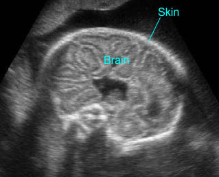
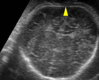
    • Severe microcephaly may be difficult to differentiate from anencephaly.
    • Microcephaly is diagnosed at a mean gestational age of 28 weeks, but may not be demonstrated until after 24 weeks, or even at late onset in the last trimester.

    Causes of microcephaly:

    • isolated microcephaly (16.7%)
    • microcephaly due to holoprosencephaly (16.7%)
    • microcephaly associated with chromosomal disorders (23.3%)
    • microcephaly as part of a genetic syndrome (20.0%)
    • microcephaly as part of multiple anomalies (23.3%)
    • microcephaly as part of intrauterine infections (i.e.: Zika virus)

    Intracranial Calcifications

    Intracranial calcifications have been rarely demonstrated prenatally. They most commonly result from intrauterine infection but are sometimes associated with intracranial tumor. The differential diagnoses of intracranial calcifications include

    • intrauterine infection (often also related to microcephaly, or ventriculomegaly, or growth restriction)
      • cytomegalovirus (most common): usually subependymal calcification
      • toxoplasmosis: usually scattered
      • varicella-zoster syndrome
      • rubella virus (very rare)
    • intracranial tumor (usually localized to tumor mass)
    • a normal second trimester ultrasound does not always exclude intracranial calcifications.

    Intracerebral hemorrhage:  
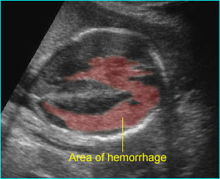
    Irregular echogenic mass in the area of lateral ventricle and cerebral mantle

    Specific abnormalities

    Some specific anomalies have a characteristic shape of the head.

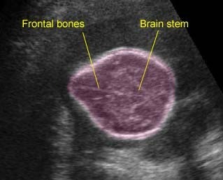
    Lemon sign: a concave deformity of the frontal bones at the level of the coronal suture, which may be associated with

    • normal fetus (1-2% of normal fetuses)
    • spina bifida (common)
    • occipital cephalocele (rare)
    • thanatophoric dysplasia (rare)
    •  long-term oligohydramnios

    Cloverleaf skull (found in 14% of cases of thanatophoric dysplasia)

    • thanatophoric dysplasia (most common)
    • homozygous achondroplasia
    • Apert’s syndrome
    • Pfeiffer’s syndrome
    • Carpenter’s syndrome

    Strawberry head shape (often associated with fetal trisomy 18).

    Read More

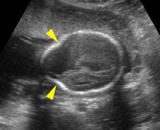
    Head-Sign: Abnormal Skull Shape

    Abnormal Skull Shape

    Deformed skull

    Fetal demise is the most common cause of deformed skull secondary to the compression effect. The severity of deformity can vary from mild Spalding’s sign and overlapping of the cranial sutures to a bizarre head shape. The diagnosis can simply be confirmed by demonstration of the fetal heartbeats. However, a deformed skull shape can be associated with other conditions. The differential diagnoses include:

    • fetal demise
    • amniotic band syndrome (bizarre and asymmetric cranial shape)
    • limb-body wall complex (bizarre and asymmetric cranial shape)
    • demineralization disorders
    • craniosynostosis (ranging from plagiocephaly to a trilobar or cloverleaf skull)
    • cephalocele.

        Fig 1, Fig 2, Fig 3

    Fig 1: Strawberry-shaped skull  Transverse scan of the skull: strawberry-shaped skull with dilated cisterna magna (arrow) (* = cerebellum)

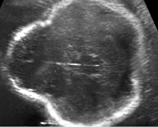
    Fig 2: Thanatophoric dysplasia  Cross-sectional scan of the skull: Cloverleaf skull; prominent parietal bone

    Fig 3: Lemon sign  Scalloping of the frontal bones (arrow) with ventriculomegaly in case of spina bifida

    Video clips of abnormal skull shape

    Lemon sign:  Indentation of the both frontal bones (arrowhead)

    Strawbery-shaped head: Strawberry head shape: the skull shape of a fetus with trisomy 18

    Specific abnormalities

    Some specific anomalies have a characteristic shape of the head.

    Lemon sign: a concave deformity of the frontal bones at the level of the coronal suture, which may be associated with

    • normal fetus (1-2% of normal fetuses)
    • spina bifida (common)
    • occipital cephalocele (rare)
    • thanatophoric dysplasia (rare)
    •  long-term oligohydramnios

    Cloverleaf skull (found in 14% of cases of thanatophoric dysplasia)

    • thanatophoric dysplasia (most common)
    • homozygous achondroplasia
    • Apert’s syndrome
    • Pfeiffer’s syndrome
    • Carpenter’s syndrome

    Strawberry head shape (often associated with fetal trisomy 18).

    Read More

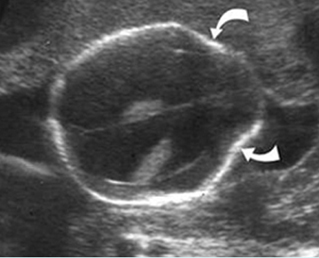
    Head-Sign: Sonolucent Skulls

    Sonolucent Skulls

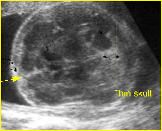
    Diffuse demineralization of the skull almost always occurs with fetal skeletal dysplasia syndromes. In the case of severe demineralization of the bony calvarium, the cranium is thin without an acoustic shadow and so poorly ossified that the intracranial structure can easily be seen. This increased visualization of the intracranial structures may be confused with such abnormalities as exencephaly due to acrania or acalvaria. Unlike exencephaly, however, there is an intact but poorly ossified skull. Careful scanning reveals concomitant limb anomalies.

          Fig 1, Fig 2

    Differential Diagnosis

    The main differential diagnoses of the sonolucent skull are as follows:

    • osteogenesis imperfecta (most common)
    • hypophosphatasia (rare)
    • achondrogenesis type I (rare)
    • acalvaria

    Fig 1: Hypophosphatasia  Cross-sectional scan of skull: thin and sonolucent calvarium (arrow = falx cerebri)

    Fig 2: Compressible skull  Poorly ossified skull associated with osteogenesis imperfecta is easily compressed (arrow) and the cerebral gyri is clearly visualized

    Video clips of sonolucent skulls

    Sonolucent skull:  The thin skull is so poorly ossified that cerebral gyri and sulci could be seen easily

    Acalvaria:  The head with intact skin but no skull, the gyri and sulci of brain can easily be seen

    Differential diagnosis

    The differential diagnoses and some sonographic features of skull defects are as follows:

    Anencephaly:

    • absent bony calvarium above the orbits
    • orbits well visualized
    • absence of supratentorial brain
    • residual brain
    • possibly spina bifida
    • polyhydramnios

    Excencephaly

    • calvarium absent
    • disorganized supratentorial brain tissue

    Amniotic band syndrome (ABS)

    • asymmetric or bizarre cephalocele
    • other deformities, including limb amputation

    Limb-body wall complex

    • asymmetric or bizarre encephaloceles
    • similar to ABS except fetal body adherent to placenta
    • usually more severe than ABS
    • associated bizarre defects of fetal body

    Cephalocele

    • midline defect
    • extracranial cyst or brain tissue
    • possibly ventriculomegaly
    • lemon sign may be present

    Open neural tube defects

    • lemon sign
    • banana sign
    •  mild ventriculomegaly
    • spinal defect

    Fetal demise

    • overlapping sutures (Spalding’s sign)
    • poor visualization of intracranial structures
    • associated findings of fetal demise

    Microcephaly

    • calvarium present
    • decreased brain tissue
    • head circumference 2-3 standard deviations below that expected for the menstrual age

    Craniosynostosis

    • complete or partial
    • deformed skull
    • possibly microcephaly
    • abnormal cephalic index.

    In addition, considerable overlap may be found in these features among abnormalities. For instance, the lemon sign was originally described with opened NTD, however, it may not be present in an opened NTD identified in the third trimester of pregnancy, and a mild lemon sign may be identified in normal fetuses.

      Read More

      Head-Sign: Skull Defects

      Skull Defects

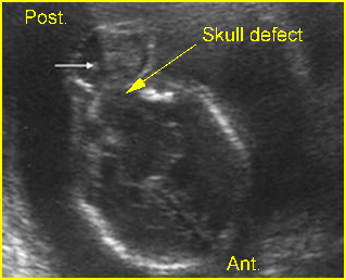
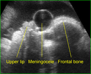
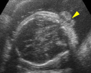
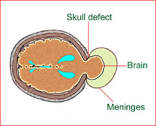
      Cranial defects vary from a small meningocele, which is very difficult to visualize, to complete absence of the cranium such as with anencephaly or exencephaly. In practice, there is no problem in the diagnosis of complete absence of the skull or a large defect, however, severe bone demineralization, such as osteogenesis imperfecta, may give the appearance of a large skull defect. Partial defects, or small cephaloceles are occasionally difficult to visualize.

      Fig 1, Fig 2, Fig 3

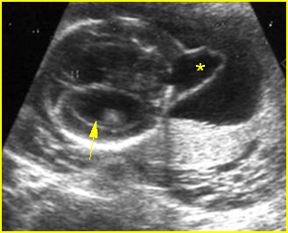
      Fig1: Schematic drawing: Occipital meningocele, protrusion of the brain tissue and meninges through the skull defect

      Fig 2: Occipital cephalocele  Transverse scan of the skull: small defect of occipital bone with meningocele (*) (arrow = ventriculomegaly)

      Fig 3: Occipital cephalocele  Transverse scan of the skull: defect of occipital bone with meningoencephalocele (arrow)

      Video clips of intracranial massive fluid collection

      Anterior meningocele:  Sagittal scan of the fetal face: frontal bone defect with midline cystic mass (meningocele)

      Occipital cephalocele:  Transverse scan the level of cerebellum: small defect of occipital bone with encephalocele

      Differential diagnosis

      The differential diagnoses and some sonographic features of skull defects are as follows:

      Anencephaly:

      • absent bony calvarium above the orbits
      • orbits well visualized
      • absence of supratentorial brain
      • residual brain
      • possibly spina bifida
      • polyhydramnios

      Excencephaly

      • calvarium absent
      • disorganized supratentorial brain tissue

      Amniotic band syndrome (ABS)

      • asymmetric or bizarre cephalocele
      • other deformities, including limb amputation

      Limb-body wall complex

      • asymmetric or bizarre encephaloceles
      • similar to ABS except fetal body adherent to placenta
      • usually more severe than ABS
      • associated bizarre defects of fetal body

      Cephalocele

      • midline defect
      • extracranial cyst or brain tissue
      • possibly ventriculomegaly
      • lemon sign may be present

      Open neural tube defects

      • lemon sign
      • banana sign
      •  mild ventriculomegaly
      • spinal defect

      Fetal demise

      • overlapping sutures (Spalding’s sign)
      • poor visualization of intracranial structures
      • associated findings of fetal demise

      Microcephaly

      • calvarium present
      • decreased brain tissue
      • head circumference 2-3 standard deviations below that expected for the menstrual age

      Craniosynostosis

      • complete or partial
      • deformed skull
      • possibly microcephaly
      • abnormal cephalic index.

      In addition, considerable overlap may be found in these features among abnormalities. For instance, the lemon sign was originally described with opened NTD, however, it may not be present in an opened NTD identified in the third trimester of pregnancy, and a mild lemon sign may be identified in normal fetuses.

        Read More

        Head-Sign: Cystic Brain Lesions

        Cystic Brain Lesions

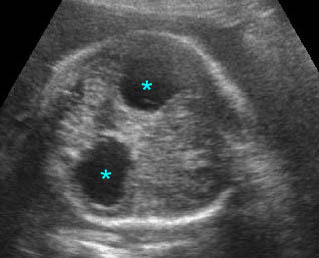
        Cystic lesions of the brain have a number of causes but ventriculomegaly and choroid plexus cyst are the most common. Usually, the location and associated abnormalities are very helpful in the differential diagnosis. For example, interhemispheric cyst is commonly related to agenesis of the corpus callosum. Likewise, ventriculomegaly and dorsal sac in the case of holoprosencephaly are often associated with other abnormalities, whereas others are mostly isolated findings. The choroid plexus cyst is always a lateral cyst located in the choroid plexus and is often isolated and not difficult to diagnose. Cystic lesions of Dandy-Walker malformations are always located in the posterior fossa.

        Fig 1, Fig 2, Fig 3, Fig 4

        Fig1: Choroid plexus cysts  Transverse scan of the skull: bilateral large choroid plexus cyst (arrow)

        Fig 2: Porencephaly Some part of cerebral tissue is absent (*)

        Fig 3: Ependymoma  Complex cystic mass (*) in the skull combined with fluid collection and partial absence of brain tissue

        Fig 4: Semilobar holoprosencephaly  Oblique coronal scan of the skull: common ventricle (*) with partial separation

        Video clips of cystic brain lesions

        Choroid plexus cyst: Transvere scan at the level of lateral ventricles: bilateral choroid plexus cyst (*) with lemon sign

        Arachnoid cyst: Transverse scan of the head: isolated cystic, circular, thin-walled brain lesion (*)

        Colpocephaly:  Transverse scan of the head: bilateral marked dilatation of occipital horn (*) of lateral ventricles

        Differential diagnosis

        The differential diagnoses of intracranial cystic lesion are as follows:

        Midline cyst

        • prominent cavum septum pellucidi (very common)
        • dorsal sac of holoprosencephaly (common)
        • interhemispheric cyst of agenesis of corpus callosum (uncommon)
        • vein of Galen aneurysm (rare)
        • arachnoid cyst (rare)
        • enlarged cavum vergae interhemispheric cystic lesions

        Lateral cyst

        • choroid plexus cyst (very common)
        • unilateral hydrocephalus (uncommon)
        • porencephaly/schizencephaly (uncommon)
        • arachnoid cyst (rare)
        • neoplastic cyst (very rare)
        • periventricular cyst formation secondary to CMV infection

        Posterior fossa cyst

        • enlarged cisterna magna (>10 mm (very common)
        • Dandy-Walker malformation (common)
        • arachnoid cyst (very rare)
        • median retrocerebellar fluid collections remain the most difficult to prognosticate, retrocerebellar cysts often being difficult to differentiate from enlarged cisterna magna and Dandy-Walker complex.
        Read More

        Head-Sign: Massive Intracranial Fluid Collections

        Massive Intracranial Fluid Collections

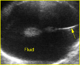
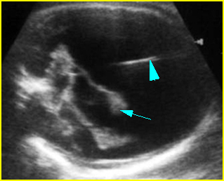
        The three common causes of massive intracranial fluid collections with an intact cranial vault are hydrocephalus, which is usually secondary to aqueductal obstruction, holoprosencephaly, and hydranencephaly. The unique features of each condition can distinguish these three entities from each other.

        Fig 1, Fig 2

        Fig1: Hydranencephaly  Transverse scan of the skull: absent brain tissue, present falx cerebri (arrow)

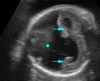
        Fig 2: Severe hydrocephalus Markedly dilated ventricles with extremely thin brain, choroid plexus dangling (arrow) (arrowhead = falx cerebri)

        Video clips of intracranial massive fluid collection

        Holoprosencephaly: Absent brain tissue (*), without falx cerebri, and fused thalamus (solid circle)

        Hydranencephaly: Transthalamic view: totally destructive brain lesion (solid circle) but preserving thalamus (*) and posterior fossa

        Severe hydrocephalus: Transverse scan of the head: markedly dilated ventricles with extremely thin cerebral tissue (solid circle = dangling choroid plexus, * = dilated third ventricle)

        Differential diagnosis

        • Hydrocephalus secondary to aqueductal stenosis (about 1 in 2000 births):
          • thin cerebral mantle with falx cerebri
          • separated thalami and dilated third ventricle
          • obvious choroid dangling sign
          • usually no midline face defects
        • Hydranencephaly (uncommon; 1.0-2.5 in 10,000 births):
          •  no cerebral mantle with falx cerebri
          • normal thalami (not separated or fused)
          • minimal choroid dangling sign
          • no midline face defects
        • Holoprosencephaly (rare; 1 in 16,000 neonates):
          • cerebral mantle with or without dorsal sac, no falx cerebri
          • fused thalami
          • minimal choroid dangling sign
          • associated midline face defects (very common).
        Read More