Facial Clefts

Facial Clefts

(See the details in the section “Specific abnormalities”.)

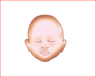
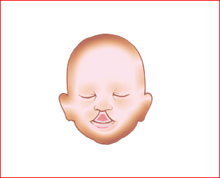
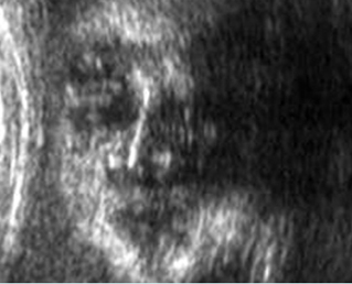
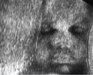
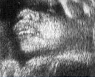
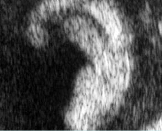
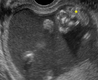
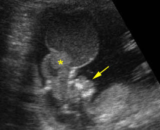
Fig 1, Fig 2

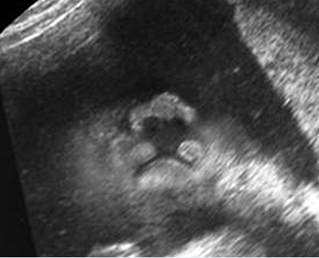
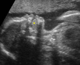
Premaxillary protrusion is an important clue to the presence of cleft lip and cleft palate and may be more conspicuous than the cleft itself.

Differential diagnosis for facial clefts

  • Artifact: normal philtrum with midline groove of the upper lip misinterpreted as cleft
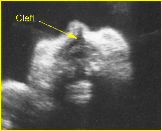
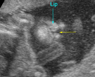
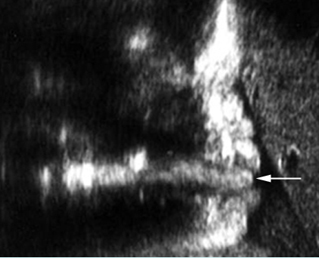
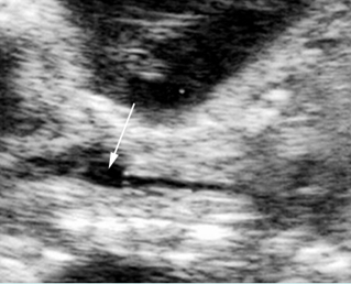
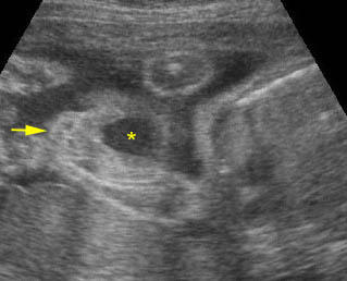
  • Lateral cleft: Fig 3, Fig 4
    • most common facial abnormalities, more common on the left side
    • paramedial location
    • bilateral in 20% of cases
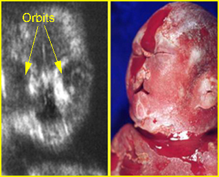
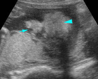
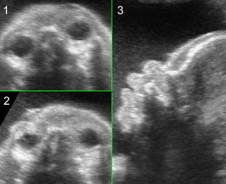
  • Midline cleft: Fig 5
    • rare
    • holoprosencephaly
    • median cleft face syndrome
  • Atypical cleft:
    • associated with amniotic band syndrome in most cases
    • asymmetric and bizarre
    • other anomalies are commonly seen.

Fig 1: Schematic drawing: acial abnormality related to holoprosencephaly: Bilateral clefts

Fig 2: Schematic drawing: Facial abnormality related to holoprosencephaly: Midline cleft

Fig 3:Paramedian cleft  Coronal view of the face: paramedian cleft lip

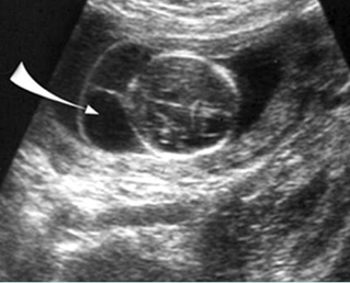
Fig 4:  Paramedian cleft  Coronal view of the face: paramedian cleft lip (arrow)

Fig 5: Median cleft lip  Coronal view of the face: median cleft lip in fetal trisomy 13

Video clips of facial clefts

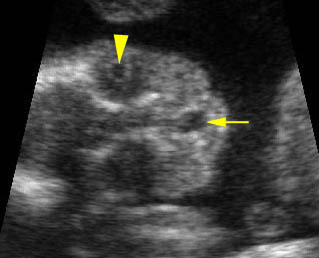
Cleft lip:  Coronal scan of the face: small paramedial cleft (arrow)

Cleft lip:  Coronal scan of the face: paramedial cleft (arrow)

Bilateral cleft lips :  Coronal scan of the face: bilateral paramedial cleft (arrow)

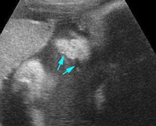
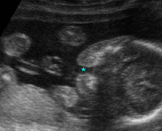
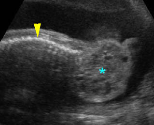
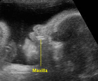
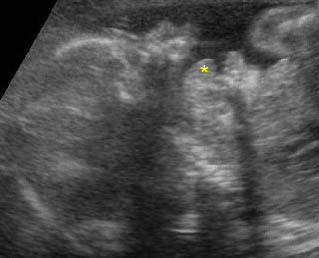
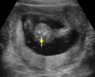
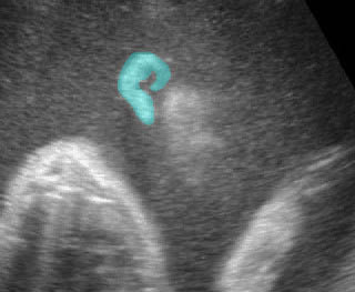
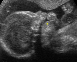
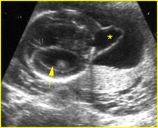
Cleft lip and palate :  Transverse scan of the face: incomplete alveolar ridge (*) of the maxilla

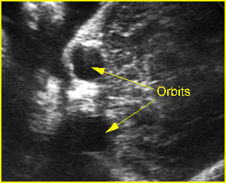
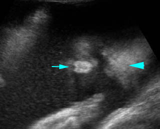
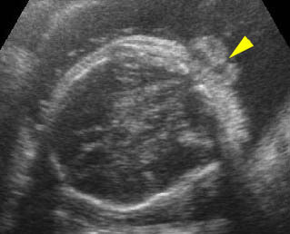
Midline cleft lip:  Coronal scan of the face: midline cleft (arrow) (arrowhead = lens in the orbit)

Read More

Hypotelorism

Hypotelorism

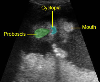
Hypotelorism, abnormally close spacing of the orbits, is an uncommon disorder, which is usually associated with many other conditions including chromosome defects. Holoprosencephaly is the most common associated malformation. It should be diagnosed when both the interocular and outer or binocular distances are below the 5th percentile, usually first diagnosable in the second trimester, but possibly in the first trimester. The extreme case, median ocular fusion or cyclopia, is almost always related to holoprosencephaly. Binocular biometric parameters may be useful sonographic markers for trisomy 13 or holoprosencephaly.

Fig 1

The differential diagnoses of hypotelorism include:

  • Holoprosencephaly (most common)
  • Median facial clefts
  • Chromosome abnormalities, especially trisomy 13
  • Meckel syndrome
  • Microcephaly.

Fig 1: Hypotelorism  Coronal view of the face: hypoteloism associated with trisomy 13

Video clips of neck masses

Hypotelorism & Proboscis:  Coronal scan of the face: arrowhead = forehead, arrow = proboscis)

Hypotelorism & Proboscis:   Coronal scan of the face showing a proboscis above the cyclopia

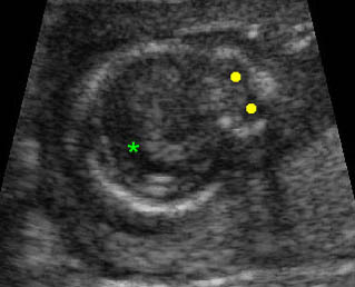
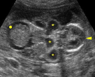
Holoprosencephaly:  Transverse scan of the head: fusion of the lateral ventricle (*), no falx cerebri, marked hypotelorism (solid circle)

Read More

Neck Masses

Neck Masses

          Fig 1, Fig 2

Differential diagnosis of common neck masses:

  • Cystic hygroma (most common; multiseptate, no solid part; posterolateral; bilateral, hydrops)
  • Cervical meningomyelocele (cystic, complex or solid; posterior midline, separation of cervical spine)
  • Occipital cephalocele (cystic, complex or solid; posterior midline, skull defect)
  • Goiter (generalized hypoechoic solid; anterior, bilateral)
  • Cervical teratoma (solid-cystic; anterolateral, unilateral)
  • Hemangioma (echogenic, variable in location, positive Doppler signals).

Fig 1: Cystic hygroma   Oblique cross-sectional scan at the level of cerebellum: anechoic cyst (arrow) with central septum

Fig 2: Cervical hemagioma   Cross-sectional scan at the neck: enlarged complex mass lateral to the cervical spine (positive Doppler signal)

Video clips of neck masses

Cervical teratoma:  Sagittal scan of the thorax and neck: solid-cystic mass (*) lateral to the neck (arrowhead = spine)

Cystic hygroma:  Coronal scan of the fetal trunk: multiple cystic areas (*) around the neck as well as ascites (solid circle)

Read More

Facial Masses

Facial Masses

Most abnormal masses of the face are located in the periorbital areas.

Fig 1, Fig 2

The major differential diagnoses for periorbital masses are as follows:

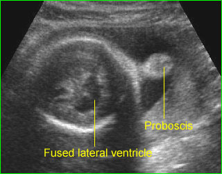
  • Proboscis: solid mass of abnormal nose formation, almost always associated with hypotelorism and holoprosencephaly.
  • Daccryocystoceles: lacrimal duct cyst, hypoechoic mass inferomedial to the fetal orbit without displacement of the globe but with synchronous eye movements.
  • Hemangioma: cystic or solid, placenta-like echogenicity, well-defined vascular space with characteristic Doppler signal.
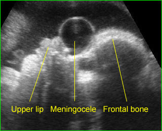
  • Anterior cephalocele: usually midline mass, displacing orbit inferiorly and laterally, anterior cranial defect.
  • Teratoma or dermoid cyst: solid or cystic or complex, occasional calcifications.
  • Anterior cystic hygroma: (rare) cystic or multicystic with loculations and septations varying in size.

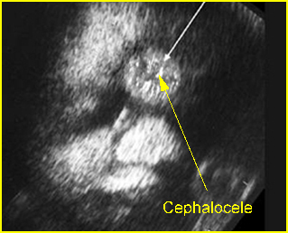
Fig 1: Anterior cephalocele  Coronal scan of the face: round solid mass above the nose (arrow)

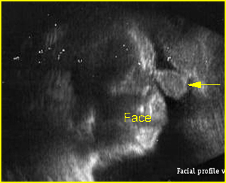
Fig 2: Proboscis  Facial profile view: proboscis (arrow), absent nose

Video clips of facial masses

Proboscis (holoprosencephaly) :  Midline solid mass at the forehead (proboscis) and fused ventricle

Anterior meningocele:  Sagittal scan of the fetal face: frontal bone defect with midline cystic mass (meningocele)

Read More

Face: Normal Examination

Normal Examination

Practically, most practitioners evaluate the face qualitatively, using three views: the coronal view, looking at bone and soft tissue from the surface well into the palate and orbits; the transverse view, looking at the mandible, maxilla, tooth buds, and orbits; and finally the sagittal view, evaluating the nasal bridge, the frontal bone, and the mandible. The measurements of various aspects of the fetal face have been studied in an attempt to quantify facial anomalies. Normative data of the fetal eye length, nose width and nostril distance, fetal upper lip and chin, binocular distance, and biparietal distance (BPD)/mandibular body length (MBL) ratio for diagnosis of micrognathia have been established. These data are expected to serve as a basis for the objective assessment of the fetal face in high-risk conditions.

Technique:

Fig 1, Fig 2, Fig 3, Fig 4, Fig 5, Fig 6, Fig 7

 Axial view

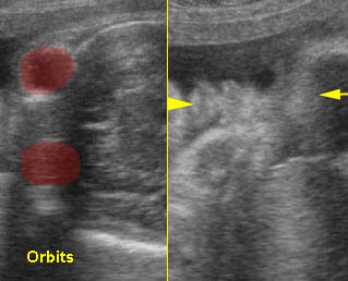
  • From the BPD plane, move the transducer down to the face slowly to obtain the biorbital view (axial orbit view). Orbital sizes and distances could be assessed. The biorbital distance can be used in estimating the gestational age and allows detection of hypo-/hypertelorism.
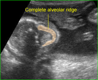
  • Move the transducer down directly to the lower level of the face. Maxilla (smooth C-shaped curve of the alveolar ridge). This is important for exclusion of cleft lip/palate. Furthermore, tooth sockets can also be assessed and the exact number after 19 weeks can be determined.
  • Normal scan: symmetry of the orbits, normal inter and intra-orbital diameter.
  • Abnormalities identified with the axial orbit view included hypotelorism, hypertelorism, microphthalmia, intra-ocular abnormalities, and periorbital mass, and with the axial maxilla view they included anterior cleft palate associated with cleft lip.

 Coronal view

  • At the biorbital view, slide and rock the transducer so that the biorbital diameter becomes parallel to the vertical line, and move the biorbital diameter line to the middle of the image.
  • Rotate the transducer 90 degrees with fine adjustment to get the coronal view; the variations of movement will reveal various images of the coronal view of upper lip, lower lip, nose, nostrils, chin, or cheeks.
  • Normal scan: normal lips, nose (nostrils), chin, eyelids, or lens should be seen. Fetal blinking is very common and is increased in frequency with vibroacoustic stimulation.
  • Abnormalities identified with this view included cleft lip and oropharyngeal mass.
  • The coronal and sagittal planes obtained from the submandibular triangle can be used well for displaying the soft and hard palates in particular.

Facial profile view (sagittal midline view)

  • At the biorbital view, slide and rock the transducer so that the biorbital diameter becomes perpendicular to the ultrasound beam, and move the biorbital diameter line to the middle of the image.
  • Rotate the transducer 90 degrees with fine adjustment to get the sagittal view; the variations of movement will reveal various images of the facial profile view, especially the relationship of the forehead, nose, lip, and chin.
  • Normal scan: normal forehead, nose, lips and chin and no abnormal masses.
  • Evaluation of the jaw is usually subjective, however mandibular measurement may be necessary in some cases.
  • Abnormalities identified with this view included micrognathia, premaxillary protrusion, proboscis, frontal bossing, flattened nasal bridge, and mass (neck, oral).

Ear

  • From the facial area, move the transducer laterally to identify the ear and make fine adjustments to reveal various views of the ear. It is easily identified as a complex soft tissue protrusion externally to the skull.
  • The location in relation to the skull and the shape of the ear should be demonstrated.
  • Abnormalities identified with this examination included ear malformation, low set ear.

Neck

  • Move the transducer downward from the axial plane of the face to the neck or just below the occipital region from the transcerebellar view; fine manipulation of the transducer is needed to visualize the axial plane of the neck.
  • Nuchal skin thickness should be routinely evaluated in the transcerebellar plane; the critical landmarks include the cavum septum pellucidi, cerebral peduncles and cerebellar hemispheres. If the transducer is not angled in the correct plane but is too steep this will produce incorrect measurements for nuchal skin thickness.
  • The cross-section of the neck appears round, containing hyperechoic spine and soft tissue structures anteriorly including esophagus, trachea, larynx and great vessels.
  • Examination of the posterior neck and the base of the skull is important to detect abnormalities such as cystic hygroma, occipital cephalocele, and cervical meningomyelocele.
  • Examination of the anterior and anterolateral neck is important to detect such abnormalities as goiter, hemangioma and teratoma. In the anterior neck, thyroid and the fluid-filled trachea and hypopharynx can normally be seen.

4D-US may be superior over 2D and 3D-US in the evaluation of complex facial activity and expression. Facial surface analysis and expressions such as eyelid and mouthing movements, smiling and scowling can be precisely observed using 4D-US.

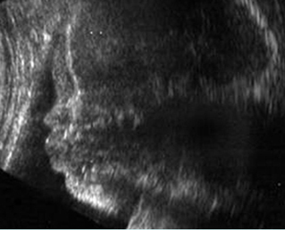
Fig 1: Normal facial profile view

Fig 2: Tongue  Facial profile view: normal tongue (arrow)

Fig 3: Normal face :  Coronal scan of the normal face

Fig 4: Normal face  Coronal scan of the face: normal face

Fig 5: Normal face :  Coronal scan of the face: nose with nostrils, lips and cheek

Fig 6: Normal ear

Fig 7: Hypolarynx  Coronal scan of the neck: anechoic loculated fluid representing hypolarynx (arrow), continuing from the trachea (arrow)

Video clips of normal face

Normal facial profile:  Rotation from biorbital view to the facial profile view (mid-sagittal view)

Normal face:  Rotation from biorbital view to coronal view of the face: normal lip (arrowhead) (arrow = forehead)

Normal facial profile : Speaking

Normal facial profile : Normal facial profile (* = tongue)

Yawn:  Coronal scan: fetal yawning (* = opening mouth, arrow = nose)

Yawn:  Facial profile: scan: fetal yawning (* = opening mouth)

Alveolar ridge:  Transverse scan of the fetal face at the level of alveolar ridge; note complete C-shape in case of no cleft palate

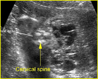
Normal neck:  Cross-sectional scan of the fetal neck (arrow = cervical spine)

Normal ear

Tongue:  Facial profile view: sucking the finger, (* = tongue)

Checklists

Axial view:

  • normal for both orbits
  • normal biorbital distance
  • normal maxilla

Facial profile view:

  • normal forehead (no bossing or flat)
  • normal nose
  • normal lips
  • normal chin (no micrognathia)

Coronal view:

  • normal upper lips
  • normal nose and nostrils
  • normal tongue (no persistent protrusion)

Ear:

  • normal shape
  • normal location

Neck:

  • no abnormal mass
  • no nuchal thickenings.

Pitfalls

  • Fetal positioning, maternal abdominal wall thickness and oligohydramnios can interfere with obtaining the optimal images of the fetal face.
  • Midline groove of the upper lip: be careful not to mistake this for cleft lip.
  • Nuchal cord: the umbilical cord wraps around the neck, and the nuchal cord is sometimes mistaken for increased thickening of the skin fold. This could be simply overcome by performing either pulse Doppler or color Doppler.
  • Non-fused amnion versus cystic neck mass: this can give the artificial appearance of a cystic mass around the occiput, therefore, one should check elsewhere within the uterus for incomplete fusion of the amnion or check the fetus when it has moved into a different anatomic position in which the neck is separate from the amnion.
  • Pseudonuchal thickening: too steep angulation and more coronal plane will artificially give the appearance of increased thickness of the skin over the posterior occiput.
Read More

Other Disorders

Arachnoid Cysts

An arachnoid cyst is the accumulation of cerebrospinal-like fluid between the meninges, arising from the arachnoid separating into two layers. The cells lining the cyst wall produce CSF, which is trapped to form a cyst. The slipstreams of CSF within arachnoid cysts may not be channeled properly leading to possible damage of the surrounding brain parenchyma.

Incidence: Rare, sporadic occurrence, accounting for 1% of the intracranial lesions. Most are located above the tentorium and 5-10% occur in the posterior fossa. It has a male preponderance and increased incidence on the left side.

Sonographic findings:

  • A well circumscribed, often circular fluid-filled space with a thin wall, located anywhere within the subarachnoid space.
  • Usually displaces adjacent brain structures.
  • Hydrocephalus secondary to a mass effect may be seen in some cases, however, most do not disturb normal brain function.
  • The differential diagnosis for asymmetric cyst is porencephaly and for the midline cyst includes an interhemispheric cyst in the case of agenesis of the corpus callosum, Dandy-walker malformation, and dorsal sac of holoprosencephaly.
  • MRI is helpful if the diagnosis is uncertain.
  • Pitfalls:
    • The cyst is sometime beneath the subcutaneous lesion mimicking meningocele.
    • They may be missed if located in the portion of the brain closest to the transducer.
  • Usually diagnosed in the second half of pregnancy, though may be detected as early as 13 weeks.

Associations: Rare.

Management: In general, isolated arachnoid cyst does not alter standard obstetric management.

Prognosis: Good, unless it is associated with other anomalies; most are asymptomatic, but they can cause neurological signs and symptoms, but good outcomes with neurosurgical intervention. Prenatal resolution can also occur.

Recurrence risk: Rare.

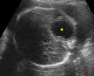
Arachnoid cyst:  Transverse scan of the head: isolated cystic, circular, thin-walled brain lesion (*)


Vein of Galen Aneurysm

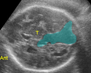
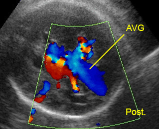
Vein of Galen aneurysm is dilatation of the vein ranging from a large single arteriovenous malformation to multiple smaller communications. It is a rare but pathognomonic midline intracranial cyst, located posterior and slightly superior to the thalami within the subarachnoid space.

Incidence: Unknown.

Sonographic findings:

  • Well-defined midline tubular cyst just posterior and superior to thalami.
  • Demonstration of the feeding vessels with arterial or venous flow by Doppler and color-flow study. Moreover, pulsation in the vein of Galen is also suggestive of fetal compromise and occurs earlier than those in umbilical vein.
  • High-output congestive heart failure, polyhydramnios, hydrops fetalis, and sequele of the aneurysm may be seen in some cases.
  • Three-dimensional color power angiography aids in visualization of the feeder vessels.
  • Differential diagnoses
    • interhemispheric cyst associated with agenesis of the corpus callosum
    • arachnoid cyst
    • dorsal sac associated with holoprosencephaly
    • the diagnosis of vein of Galen aneurysms is rapidly established with color and pulsed Doppler.
  • Pitfalls:
    • A quadrigeminal cistern, a normal cystic structure posterior to the third ventricle, may be mistaken for the aneurysm, however, Doppler ultrasound will show no flow.
    • Centrally placed arachnoid cyst can be confused with the aneurysm, however, Doppler ultrasound will show no flow.
  • Usually first diagnosable in the third trimester, but possible as early as 14 weeks.

Associations: Most cases occur as isolated anomalies but cardiac defects, cystic hygroma and hydrocephalus may be seen in some cases. Pulmonary hypertension, and high-output heart failure or hydrops fetalis can be a consequence of the aneurysms.

Management: Termination of pregnancy may be offered, especially in the case of hydrops. In continuing pregnancies, serial sonographic monitoring to identify early signs of hydrops is indicated. Elective cesarean section when fetal lung maturity is attained may improve prognosis.

Prognosis: Poor in cases of heart failure or hydrocephalus, with numerous feeder vessels, a wide draining vein, and steal retrograde aortic flow; better in selected cases without suprasystemic pulmonary hypertension with appropriate surgical correction such as a staged endovascular technique or embolization. The malformations can also spontaneously disappear in rare cases.

Recurrence risk: Rare.

Aneurysm of vein of Galen:  Midline tubular cystic space, predominantly located posteriorly (Ant: anterior; T: thalamus)

Aneurysm of vein of Galen (AVG):  Midline tubular cystic space with circulating blood flow (Post: posterior)

Intracranial Hemorrhage In Utero

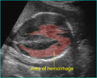
Fetal intracranial hemorrhage has rarely been observed. Such hemorrhage usually originates from germinal matrix with rupture into the ventricles. This may be due to the extreme capillary permeability of the vascular germinal matrix, which makes it susceptible to hemorrhage and often related to maternal factors, such as preeclampsia, or bleeding disorders. Germinal matrix or intraventricular hemorrhage is commonly classified in four grades as follows: Grade I: limited to subependymal matrix, Grade II: intraventricular extension, but without ventriculomegaly, Grade III: intraventricular extension, with ventriculomegaly, Grade IV: intraparenchymal extension.

Incidence: About 1 in 1000 pregnancies, very common in premature infants following delivery, may be as high as 40% among infants of <32 weeks.

 Sonographic findings:

  • The sonographic appearance varies with the location, size, and age of hemorrhage.
  • An echogenic clot filling the ventricles followed by ventriculomegaly. Once the hemorrhage resolves, only ventriculomegaly may be evident.
  • Hydrocephalus with mixed echogenic debris layering within the ventricles.
  • Large hemorrhages may produce a marked mass effect.
  • Subdural hematomas appear as echogenic or complex fluid collections just beneath the cranium, displacing and distorting the brain.
  • Occasionally, mixed echogenic, enlarged choroid plexus results from hemorrhage into the choroid plexus.
  • Porencephalic cyst following the hemorrhage may be seen.
  • Mostly occurs in the beginning of the third trimester of pregnancy.
  • The primary differential diagnosis includes intracranial neoplasm or infection.
  • MRI is helpful for differentiating intracranial echogenicities suspected of hemorrhage.

Associations: No specific anomaly.

Prognosis: Depends on the extent of hemorrhage and intraparenchymal hemorrhage. The outcome is usually poor, especially for those fetuses affected by higher-grade intraventricular hemorrhages, whereas it is better in the subgroup with intraventricular hemorrhage.

Intracerebral hemorrhage:  Irregular echogenic mass in the area of lateral ventricle and cerebral mantle

Intracranial Neoplasms

Fetal intracranial neoplasms are rare and most occur in the perinatal period. Teratoma is the most common tumor detected antenatally, accounting for 62% of cases.

Incidence: Approximately 0.34 per 1 million live births.

Sonographic findings:

  • Usually irregular in appearance.
  • Mostly echogenic mass with occasional cystic portions with or without distortion of normal symmetrical intracranial structures, calcifications.
  • Located anywhere within the brain.
  • Craniomegaly, obstructive hydrocephaly seen in some cases.
  • Associated polyhydramnios and hydrops fetalis occasionally seen.
  • Infrequently abnormal cerebral Doppler flow velocimetry.
  • Prenatal ultrasonography had high specificity in the diagnosis of brain neoplasms but the accuracy in predicting the histological type was limited.
  • Most diagnosed in late second and third trimesters.

 Association: No.

Management: Termination of pregnancy can be offered when diagnosed before viability.

Prognosis: Usually poor, though postnatal successful resection has been reported.

Recurrence risk: Rare.

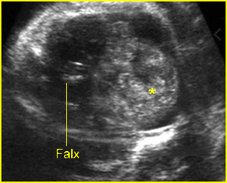
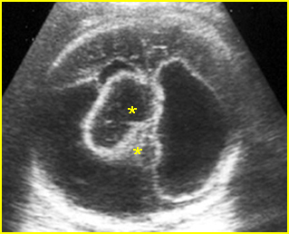
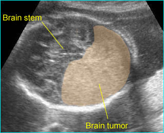
Fig 1: Fetal brain tumor   Abnormal complex mass (*) with heterogeneous echo in the skull

Fig 2: Ependymoma   Complex cystic mass (*) in the skull combined with fluid collection and partial absence of brain tissue

Video clips of cerebral tumors

Solid brain tumor:  Large homogeneous echogenic mass in the occipital region

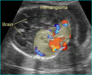
Hemangioma of subarachnoid:  Complex solid-cystic mass in the occipital region with high vascularization

Read More

Cephalocele

Cephalocele

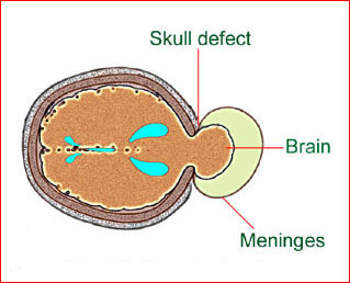
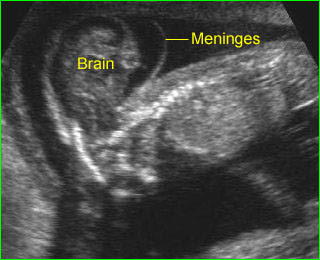
Cephalocele is a protrusion of intracranial contents through a bony defect of the skull. Most cephaloceles are midline and occipital extracranial masses. If only meninges protrude, it is called meningocele. Cephaloceles may be a part of several syndromes, especially Meckel syndrome (cephalocele, polycystic kidneys, and polydactyly) or trisomy 13, and are often associated with other malformations.

Incidence: 1-5 in 10,000 births, which varies with ethnic group and geographic region. Occipital cephaloceles are most common in the West, whereas anterior cephaloceles are much more common in Southeast Asia (1:5000 in Thailand compared with 1:35,000-40,000 in Europe).

Sonographic findings:

Fig 1, Fig 2, Fig 3, Fig 4

  • Skull defect, usually in the occipital area (90% of cases), best visualized on the standard transcerebellar and transthalamic view.
  • Skull defect of anterior cephalocele is more difficult to visualize and may present as extracranial mass with hypertelorism and the cephalocele may best be seen on the facial profile view.
  • Skull defect in an asymmetric area is often related to amniotic band syndrome and other associated anomalies may be seen.
  • Extracranial mass protruding through the defect
    • meningocele, an entirely cystic mass
    • a solid mass, often showing gyri and sulci (encephalocele)
    • a solid cystic mass (meningoencephalocele).
  • Microcephaly, commonly seen.
  • Possibly ventriculomegaly.
  • Polyhydramnios in some cases.
  • The lemon-shaped appearance of the bony calvarium that occurs with open NTD and with the Arnold-Chiari type II malformation.
  • Associated anomalies of specific syndrome, especially Meckel syndrome (cephalocele, polycystic kidneys, and polydactyly) , Waker-Warburg syndrome (cephalocele, ocular malformations, Dandy-Walker malformations), and Joubert syndrome.

Fig 1: Schematic drawing: Occipital meningocele, protrusion of the brain tissue and meninges through the skull defect

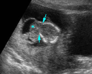
Fig 2: Occipital cephalocele  Transverse scan of the skull: small defect of occipital bone with meningocele (*) (arrow = ventriculomegaly)

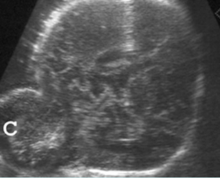
Fig 3: Occipital cephalocele  Transverse scan of the skull: defect of occipital bone with meningoencephalocele (C)

Fig 4: Anterior cephalocele   Coronal scan of the face: round solid mass above the nose (arrow)

Video clips of cephalocele

Anterior meningocele:   Sagittal scan of the fetal face: frontal bone defect with midline cystic mass (meningocele)

Anterior cephalocele:  Transverse scan of the head: frontal skull defect (arrow) with protrusion of brain and meninges (*)

Occipital cephalocele:  Transverse scan the level of cerebellum: small defect of occipital bone with encephalocele

Occipital cephalocele:  Transverse scan of the head: occipital skull defect with meningoencephalocele and mild ventriculomegaly

  • Pitfalls:
    • A scalp hemangioma, cephalhematoma, epidermal scalp cyst, caput succedaneum, or even the normal fetal ear can be mistaken for a cephalocele.
  •  The primary differential diagnosis of occipital cephaloceles includes
    • cystic hygroma (most common); usually septate, cystic, no skull defect, often related to hydrops fetalis
    • cervical meningocele
    • cervical teratoma
    • cranial hemangioma
    •  iniencephaly (very rare).
  • Usually diagnosable after 11-14 weeks (transvaginal ultrasound) .

Associations: Ventriculomegaly (most common), Meckel syndrome, amniotic band syndrome, and limb-body wall complex, chromosome abnormalities (14% of cases).

Management: Termination of pregnancy can be offered for a severe case diagnosed before viability. For the less severe form, a careful search for other anomalies and karyotyping is indicated. The fetus should be delivered and have surgical correction in a tertiary center. A cesarean section could improve the prognosis by avoiding birth trauma and contamination of brain tissue with vaginal bacteria.

Prognosis: Poor in most cases, but good for small, cystic isolated meningocele. Neurological outcome depended on the occurrence or not of hydrocephalus, while the intelligence level was mainly related to the absence of cerebral tissue within the sac.

Recurrence risk: About 2-4%. Periconceptional folic acid supplementation can significantly decrease the recurrent risk.

Read More

Exencephaly

Exencephaly (Acrania)

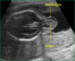
Exencephaly is a developmental abnormality characterized by partial or complete absence of the cranial vault with relatively complete but heterogeneous and disorganized brain. It is an embryonic precursor of anencephaly. Some cases are an amniotic band sequence. The developing brain exposed to amniotic fluid is gradually destroyed and disappears or becomes residual tissue (angioma stroma).

Sonographic findings:

  • Absence of the calvarial vault with brain tissue, relatively normal in amount, floating in amniotic fluid. Fig 1, Fig 2
  • Frog-eye appearance face on the coronal view. Fig 3
  • Abnormally-shaped brain with distorted specific structures such as the ventricles or choroid plexus and the visualization of surrounding membrane instead of skull.
  • The first trimester exencephalic fetus may have a wide and bilobed head, a so-called Mickey Mouse head.
  • Most diagnosed in the first trimester because of the rapid necrosis of brain tissue when exposed to amniotic fluid, subsequently leading to anencephaly.
  • Echogenic amniotic fluid is found in nearly 90% of cases of acrania/anencephaly in the first trimester.
  • The primary differential diagnosis includes sonolucent skulls such as osteogenesis imperfecta type II, hypophosphatasia.
  • Pitfalls: An irregular fetal head outline in the first trimester can simply be overlooked.
  • Diagnosable from 10 to 14 weeks of gestation, usually can be detected during NT scanning.

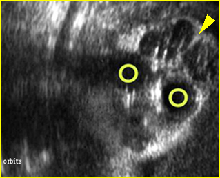
Fig 1: Exencephaly  Coronal scan of the face: significant brain tissue (arrowhead) above the orbits (circle) without covering skull

Fig 2: Exencephaly  Free floating large brain mass without covering skull and skin

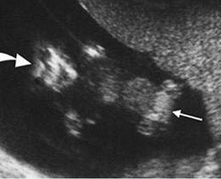
Fig 3: Exencephaly/anencepahly  Coronal scan of the fetus at 11 weeks: no skull above the orbits (large arrow), omphalocele (small arrow)

Video clips of exencephaly

Exencephaly:  Coronal scan of the fetal face at 12 weeks: frog-face appearance with free-floating brain (*) above the orbits

Exencephaly:  Sagittal scan of the fetus at 12 weeks: free-floating brain with meningeal coverings (*) without skull (arrow = face)

Exencephaly:  Free-floating brain in the amniotic fluid without cranium

Associations: Spina bifida, non-specific anomalies, chromosome abnormality is found in about 2% of fetuses with acrania.

Management: Termination of pregnancy should always be offered.

Prognosis: Uniformly lethal.

Recurrence risk: About 2-4%. Periconceptional folic acid supplementation can significantly decrease the recurrent risk

Read More
FetalEchoXI

Fetal Echocardiography XI

Dowload ไฟล์ exel เพื่อลงทะเบียน คลิกที่นี่

Download เอกสารประกอบการเข้าร่วมประชุมโดยไม่ถือเป็นวันลา คลิกที่นี่

21-23 กันยายน 2563

กรุณาลงทะเบียนโดยกรอกข้อมูลในไฟล์ exel แล้วส่ง submit ทาง e-mail ที่: mfm.cmu@gmail.com

Please register Fetal Echo X (using the downloaded form from the link above and sending via e-mail: mfm.cmu@gmail.com

FetalEchoXI

โครงการอบรมเชิงปฏิบัติการ
“การตรวจโรคหัวใจทารกในครรภ์ด้วยอัลตราซาวด์”
Fetal Echocardiography XI: Practical Points


MFMslogoMUA

วันที่ 22-23 กันยายน 2563
ณ ห้องประชุมชั้น 15 อาคารสุจิณโณ
คณะแพทยศาสตร์ มหาวิทยาลัยเชียงใหม่

1. หลักการและเหตุผล

ปัญหาสำคัญในการวินิจฉัยก่อนคลอดของโรคหัวใจพิการโดยกำเนิดสำหรับทารกในครรภ์ นับเป็นหนึ่งในเรื่องร้อนที่ท้าทาย (hot issue) ในสาขาวิชาเวชศาสตร์มารดาและทารก เนื่องจากเป็นความพิการโดยกำเนิดที่พบได้บ่อยที่สุด มีความชุกถึงประมาณร้อยละ 1 ของการคลอด เป็นสาเหตุสำคัญของการตายปริกำเนิดที่สัมพันธ์กับภาวะพิการโดยกำเนิดบ่อยที่สุดเช่นเดียวกัน และเป็นความพิการโดยกำเนิดที่พลาดจากการวินิจฉัยก่อนคลอดได้บ่อยที่สุด ทีมงานเวชศาสตร์มารดาและทารก ภาควิชาสูติศาสตร์และนรีเวชวิทยา คณะแพทยศาสตร์ มหาวิทยาลัยเชียงใหม่ ได้ทำการศึกษาค้นคว้าด้านการวินิจฉัยก่อนคลอดของโรคหัวใจพิการโดยกำเนิด (fetal echocardiography) อย่างต่อเนื่องมากว่า 20 ปี(1-57) และเป็นศูนย์กลางการฝึกอบรมแพทย์ต่อยอดเฉพาะทางเวชศาสตร์มารดาและทารก ได้เล็งเห็นความสำคัญของการมีทักษะในด้านนี้ ซึ่งนอกเหนือจากการมีประสบการณ์ ทักษะ ความคุ้นเคย และมีหลักการในการตรวจค้นหาอย่างเป็นระบบแล้ว ยังจำเป็นต้องมีการศึกษาต่อยอดอย่างต่อเนื่อง แบ่งปันประสบการณ์ เทคนิค และปรับตัวเรียนรู้ไปตามวิทยาการและเทคโนโลยีด้านนี้อย่างต่อเนื่อง ทางทีมงานมีความคาดหวังที่จะช่วยกันส่งเสริมความรู้และทักษะเชิงลึกด้านการตรวจหัวใจทารกในครรภ์ ให้มีประสิทธิภาพมากยิ่งขึ้น เพื่อการวินิจฉัยก่อนคลอดและวางแผนการดูแลรักษาตั้งแต่อายุครรภ์ยังน้อย หรือส่งตัวผู้ป่วยไปสู่การดูแลรักษาในสถานที่ที่มีความพร้อมมากกว่าเป็นต้น หรือแม้แต่การให้โอกาสทางเลือกในการยุติการตั้งครรภ์ตั้งแต่อายุครรภ์ยังน้อย (สำหรับรายที่ทารกมีความพิการชนิดรุนแรงที่อาจไม่สามารถมีชีวิตรอดได้) ดังนั้นทีมงานจึงได้จัดการประชุมวิชาการด้าน “การตรวจอัลตราซาวด์หัวใจทารกในครรภ์ : fetal echocardiography” อย่างต่อเนื่อง ในการจัดประชุมครั้งนี้จะเน้นเทคนิค เคล็ดลับสำคัญในการตรวจ เพื่อการวินิจฉัยโรคหัวใจทารกในครรภ์อย่างถูกต้อง และมั่นใจ ซึ่งเป็นการประยุกต์เทคโนโลยีที่มีอยู่ทั่วไปให้เกิดประโยชน์สูงสุด โดยมุ่งหวังให้ผู้เข้ารับการฝึกอบรมเพิ่มพูนทักษะการประยุกต์ใช้ให้มีความสามารถในการวินิจฉัยโรคหัวใจพิการโดยกำเนิดได้ก่อนคลอดด้วยความมั่นใจสูง รวมทั้งฟื้นฟูความรู้และเทคนิคในการตรวจใหม่ ๆ ของระบบหัวใจและหลอดเลือดซึ่งมีความก้าวหน้าไปอย่างรวดเร็ว และเนื่องจากทักษะสำคัญของการวินิจฉัยคือการตรวจหัวใจทารกในครรภ์ ซึ่งต้องอาศัยทักษะสูงกว่าการตรวจทั่วไป และแลกเปลี่ยนความรู้ในเชิงลึกของศาสตร์ด้านนี้กับผู้มีประสบการณ์

2. วัตถุประสงค์ เพื่อให้ผู้เข้ารับการอบรม

เพื่อเพิ่มทักษะ อัพเดตความรู้ และแลกเปลี่ยนประสบการณ์ในการตรวจวินิจฉัยหัวใจทารกในครรภ์ และ ตรวจวินิจฉัยโรคหัวใจพิการโดยกำเนิดให้ได้เร็วขึ้น โดยคาดหวังว่าผู้ฝึกอบรมสามารถวินิจฉัยโรคหัวใจพิการโดยกำเนิดตั้งแต่ระยะเริ่มแรก และสามารถประยุกต์ใช้อัลตราซาวด์ในการคัดกรองและวินิจฉัยหัวใจทารกผิดปกติได้อย่างมีระบบด้วยความมั่นใจมากขึ้น

3. ผู้เข้าร่วมโครงการ

3.1 วิทยากร 11 คน
3.2 สูติแพทย์ แพทย์ต่อยอดสาขาเวชศาสตร์มารดาและทารก และผู้สนใจ 60 คน
3.3 แพทย์ประจำบ้านสาขาสูติศาสตร์และนรีเวชวิทยา คณะแพทยศาสตร์ มหาวิทยาลัยเชียงใหม่ 60 คน
3.4 เจ้าหน้าที่ 6 คน

รวม 127 คน

4. วิทยากร

4.1 รองศาสตราจารย์ นายแพทย์วัชระ จามจุรีรักษ์
4.2 ศาสตราจารย์ นายแพทย์ธีระ ทองสง
4.3 รองศาสตราจารย์ นายแพทย์ชเนนทร์ วนาภิรักษ์
4.4 ศาสตราจารย์ แพทย์หญิงสุพัตรา ศิริโชติยะกุล
4.5 รองศาสตราจารย์ ดร. นายแพทย์วีรวิทย์ ปิยะมงคล
4.6 รองศาสตราจารย์ แพทย์หญิงเฟื่องลดา ทองประเสริฐ
4.7 รองศาสตราจารย์ แพทย์หญิง เกษมศรี ศรีสุพรรณดิฐ
4.8 รองศาสตราจารย์ แพทย์หญิงสุชยา ลือวรรณ
4.9 ผู้ช่วยศาสตราจารย์ แพทย์หญิงกุณฑรี ไตรศรีศิลป์
4.10 ผู้ช่วยศาสตราจารย์ นายแพทย์ ภูดิศ เจต๊ะวรรณ
4.11 อาจารย์ แพทย์หญิง ศิรินาถ ศิริเลิศศ

5. สถานที่

ห้องประชุมชั้น 15 อาคารสุจิณโณ คณะแพทยศาสตร์ มหาวิทยาลัยเชียงใหม่

6. ระยะเวลา

วันที่ 26-27 กันยายน 2562

7. ผู้รับผิดชอบโครงการ

หน่วยเวชศาสตร์มารดาและทารก ภาควิชาสูติศาสตร์และนรีเวชวิทยา
คณะแพทยศาสตร์ มหาวิทยาลัยเชียงใหม่

8. การลงทะเบียน

ลงทะเบียนโดยไม่เก็บค่าลงทะเบียน

9. งบประมาณ

9.1 ประมาณการรายรับ
• ได้รับการสนับสนุนจากโครงการวิจัยพัฒนาศักยภาพการวิจัยเชิงสถาบัน 100,000.- บาท

9.2 ประมาณการรายจ่าย
• ของที่ระลึกวิทยากร 3,000.- บาท
• ค่าอาหารว่างและอาหารกลางวัน 60,000.- บาท
• ค่าเอกสารและซีดีมัลติมีเดียประกอบการอบรม (130.- x 130 ชุด) 30,000.- บาท
• ค่าแผ่นพับ (10.- x 500 แผ่น) 5,000.- บาท
• ค่าแสตมป์และซองจดหมาย 2,000.- บาท

รวม 100,000.- บาท

10. ผลที่คาดว่าจะได้รับ

ผู้ฝึกอบรมสามารถประยุกต์ใช้อัลตราซาวด์ ในการตรวจประเมินโครงสร้างและการทำงานของหัวใจทารกในครรภ์ได้อย่างเป็นระบบและมีความมั่นใจมากขึ้น และสามารถวินิจฉัยก่อนคลอดของโรคหัวใจพิการโดยกำเนิดตั้งแต่อายุครรภ์ยังน้อยด้วยความมั่นใจมากขึ้น

11. เอกสารอ้างอิง
  1. Tongsong T, Srisomboon J, Wanapirak C, Sirichotiyakul S, Pongsatha S, Polsrisuthikul T. Pregnancy outcome of threatened abortion with demonstrable fetal cardiac activity: a cohort study. J Obstet Gynaecol (Tokyo 1995) 1995;21:331-5.
  2. Tongsong T, Wanapirak C, Sirichotiyakul S. Placental thickness at mid-pregnancy as a predictor of Hb Bart’s disease. Prenat Diagn 1999;19:1027-30.
  3. Tongsong T, Wanapirak C, Sirichotiyakul S, Piyamongkol W, Chanprapaph P. Fetal sonographic cardiothoracic ratio at midpregnancy as a predictor of Hb Bart disease. J Ultrasound Med 1999;18:807-11.
  4. Tongsong T, Sirichotiyakul S, Sittiwangkul R, Wanapirak C. Prenatal sonographic diagnosis of cardiac hemangioma with postnatal spontaneous regression. Ultrasound Obstet Gynecol 2004;24:207-8.
  5. Tongsong T, Sirichotiyakul S, Sukpan K, Sittiwangkul R. Prenatal features of a truncus arteriosus with pulmonary atresia and pulmonary circulation derived from the ductus arteriosus. J Ultrasound Med 2004;23:1221-4.
  6. Tongsong T, Sittiwangkul R, Wanapirak C, Chanprapaph P. Prenatal diagnosis of isolated tricuspid valve atresia: report of 4 cases and review of the literature. J Ultrasound Med 2004;23:945-50.
  7. Tongsong T, Tatiyapornkul T. Cardiothoracic ratio in the first half of pregnancy. J Clin Ultrasound 2004;32:186-9.
  8. Tongsong T, Wanapirak C, Sirichotiyakul S, Chanprapaph P. Sonographic markers of hemoglobin Bart disease at midpregnancy. J Ultrasound Med 2004;23:49-55.
  9. Tongsong T, Chanprapaph P, Khunamornpong S, Sirichotiyakul S. Sonographic features of Ebstein anomaly associated with hydrops fetalis: a report of two cases. J Clin Ultrasound 2005;33:149-53.
  10. Tongsong T, Chanprapaph P, Sittiwangkul R, Sirichotiyakul S. Rupture of fetal ductus arteriosus aneurysm. Obstet Gynecol 2005;105:1275-8.
  11. Tongsong T, Iamthongin A, Wanapirak C, Piyamongkol W, Sirichotiyakul S, Boonyanurak P, et al. Accuracy of fetal heart-rate variability interpretation by obstetricians using the criteria of the National Institute of Child Health and Human Development compared with computer-aided interpretation. J Obstet Gynaecol Res 2005;31:68-71.
  12. Tongsong T, Khunamornpong S, Wanapirak C, Sirichotiyakul S. Prenatal sonographic diagnosis of truncus arteriosus associated with holoprosencephaly. J Clin Ultrasound 2005;33:193-6.
  13. Tongsong T, Sittiwangkul R, Khunamornpong S, Wanapirak C. Prenatal sonographic features of isolated hypoplastic left heart syndrome. J Clin Ultrasound 2005;33:367-71.
  14. Tongsong T, Sittiwangkul R, Wanapirak C, Sirichotiyakul S. Prenatal diagnosis of transposition-like double-outlet right ventricle with mitral valve atresia in heterotaxy syndrome. J Clin Ultrasound 2005;33:197-200.
  15. Piyamongkol W, Trungtawatchai S, Chanprapaph P, Tongsong T. Comparison of the manual stimulation test and the nonstress test: a randomized controlled trial. J Med Assoc Thai 2006;89:1999-2002.
  16. Tongsong T, Chanprapaph P, Sittiwangkul R, Khunamornpong S. Antenatal diagnosis of double outlet of right ventricle without extracardiac anomaly: a report of 4 cases. J Clin Ultrasound 2007;35:221-5.
  17. Srisupundit K, Piyamongkol W, Tongsong T. Identification of fetuses with hemoglobin Bart’s disease using middle cerebral artery peak systolic velocity. Ultrasound Obstet Gynecol 2009;33:694-7.
  18. Tongsong T, Tongprasert F, Srisupundit K, Luewan S. High fetal splenic artery peak velocity in fetuses with hemoglobin Bart disease: a preliminary study. J Ultrasound Med 2009;28:13-8.
  19. Tongsong T, Tongprasert F, Srisupundit K, Luewan S. Venous Doppler studies in low-output and high-output hydrops fetalis. Am J Obstet Gynecol 2010;203:488.e1-6.
  20. Tongsong T, Tongprasert F, Srisupundit K, Luewan S. The complete three-vessel view in prenatal detection of congenital heart defects. Prenat Diagn 2010;30:23-9.
  21. Jatavan T, Luewan S, Tongsong T. Outcomes of pregnancy complicated by heart disease at Maharaj Nakorn Chiang Mai Hospital. J Med Assoc Thai 2011;94:1159-63.
  22. Luewan S, Tongprasert F, Piyamongkol W, Wanapirak C, Tongsong T. Fetal liver length measurement at mid-pregnancy among fetuses at risk as a predictor of hemoglobin Bart’s disease. J Perinatol 2011;31:157-60.
  23. Luewan S, Yanase Y, Tongprasert F, Srisupundit K, Tongsong T. Fetal cardiac dimensions at 14-40 weeks’ gestation obtained using cardio-STIC-M. Ultrasound Obstet Gynecol 2011;37:416-22.
  24. Tongprasert F, Srisupundit K, Luewan S, Sirichotiyakul S, Piyamongkol W, Wanapirak C, et al. Reference ranges of fetal aortic and pulmonary valve diameter derived by STIC from 14 to 40 weeks of gestation. Prenat Diagn 2011;31:439-45.
  25. Tongsong T, Piyamongkol W, Tongprasert F, Srisupundit K, Luewan S. Fetal splenic artery peak velocity (SPA-PSV) at mid-pregnancy as a predictor of Hb Bart’s disease. Ultraschall Med 2011;32 Suppl 1:S41-5.
  26. Tongsong T, Wanapirak C, Piyamongkol W, Sirichotiyakul S, Tongprasert F, Srisupundit K, et al. Fetal ventricular shortening fraction in hydrops fetalis. Obstet Gynecol 2011;117:84-91.
  27. Traisrisilp K, Tongprasert F, Srisupundit K, Luewan S, Tongsong T. Reference ranges for the fetal cardiac circumference derived by cardio-spatiotemporal image correlation from 14 to 40 weeks’ gestation. J Ultrasound Med 2011;30:1191-6.
  28. Luewan S, Srisupundit K, Tongprasert F, Tongsong T. Normal reference ranges of inferior vena cava doppler indices from 14 to 40 weeks of gestation. J Clin Ultrasound 2012;40:214-8.
  29. Tongprasert F, Srisupundit K, Luewan S, Wanapirak C, Tongsong T. Normal reference ranges of ductus venosus Doppler indices in the period from 14 to 40 weeks’ gestation. Gynecol Obstet Invest 2012;73:32-7.
  30. Jatavan T, Tongsong T. Comparison of fetal cardiac spatiotemporal image correlation segmental analysis between cardiac- and body-based scrolling. J Ultrasound Med 2013;32:2125-9.
  31. Luewan S, Tongprasert F, Srisupundit K, Tongsong T. Fetal myocardial performance (Tei) index in fetal hemoglobin Bart’s disease. Ultraschall Med 2013;34:355-8.
  32. Mekjarasnapha M, Traisrisilp K, Luewan S, Srisupundit K, Tongprasert F, Tongsong T. Reference ranges for fetal septum primum excursion from 14 to 40 weeks’ gestation. J Ultrasound Med 2013;32:1729-34.
  33. Siwawong W, Tongprasert F, Srisupundit K, Luewan S, Tongsong T. Fetal cardiac circumference derived by spatiotemporal image correlation as a predictor of fetal hemoglobin Bart disease at midpregnancy. J Ultrasound Med 2013;32:1483-8.
  34. Tongsong T, Luewan S, Srisupundit K, Jatavan T. Hemodynamic assessment of indomethacin-induced fetal heart failure in high-output state. J Clin Ultrasound 2013;41:438-40.
  35. Traisrisilp K, Tongprasert F, Srisupundit K, Luewan S, Tongsong T. Reference ranges of ductus arteriosus derived by cardio-spatiotemporal image correlation from 14 to 40 weeks of gestation. Gynecol Obstet Invest 2013;76:25-31.
  36. Luewan S, Tongprasert F, Srisupundit K, Tongsong T. Inferior vena cava Doppler indices in fetuses with hemoglobin Bart’s hydrops fetalis. Prenat Diagn 2014;34:577-80.
  37. Luewan S, Tongprasert F, Srisupundit K, Traisrisilp K, Tongsong T. Reference ranges of myocardial performance index from 12 to 40 weeks of gestation. Arch Gynecol Obstet 2014;290:859-65.
  38. Thathan N, Traisrisilp K, Luewan S, Srisupundit K, Tongprasert F, Tongsong T. Screening for hemoglobin Bart’s disease among fetuses at risk at mid-pregnancy using the fetal cardiac diameter to biparietal diameter ratio. BMC Pregnancy Childbirth 2014;14:230.
  39. Tongprasert F, Srisupundit K, Luewan S, Tongsong T. Comparison of cardiac troponin T and N-terminal pro-B-type natriuretic peptide between fetuses with hemoglobin Bart’s disease and nonanemic fetuses. Prenat Diagn 2014;34:864-9.
  40. Luewan S, Tongprasert F, Srisupundit K, Tongsong T. Fetal cardiac Doppler indices in fetuses with hemoglobin Bart’s disease at 12-14weeks of gestation. Int J Cardiol 2015;184:614-6.
  41. Tongsong T, Tongprasert F, Srisupundit K, Luewan S, Traisrisilp K. Cardio-STIC (spatio-temporal image correlation) as genetic ultrasound of fetal Down syndrome. J Matern Fetal Neonatal Med 2015;28:1943-9.
  42. Traisrisilp K, Tongprasert F, Srisupundit K, Luewan S, Sukpan K, Tongsong T. Prenatal differentiation between truncus arteriosus (Types II and III) and pulmonary atresia with ventricular septal defect. Ultrasound Obstet Gynecol 2015;46:564-70.
  43. Jatavan P, Kemthong W, Charoenboon C, Tongprasert F, Sukpan K, Tongsong T. Hemodynamic studies of isolated absent ductus venosus. Prenat Diagn 2016;36:74-80.
  44. Jatavan P, Tongprasert F, Srisupundit K, Luewan S, Traisrisilp K, Tongsong T. Quantitative Cardiac Assessment in Fetal Tetralogy of Fallot. J Ultrasound Med 2016;35:1481-8.
  45. Sirilert S, Tongprasert F, Srisupundit K, Luewan S, Tongsong T. Fetal septum primum excursion (SPE) and septum primum excursion index (SPEI) as sonomarkers of fetal anemia: using hemoglobin Bart’s fetuses as a study model. Prenat Diagn 2016;36:680-5.
  46. Srisupundit K, Tongprasert F, Luewan S, Traisrisilp K, Jatavan P, Tongsong T. Effect of cordocentesis on fetal myocardial performance. Prenat Diagn 2016;36:871-4.
  47. Tongsong T, Luewan S, Jatavan P, Tongprasert F, Sukpan K. A Simple Rule for Prenatal Diagnosis of Total Anomalous Pulmonary Venous Return. J Ultrasound Med 2016;35:1601-7.
  48. Tongsong T, Tongprasert F, Srisupundit K, Luewan S, Traisrisilp K. Ventricular Diastolic Function in Normal Fetuses and Fetuses with Hb Bart’s Disease Assessed by Color M-Mode Propagation Velocity using Cardio-STIC-M (Spatio-Temporal Image Correlation M-Mode). Ultraschall Med 2016;37:492-6.
  49. Traisrisilp K, Charoenkwan P, Tongprasert F, Srisupundit K, Tongsong T. Hemodynamic assessment of hydrops foetalis secondary to transient myeloproliferative disorder associated with foetal Down syndrome: A case report and literature review. J Obstet Gynaecol 2016;36:861-4.
  50. Tongprasert F, Sittiwangkul R, Jatavan P, Tongsong T. Prenatal Diagnosis of Aortopulmonary Window: A Case Series and Literature Review. J Ultrasound Med 2017;36:1733-8.
  51. Tongprasert F, Srisupundit K, Luewan S, Traisrisilp K, Jatavan P, Tongsong T. Fetal isovolumetric time intervals as a marker of abnormal cardiac function in fetal anemia from homozygous alpha thalassemia-1 disease. Prenat Diagn 2017;37:1028-32.
  52. Wanapirak C, Sirichotiyakul S, Luewan S, Srisupundit K, Tongprasert F, Tongsong T. Appearance of Abnormal Cardiothoracic Ratio of Fetuses with Hemoglobin Bart’s Disease: Life Table Analysis. Ultraschall Med 2017;38:544-8.
  53. Jatavan P, Chattipakorn N, Tongsong T. Fetal hemoglobin Bart’s hydrops fetalis: pathophysiology, prenatal diagnosis and possibility of intrauterine treatment. J Matern Fetal Neonatal Med 2018;31:946-57.
  54. Rueangdetnarong H, Sekararithi R, Jaiwongkam T, Kumfu S, Chattipakorn N, Tongsong T, et al. Comparisons of the oxidative stress biomarkers levels in gestational diabetes mellitus (GDM) and non-GDM among Thai population: cohort study. Endocr Connect 2018;7:681-7.
  55. Tongprasert F, Sittiwangkul R, Lerthiranwong T, Tongsong T. Prenatal sonographic monitoring of progressive cardiac damages caused by anti-Ro antibodies: A case report. J Clin Ultrasound 2018;46:347-50.
  56. Tongsong T, Tongprasert F, Srisupundit K, Luewan S, Traisrisilp K, Jatavan P. Fetal Cardiac Remodeling in Response to Anemia: Using Hemoglobin Bart’s Disease as a Study Model. Ultraschall Med 2018.
  57. Sirilert S, Tongprasert F, Srisupundit K, Tongsong T, Luewan S. Z Score Reference Ranges of Fetal Cardiothoracic Diameter Ratio. J Ultrasound Med 2019;38:999-1007.


FetalEchoXI

ตารางกำหนดการประชุม

Pre-Seminar course: วันจันทร์ที่ 21 กันยายน 2563: 9.00-16.00 น. โดยคณาจารย์

Pre-conference Course: โดยคณาจารย์

  • Cardiac Embryology (8.30-10.00 น.) : นพ. ธีระ ทองสง
  • The benefits of Colour M-mode in a normal fetal heart (10.00-10.30) : นพ. วัชระ จามจุรีรักษ์
  • Standard cardiac exam (10.45-12.00 น.) :  พญ. สุพัตรา ศิริโชติยะกุล
  • Physics of ultrasound (13.00-14.00 น.) : นพ. ธีระ ทองสง
  • Image optimization (ปรับภาพ) (14.00-15..00 น.) : พญ. สุชยา ลือวรรณ
  • Hands-on live demonstration : พญ. สุชยา ลือวรรณ นพ. ธีระ ทองสง นพ. วัชระจามจุรีรักษ์

{tabulizer:style[gr.alterora.elemental_1_yellow_green.css]}

วันอังคารที่ 22 กันยายน 2563
08.15 – 08.45 น. ลงทะเบียน – Spot Diagnosis ศ. นพ. ธีระ ทองสง
08.45 – 09.00 น. Opening Ceremony คณบดี / หัวหน้าภาควิชาฯ
09.00 – 10.00 น. Right heart anomaly ศ. พญ. สุพัตรา ศิริโชติยะกุล
10.00 – 10.15 น. ———————-พัก———————–
10.15 – 11.00 น. Tip & Tricks XI : 3VV-3VT view ศ. นพ. ธีระ ทองสง
11.00 – 12.00 น. Cardiac function : Brain-heart relationship ผศ. ดร. นพ. ภูดิศ เจต๊ะวรรณ
12.00 – 13.00 น. ———รับประทานอาหารกลางวัน———-
13.00 – 13.45 น. First trimester scan รศ. พญ. สุชยา ลือวรรณ
13.45 – 14.30 น. Amazing five-chamber view ศ. นพ. ธีระ ทองสง
14.30 – 14.45 น. ———————-พัก———————–
14.45 – 15.45 น. Genetic aspect of congenital heart ผศ. พญ. กุณฑรี ไตรศรีศิลป์ หมื่นพินิจ
15.45 – 16.30 น. Case presentation I: Echo-patho correlation อ. พญ. ศิรินาถ ศิริเลิศ / รศ. พญ. กรกนก สุขพันธ์

{tabulizer:style[gr.alterora.elemental_1_orange.css]}

วันพุธที่ 23 กันยายน 2563
08.15 – 9.15 น. Case presentation II เฟลโล่ / ทีมงาน
09.15 – 10.15 น. DORV/TGA/TOF รศ. พญ. สุชยา ลือวรรณ
10.15 – 10.30 น. ———————-พัก———————–
10.30 – 11.00 น. Truncus arteriosus ศ นพ. ธีระ ทองสง
11.00 – 12.00 น. Fetal heart failure รศ. พญ. เกษมศรี ศรีสุพรรณดิฐ
12.00 – 13.00 น. ———รับประทานอาหารกลางวัน———-
13.00 – 14.00 น. Arrhythmia I รศ. พญ. เฟื่องลดา ทองประเสริฐ
14.00 – 14.45 น. Arrhythmia II รศ. พญ. เฟื่องลดา ทองประเสริฐ
14.45 – 15.00 น. ———————-พัก———————–
15.00 – 15.45 น. Left heart anomalies ศ. นพ. ธีระ ทองสง
15.45 – 16.15 น. Bizarre clip quiz รศ. นพ. ชเนนทร์ วนาภิรักษ์
16.15 – 16.30 น. Closing Ceremony ร.ศ. พ.ญ. พรรณี ศิริวรรธนาภา/รศ. นพ. ชเนนทร์ วนาภิรักษ์


Read More