

Facial Clefts
(See the details in the section “Specific abnormalities”.)
Fig 1, Fig 2
Premaxillary protrusion is an important clue to the presence of cleft lip and cleft palate and may be more conspicuous than the cleft itself.
Differential diagnosis for facial clefts
- Artifact: normal philtrum with midline groove of the upper lip misinterpreted as cleft
- Lateral cleft: Fig 3, Fig 4
- most common facial abnormalities, more common on the left side
- paramedial location
- bilateral in 20% of cases
- Midline cleft: Fig 5
- rare
- holoprosencephaly
- median cleft face syndrome
- Atypical cleft:
- associated with amniotic band syndrome in most cases
- asymmetric and bizarre
- other anomalies are commonly seen.
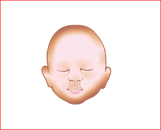
Fig 1: Schematic drawing: acial abnormality related to holoprosencephaly: Bilateral clefts
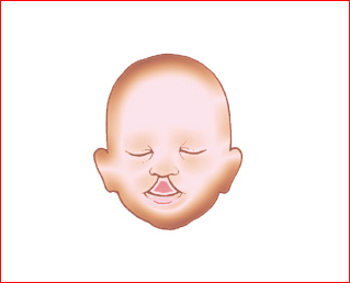
Fig 2: Schematic drawing: Facial abnormality related to holoprosencephaly: Midline cleft
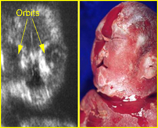
Fig 3:Paramedian cleft Coronal view of the face: paramedian cleft lip
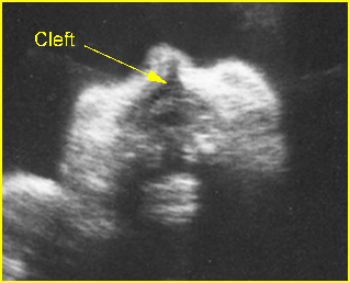
Fig 4: Paramedian cleft Coronal view of the face: paramedian cleft lip (arrow)
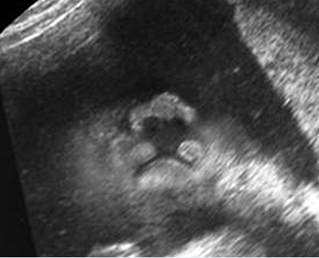
Fig 5: Median cleft lip Coronal view of the face: median cleft lip in fetal trisomy 13
Video clips of facial clefts
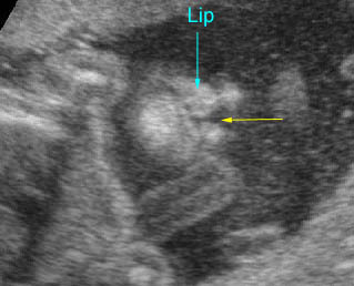
Cleft lip: Coronal scan of the face: small paramedial cleft (arrow)
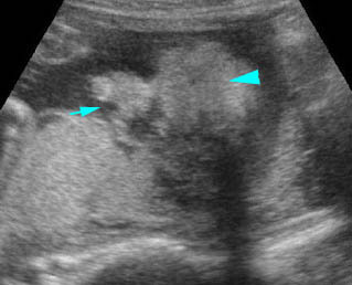
Cleft lip: Coronal scan of the face: paramedial cleft (arrow)
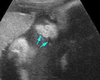
Bilateral cleft lips : Coronal scan of the face: bilateral paramedial cleft (arrow)
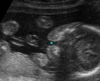
Cleft lip and palate : Transverse scan of the face: incomplete alveolar ridge (*) of the maxilla
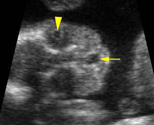
Midline cleft lip: Coronal scan of the face: midline cleft (arrow) (arrowhead = lens in the orbit)