Double Bubbles

Double Bubbles

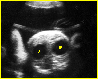
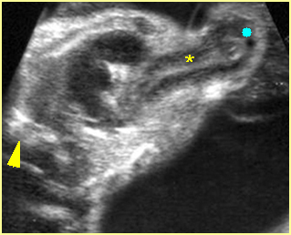
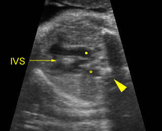
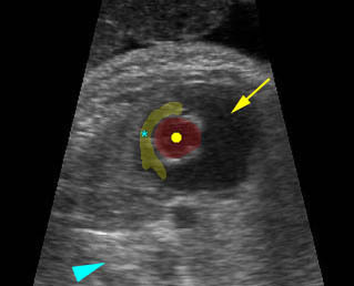
Differential diagnosis of the appearance of double bubbles at the upper abdomen includes:

  • Duodenal obstruction (connection between proximal duodenum and stomach)
    • duodenal atresia
    • duodenal stenosis
    • duodenal web
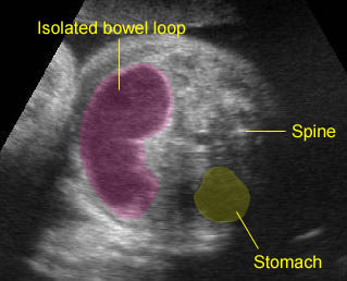
  • Bowel duplication
  • Choledocal cyst
  • Hepatic cyst
  • Ovarian cyst (with stomach bubble)
  • Renal cyst (with stomach bubble)

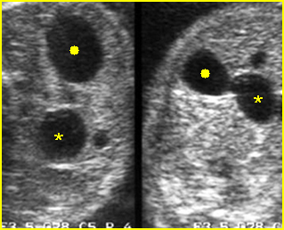
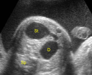
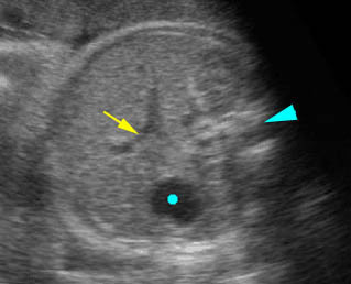
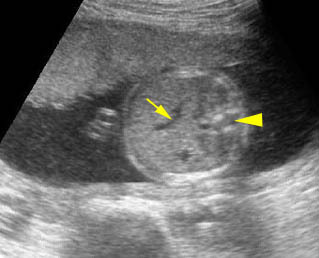
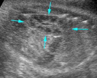
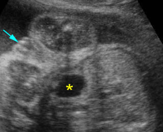
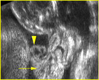
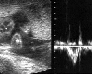
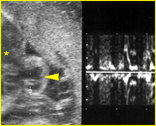
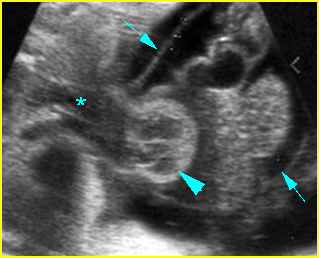
Fig 1, Fig 2, Fig 3, Fig 4

Continuity with the stomach should be demonstrated to distinguish a distended duodenum from other cystic masses in the right upper quadrant, such as choledocal cyst, or duplication cyst. In the case of congenital pyloric atresia, an intermittent ‘double bubble’ sign can be visualized. The double bubble sign disappears during gastric peristalsis. However, a whole stomach configuration should be delineated by continuous observation covering periods when gastric peristalsis is active as well as quiet.

Fig 1:  Schematic drawing of duodenal atresia, The fluid-filled stomach (St) and duodenum (D) cause double bubble sign on ultrasound examination

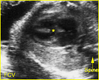
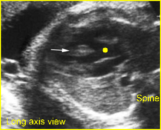
Fig 2:  Duodenal atresia  Cross-sectional scan of the abdomen: Double bubble sign: two cystic masses (* = proximal duodenum, solid circle = stomach) in the upper abdomen

Fig 3:  Duodenal atresia  Cross-sectional scan of the abdomen: Double bubble sign: two cystic masses (* = stomach, solid circle = proximal duodenum ) in the upper abdomen

Fig 4:  Duodenal atresia  Cross-sectional scan of the abdomen: Double bubble sign: two cystic masses (* = proximal duodenum, solid circle = stomach) with connection together in the upper abdomen

Video clips of double bubbles

Duodenal atresia: Double bubble sign with continuation representing stomach (St) and duodenum (D) (Sp = spine)

Bowel duplication:  Sausage cystic mass secondary to bowel duplication; isolated bowel loop with no other visible bowel loops

Read More

Absent Stomach

Absent Stomach

The stomach should be visualized on the standard axial views of the abdominal circumference. Failure to visualize the stomach in the normal location after 14 weeks of gestation is suggestive of abnormalities. However, absent stomach bubble can be identified in 1.4% of all fetal sonographic surveys and it may be found in up to 1% of normal fetuses on an initial scan.

     Fig 1, Fig 2

Differential diagnosis of absent stomach bubble

  • Transient non-visualization of the stomach in normal fetuses
  • Esophageal atresia + tracheoesophageal fistula
  • Diaphragmatic hernia
  • Situs inversus
  • Oligohydramnios from any cause, i.e. renal agenesis
  • Facial clefts (ineffective swallowing)
  • Central nervous system disorders.

A three-sectional view of the neck and upper chest is useful for in utero detection of an esophageal pouch that may enhance the prenatal diagnosis of early amniocentesis (EA)

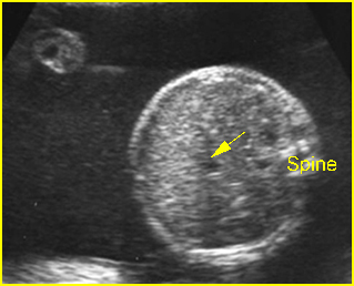
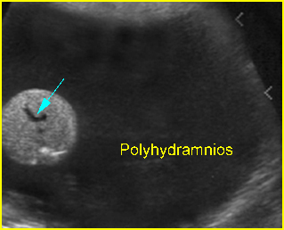
Fig 1:  Esophageal atresia  Cross-sectional scan of the abdomen: absent stomach with marked polyhydramnios (arrow = umbilical vein)

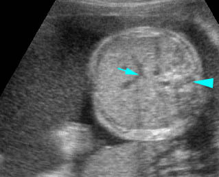
Fig 2:  Esophageal atresia   Cross-sectional scan of the abdomen: absent stomach at the level of umbilical vein complex (arrow) with marked polyhydramnios

Video clips of absent stomach

Esophageal atresia:  Cross-sectional scan of the abdomen: persistently absent stomach (arrow = umbilical vein, arrowhead = spine)

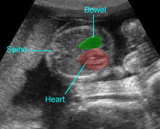
Diaphragmatic hernia:  Cross-sectional scan: The stomach is herniated into the chest, not seen in the abdomen, with cardiac displacement

Read More

Normal Abdomen

Normal Abdomen

Normal Examination

Routine screening often provides clues to the presence of a fetal GI disorder. Recognition of various signs such as ascites or intra-abdominal cysts, etc. requires a familiarity with the appearance of the structures normally found in the fetal abdomen. Guidelines proposed by the American Institute of Ultrasound in Medicine include five views of the abdominal structures: abdominal circumference, stomach, renal area, bladder, and cord insertion. These include the stomach, gallbladder, urinary bladder, and portal vein, etc. Routine evaluation of these structures can identify most fetuses with abdominal anomalies.

Axial scans:

  • In general, the fetal abdomen has a round shape on axial scan. However, pressure from the uterine wall and fetal extremities often distorts the abdominal contour in late pregnancy.
  • Abdominal circumference (AC): The AC is a measure of fetal girth, including soft tissue of the abdominal wall, as well as a measure of internal organs, primarily the liver. It is measured on an axial plane at the level of the stomach and the bifurcation of the main portal vein into the right and left branches. It should be as round as possible and in a perpendicular plane to the spine. Ribs should show a symmetric covering of the AC contours. The AC, although useful as an adjunctive parameter for fetal dating, is of greatest value in the evaluation of fetal growth in the latter part of pregnancy. Normal measurements of the liver, spleen, pancreas, stomach, gallbladder, and intestine have been established. Comprehensive reference ranges for first trimester fetal biometry have also been established. AC is the most important parameter in the estimation of fetal weight. However, careful measurement is always required. The accuracy is better among experienced sonographers. AC quality criteria and adherence to protocols and audit are important. Failure to meet quality criteria results in clinically significant errors.
  • Pseudoascites: In early pregnancy, intra-abdominal organs are not clearly separated from the abdominal wall but near term, the echogenic rim of subcutaneous fat may be separated from intra-abdominal contents by a lucent band, sometimes called pseudoascites, which represents the musculature of the abdominal wall.
  • Esophagus: The normal esophagus can be visualized in the thorax in the latter half of pregnancy when carefully sought. It can be observed as either two or more parallel echogenic lines.
  • Stomach: In the fetal left upper quadrant, the fluid-filled stomach can be visualized in nearly all normal fetus from the beginning of the second trimester. It is round or oval on transverse scan and elongated on sagittal scan. If it is well distended, the fundus, body, antrum and incisura angularis (a notch at the lesser curvature between the body and antrum) can be identified. Failure to visualize the stomach after 14 weeks is suggestive of esophageal atresia.
  • Small bowels: The small bowel is normally visualized as an ill-defined area of increased but relatively inhomogeneous echogenicity at the lower central abdomen. Additionally, it also undergoes continuous, active peristalsis throughout late pregnancy. Distinguishing small bowel from large bowel is possible after midpregnancy, and becomes more obvious with advancing gestational age. Individual segments of normal small bowel should not exceed approximately 7 mm in diameter or 15 mm in length and are usually considerably smaller than these values. The small bowel is often seen in active peristalsis and changes in configuration during real-time observation. Hyperechogenic bowel is potentially associated with medically significant outcomes, including multiple anomalies, fetal death, viral infection and aneuploidy.
  • Large bowel: The colon appears as a continuous tubular structure located in the periphery, filled with hypoechoic meconium. It progressively enlarges with meconium from 3 to 5 mm at 20 weeks to up to 20 mm at term. By the middle of the third trimester, colonic haustra can be identified in nearly all fetuses. The colon exhibits far less peristalsis than the small intestine. Defecation in utero is a normal function. A normal range of fetal bowel (descending colon and rectum) diameters from the early second trimester of pregnancy onwards has been established.
  • Spleen: The spleen can be seen in the left upper abdomen, posterior to the stomach and lateral and superior to the left kidney, separated from the lung by the hypoechoic band of the diaphragm. Although not always easily visualized, it can usually be seen throughout the second half of gestation. It is homogeneous in appearance and slightly less echogenic than the liver, similar to the kidney. The nomogram for fetal spleen dimensions has been established and may be useful in detecting growth disorders of the fetal spleen. Additionally, color Doppler and three-dimensional power Doppler ultrasound can facilitate the antenatal diagnosis of splenomegaly and can help to delineate the spleen from the similar-looking neighboring liver.
  • Pancreas: The pancreas, although often overlooked, is sometimes visualized in the third trimester, especially with the fetus in a supine position. It lies posterior to the stomach and appears as a band of tissue with rather homogeneous echoes between the splenic and superior mesenteric artery.
  • Liver: The liver is the dominant organ in the upper fetal abdomen, the area for standard abdominal circumference measurements. It spans the entire width of the abdomen throughout pregnancy. Except for anechoic vessels running through the parenchyma, the liver is homogeneous. The right lobe is all of the hepatic tissue lying to the right of the gallbladder middle hepatic vein, and inferior vena cava. Fetal liver measurements are usually obtained in a longitudinal plane, from the dome of the right hemidiaphragm to the tip of the right lobe. The liver progressively increases in size throughout a normal gestation. The normal range of fetal liver sizes for each week of pregnancy from 20 weeks of gestation to term has been established.
  • Gallbladder: The gallbladder appears as a fluid-filled, teardrop-shaped structure inferior to the liver on the right. The length and width can be easily measured. Nonvisualization of the fetal gallbladder in early pregnancy is uncommon but is significantly associated with other fetal anomalies, for example extrahepatic biliary atresia.
  • Hepatic veins converge near the superior central portion of the liver to join the inferior vena cava. In the same region, the ductus venosus can occasionally be identified on transverse scan forming a communication between the left portal vein and the inferior vena cava.
  • The umbilical vein enters the mid-abdomen and courses superiorly near the anterior abdominal wall to the liver, where it turns posteriorly through the intersegmental fissure to terminate in the left portal vein. Two umbilical arteries join the umbilical vein at the umbilicus and just inferior to it. They arise from the iliac vessels bilaterally below the aortic bifurcation.
  • Kidneys: The kidneys can usually be visualized in the late first trimester in more than 90% of cases. The kidneys appear on either side of the spine. At the beginning of the second trimester, they are sonolucent, and are round on transverse scans and bean-shaped on longitudinal scans. By midpregnancy, an echogenic outer rim of the renal capsule is seen. The central collecting system becomes echogenic toward the end of the second trimester as renal sinus fat develops. By the third trimester, hypoechoic medullary pyramids can be seen radiating from the renal sinus. The anteroposterior diameter of the renal pelvis is the simplest and most sensitive technique for prenatal diagnosis of congenital hydronephrosis, allowing identification of 100% of cases. Postnatal follow-up studies are warranted if the anteroposterior pelvic diameter on transverse scan is >4 mm before 33 weeks or >7 mm after 33 weeks. Throughout pregnancy, the ratio of the kidney circumference to the abdominal circumference remains relatively constant at 0.27-0.30. Nomograms of fetal renal size are established and may be useful in the evaluation of fetal growth restriction or renal pathology.
  • Adrenal glands: The fetal adrenal glands, located superior to the upper renal poles, are often visualized when searching for the kidneys, especially in the third trimester. The gland characteristically has two long limbs that are relatively anechoic (thought to be the fetal zone of the cortex) and a central echogenic line in each limb (presumed medulla). They have a specific size, shape and echogenicity, appearing as hypoechoic crescents or chevrons with a faint echogenic band through the middle. The echo pattern is so characteristic that the cortex and medulla can be appreciated separately. Fetal adrenal gland volume, especially using 3D ultrasound, may be useful in prenatal diagnosis of congenital adrenal hyperplasia or fetal growth.
  • Bladder: The bladder is a cystic intra-abdominal structure that can be seen from late first trimester. It is located between two echogenic iliac crests on transverse scan and fills the small fetal pelvis on sagittal scan. The bladder wall is thin and usually invisible when the bladder is distended. Changes in the volume of the bladder with time differentiate it from other pathologic cysts. The fetal urethra is usually invisible, however, it may occasionally be visualized as an echogenic line extending the length of an erect penis.
  • Fetal gender: The external genitalia can usually be imaged from early in the second trimester onwards. Gender should always be determined in certain circumstance such as twins pregnancy, suspicion of lower urinary tract obstruction, fetal risk for X-linked disorders, etc. Male genitalia are readily seen. The penis and scrotum are most obvious. Testes may be seen in the scrotal sac, sometimes in the third trimester. Hydrocele, mostly insignificant, observed during pregnancy is common. Female gender should be assigned only by identification of the major and minor labia. Assigning female gender due to an inability to see a penis will result in many diagnostic errors. In approximately 3% of cases fetal gender is incorrectly assigned. This inaccuracy often is due to the fact that the labial hypertrophy during pregnancy may be misinterpreted as the male scrotum.
  • Polyhydramnios: Although polyhydramnios can occur in a variety of disorders and is often a non-specific finding of unknown etiology, it is significantly related to GI obstruction.
  • Oligohydramnios: Oligohydramnios, which is not secondary to fetal growth restriction or rupture of membrane, is often associated with urinary tract abnormalities.

Abdominal Wall

Visualization of cord insertion and the adjacent anterior abdominal wall must be included in routine obstetric scanning as recommended by the guidelines adopted by the American Institute of Ultrasound in Medicine. Normal cord insertion and the adjacent abdominal wall exclude the majority of abdominal wall defects.

Fig 1:  Normal fetal abdomen:  Transverse scan: arrow = umbilical vein, solid circle = stomach, arrowhead = spine

Fig 2:  Normal fetal abdomen:  Transverse scan: arrow = umbilical vein, arrowhead = spine

Video clips of normal fetal abdomen

Normal fetal abdomen:  Transverse scan: arrow = umbilical vein, solid circle = stomach, arrowhead = spine

Normal fetal abdomen:  Transverse scan: arrow = umbilical vein, arrowhead = spine

Normal kidney:  Sagittal scan of the fetal kidney: arrows = outline of the kidney consisting of several hypoechoic pyramids

Female genitalia:  Scan focus on the perineum: arrow = labia minora, * = bladder

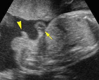
Male genitalia:  From cross-section to mid-sagittal scan of the fetal trunk: arrow = cord insertion, arrowhead = penis

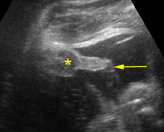
Male genitalia:  Scan focus on the scrotum (*) and urinating penis (arrow)

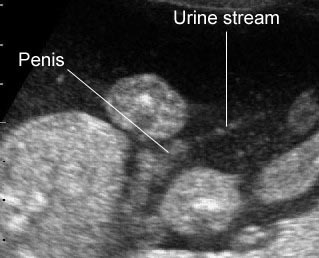
Voiding:  Male genitalia with urination

Technique

  • On routine ultrasound examination in pregnancy, the demonstration of the abdominal cord insertion and the integrity of the anterior abdominal wall as well as the stomach and bladder must always be studied.
  • The serial transverse scans from one end of the abdomen to the other end may be performed first and the fetal abdomen wall can be viewed continuously.
  • On these serial scans, one should observe the smooth contour of the abdominal wall, normal cord insertion and umbilical location, fluid-filled stomach at the left upper abdomen, and fluid-filled urinary bladder at the mid-lower abdomen. It is extraordinarily important that cord insertion is imaged, due to the fact that many of the small omphaloceles which are often associated with chromosomal defects can easily be missed. The number of umbilical vessels can also be counted at the umbilicus. With color flow, both umbilical arteries at both sides of the bladder can be easily determined.
  • On the inner aspect of the abdominal wall, a thin, 1-3 mm anechoic zone, representing the muscle tissue of the abdominal wall, runs continuous with the rib.
  • Demonstration of the fetal abdominal wall may be very difficult in certain circumstances. For example, the extremities are tight to the abdominal wall, and the fetus may be in a prone position, especially when combined with oligohydramnios. In these situations, the location of the umbilicus, often invisible, may be assessed by identifying the umbilical vein in the upper abdomen and following it caudally until it approaches the abdominal wall and finally disappears from the abdomen. This point of disappearance should be at or just below the umbilicus.
  • Sagittal views can also be helpful in following the umbilical vein and studying the contour of the abdominal wall. When possible, visualization of the proximal umbilical cord with enumeration of the vessels is ideal to rule out cystic lesions of the umbilical cord, such as a patent urachus.
  • Fetal abdominal fat layer measurement may be useful for growth evaluation especially in diabetic mothers.

Checklists

On routine screening ultrasound, be sure that all of the followings are seen:

  • Intact abdominal wall
  • Fluid-filled stomach
  • No ascites
  • No abnormal mass
  • Bladder
  • Both kidneys with normal size, site and echogenicity
  • Renal pelvis not dilated
  • Bladder/ureter not dilated
  • No renal cyst
  • Normal amniotic fluid

Pitfalls

  • Absent stomach: Inability to visualize the stomach in the second and third trimesters of pregnancy requires a repeated examination. About half of the cases will be proven to have a normal stomach on the repeated examination overtime (several hours or days later).
  • Gastric pseudomass: A well-defined echogenic focus within the fetal stomach can normally be visualized in about 1% of sonographic evaluations. This is a pseudomass of cellular debris which mostly resolves spontaneously and should not be confused with an abnormality.
  • Dilated colon: Unless correctly recognized, a normal enlarged colon is often mistaken for dilated small bowel, cysts or other pathological processes including renal cysts and pelvic masses. This potential pitfall is especially likely when the meconium, which is usually hypoechoic compared to the bowel wall, has a more sonolucent appearance.
  • Spleen: The spleen is another upper abdominal organ that is often overlooked or mistaken for an abnormal solid left-sided mass.
  • Gallbladder: The gallbladder is often overlooked or misrepresented sonographically (under the assumption that it represents an intrahepatic vein). Although it is a sonolucent structure, its ovoid/conical shape and lack of flow on Duplex or color Doppler, as well as its location inferior and to the right of the intrahepatic segment of the umbilical vein, should help distinguish it.
  • Physiologic omphalocele: The bowel normally herniates into the proximal umbilical cord in the 8th gestational week but returns to the abdomen during the 12th week. However, the large herniated mass (>7 mm) regardless of gestational age should be considered abnormal.
  • Pseudo-omphalocele: Oblique views of the fetus can give the false impression of an omphalocele, especially when the fetal limbs are tight to the abdomen and oligohydramnios. Compression of the fetal abdomen between the walls of the uterus or placenta can result in an appearance that mimics that of an omphalocele on sonography. This problem may simply be solved by obtaining all three proper planes. Furthermore, the differentiation can be made by demonstration of the thin covering membrane of the omphalocele, instead of a normal abdominal wall with subcutaneous fat in a pseudo-omphalocele.
  • Pseudo-ascites: The thin anechoic zone of the fetal abdominal wall musculature which runs continuous with the rib can be mistaken for ascites. This anechoic zone is smooth and not crescent-shaped, as is ascites when bounded by bowel.
  • A normal umbilical cord can be mistaken for gastroschisis especially when the umbilical cord is bunched and close to the abdomen. This problem can simply be solved by the demonstration of the flow with color Doppler. Nomograms of the umbilical cord and Wharton’s jelly area have been generated.
  • Localized deposition of Wharton’s jelly at the umbilicus can be mistaken for a small omphalocele, but the mass is hyperechoic, not anechoic, as with bowel in an omphalocele, and the vessels run through it, not around it. Omphaloceles with predominately liver contents are also hyperechoic, but these defects are larger.
  • A cystic mass near the cord insertion, such as omphalomesenteric cysts or allantoic cysts, may be confused with abdominal wall defects.
Read More

Pentalogy of Cantrell

Pentalogy of Cantrell

Pentalogy of Cantrell is an association of cleft sternum, diaphragmatic hernia, midline abdominal wall defect, ectopia cordis, and cardiac defects.

Incidence: Unknown.

Sonographic findings:

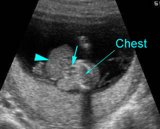
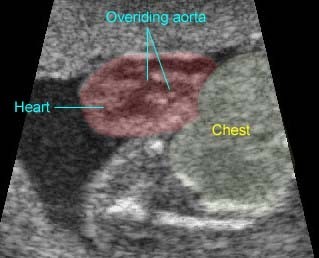
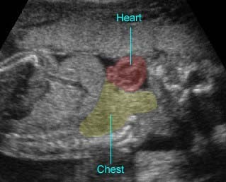
  • The heart is located outside the body due to a defect in the chest or abdominal walls. Fig 1, Fig 2, Fig 3, Fig 4
  • Omphalocele, typically high or supraumbilical. Fig 5
  • Diaphragmatic hernia. Fig 6
  • Cardiac abnormalities. Fig 5
  • Pericardial or pleural effusion associated with an omphalocele is often suggestive of diaphragmatic hernia, an important clue to pentalogy of Cantrell.
  • Usually first diagnosable in late first trimester.
  • The additional information gained by complementary three-dimensional ultrasound can be useful for more efficient counseling and postnatal planning.

Fig 1:  Ectopia cordis  Sagittal scan at 9 gestational weeks: the heart (arrowhead) protruding outside of the chest

Fig 2:  Ectopia cordis  Sagittal scan at 13 gestational weeks: the heart (arrowhead) protruding outside of the chest with omphalocele (arrow)

Fig 3:  Ectopia cordis  Cross-sectional scan of the free floating heart with pulsation at the four-chamber view

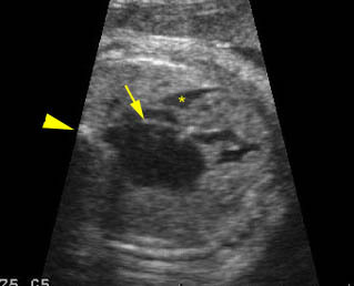
Fig 4:  Ectopia cordis  Cross-sectional scan of the chest: the pulsating heart partially protruding outside of the chest (arrowhead) (* = chest)

Fig 5:  Ectopia cordis (Pentalogy of Cantrell)  Cross-sectional scan of the chest: the heart (arrowhead) partially protruding outside, with omphalocele containing bowel and liver (arrow); note parallel great arteries (*) with transposition

Fig 6:  Ectopia cordis (Pentalogy of Cantrell)  Cross-sectional scan of the chest: the heart (solid circle) partially protruding outside; note parallel great arteries (*) with transposition (arrowhead = spine)

Video clips of fetal cardiac arrhythmias

Pentalogy of Cantrell :  Cross-sectional scan of lower chest: liver (arrowhead) and heart (arrow) located outside

Ectopia Cordis :  Ectopia cordis: the heart outside the chest; note the overriding aorta

Ectopia Cordis :  Ectopia cordis: sagittal scan of the fetal trunk showing the heart pulsating outside the chest

Associations: Genital defects in both conditions; renal, skeletal, neural tube, intestinal, cardiovascular and omphaloceles defects in cloacal exstrophy.

Management: Termination of pregnancy can be offered.

Prognosis: Fatal, though some cases may survive with full surgical repair, with excellent prognosis for cleft sternum alone.

Recurrence risk: Sporadic (rare recurrence).

Read More

Fetal Cardiac Arrhythmias

Fetal Cardiac Arrhythmias

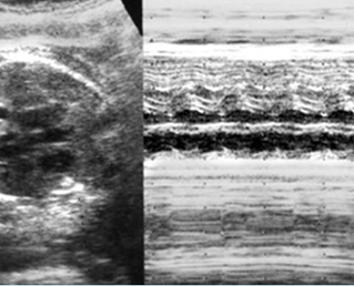
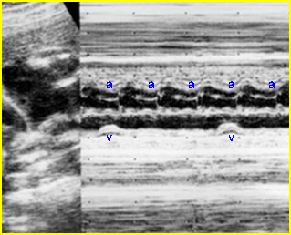
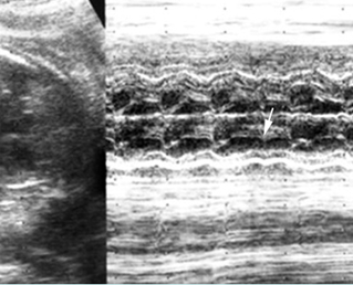
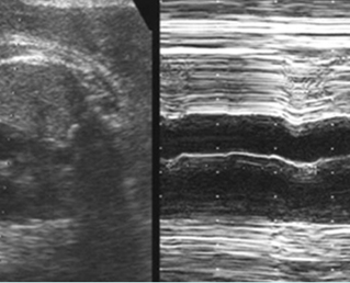
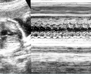
Fetal arrhythmia is abnormal fetal heartbeats, including an irregular rhythm, tachycardia and bradycardia. Utilizing M-mode echocardiography, both fetal atrial and ventricular contractions can be evaluated for regularity and their relationship to each other in time. The most common important fetal arrhythmias are: (1) supraventricular tachycardias; and (2) severe bradyarrhythmias, associated with complete heart block. These may be associated with the development of hydrops fetalis.

Incidence: 1-2% of pregnancies. In clinical practice, these are commonly seen in fetuses whose mothers have a high intake of caffeine. Cessation of that intake often eliminates this arrhythmia.

  Fig 1, Fig 2, Fig 3, Fig 4, Fig 5

Irregular rhythms:

  • The most common type is premature atrial contractions.
  • They may be associated with maternal drug use, including hydralazine, nifedipine, as well as caffeine.
  • Diagnosis: With M-mode or Doppler ultrasound, the cursor is placed through the atrial wall and ventricular wall, and the fetal heart rate and regularity can be evaluated. Premature atrial contractions may be either conducted to the ventricles or blocked, both resulting in an increased ventricular rate.
  •  Cardiac and other associated anomalies are rare.
  • Slightly increased risk of developing supraventricular tachycardia.
  • Prognosis: Almost always benign and requires no treatment. Those without persistent irregularities can be followed up with routine prenatal care.

Tachycardia

  • Tachycardia refers to a fetal heart rate faster than 160 bpm.
  • With sinus tachycardia, the impulse originates from the sinus node, usually ranges between 180 and 200 bpm and is commonly related to changes in autonomic tone, including those caused by maternal fever or drugs.
  • Supraventricular tachycardia (SVT) , a re-entry tachycardia involving an accessory pathway between the atrium and the ventricle, is the most common type of sustained tachycardia with a rate of 220-280 bpm and a 1:1 association between the atrial and ventricular contractions.
  • Atrial flutter, a single re-entry circuit within the atria, refers to a rate that ranges from 300 to 600 bpm, with 2:1 atrioventricular block.
  • Ventricular tachycardia is a rare arrhythmia associated with atrioventricular dissociation, with the ventricular rate (150-250 bpm) faster than the atrial rate.
  • Hydrops fetalis can occur in approximately 50% of fetuses with sustained tachycardia.
  • The fetuses with sustained tachycardia need in utero treatment. The hydropic fetuses with unsuccessful in utero therapy may need early delivery.
  • Prognosis: The survival rate of the fetuses with SVT depends on the development of hydrops. 50-80% of those with hydrops can be in utero controlled with currently available drugs such as sotolol, flecainide, and amiodorane; however, these fetuses may have already developed ischemic cerebral complications that may not be completely resolved. In about 50% of fetuses with tachycardia, the condition persists or recurs postnatally and needs long-term treatment.

Bradycardia

  • Bradycardia is defined as a ventricular rate <110 bpm, often associated with extracardiac causes, such as fetal distress, maternal drugs, or cord compression.
  • Sinus bradycardia, typically 70-110 bpm, in which the impulse originates at the sinus node with 1:1 atrioventricular association, is usually well tolerated.
  • Second degree heart block refers to incomplete penetration of the sinus beat through the AV node and bundle of His.
  • Complete heart block (third degree), a complete dissociation between atrial and ventricular contraction, probably accounts for 80% of cases. About 50% are associated with structural cardiac defects, especially heterotaxy or corrected transposition, and most of the remainder are associated with maternal autoantibodies, such as anti-SSA and SSB.
  • Hydrops fetalis can be seen in about one-third of cases in which the ventricular rate is usually <55 bpm.
  • In utero treatment: A beta agonist, such as salbutamol, can increase the fetal heart rate significantly and improve ventricular function or even reverse hydrops fetalis. Furthermore, dexamethasone can also successfully be used to reverse the heart block to a lesser degree.
  • The prognosis depends on the presence of structural cardiac defects or hydrops. The prognosis is generally poor in those with long QT syndrome manifested as intermittent fetal bradycardia and tachycardia with AV dissociation, though some cases have a favorable outcome.

Conditions commonly associated with conduction defects that produce arrhythmias

  • Tetralogy of Fallot
  • Double-outlet right ventricle
  • Transposition of the great vessels
  • Pulmonic stenosis
  • Total anomalous pulmonary venous return.
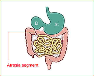
Fig 1:  Normal fetal heart rate  M-mode: normal fetal heart rate

Fig 2:  Congenital heart block  M-mode: complete heart block, normal atrial activity (a) with irregular independent ventricular activity (v)

Fig 3:  Congenital heart block  M-mode: occasional heart block (arrow)

Fig 4:  Fetal bradycardia   M-mode: fetal heart rate of 30 beat per minute

Fig 5:  Fetal supraventricular tachycardia  M-mode: regular fetal heart rate of 240 beat per minute

Video clips of fetal cardiac arrhythmias

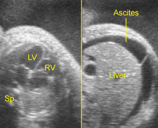
SVT (Supraventricular tachcardia) with hydrops fetalis: Normal four-chamber view with marked tachycardia resulting in hydrops fetalis (LV = left ventricle, RV = right ventricle, Sp = spine)

Cardiomegaly / Bradycardia:  Four-chamber view: generalized enlargement of the heart with pericardial effusion related to Hb Bart’s disease with bradycardia (arrowhead = spine)

HLHS / Arrhythmias:  Four-chamber view: very small left heart with thickened ventricular wall with irregular heart beats, arrow = flap of foramen ovale, * = pericar-dial effusion, arrowhead = spine

Cardiac arrhythmia:  Irregular fetal heart beat and bradycardia

Read More

Double-Outlet Right Ventricle (DORV)

Double-Outlet Right Ventricle (DORV)

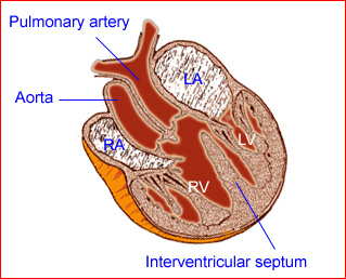
DORV is a cardiac abnormality characterized by most of the aorta and pulmonary trunk being connected to the right ventricle.

Fig 1

Incidence: 0.03-0.07 in 1000 live births, accounting for 2% of structural cardiac defects.

Sonographic findings:

  • Alignment of the two vessels totally or predominantly from the right ventricle.
  • The presence in most cases of bilateral coni (subaortic and subpulmonary).
  • The directions of the two vessels are not perpendicular to each other as usual; therefore, differentiation from truncus arteriosus or TOF may be difficult in some cases.
  • Abnormal cardiac axis is common.
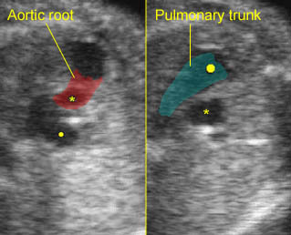
  • Four-dimensional ultrasonography using color Doppler spatiotemporal image correlation is helpful in diagnosis.
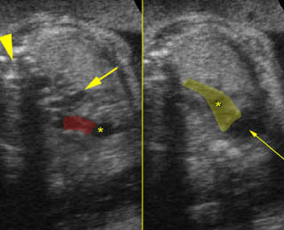
  • Thymic hypoplasia or aplasia may be noted in a case related to 22q11.2 deletion.
  • Pitfalls: Although most cases can be identified antenatally as having a CHD, it is difficult to distinguish from other conotruncal abnormalities.
  • Usually diagnosed after 16-18 weeks, but possible after 12-14 weeks with transvaginal ultrasound in some cases.

Fig 1:  Schematic drawing of double outlets of right ventricle with VSD (LA = left atrium, LV = left ventricle, RA = right atrium, RV = right ventricle)

Video clips of Double-Outlet Right Ventricle (DORV)

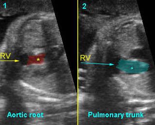
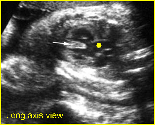
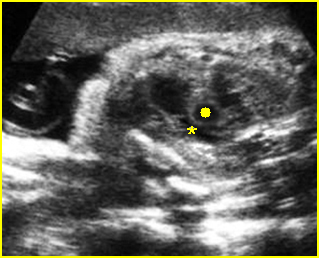
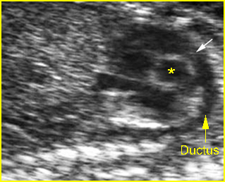
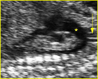
Double outlet of right ventricle:  Long-axis view: pulmonary (solid circle) and aortic root (*) arising from right ventricle

Double outlet of right ventricle: 
Long-axis view:
– aortic root (*) arising from right ventricle pulmonary (arrow = aorta, arrowhead = spine)
– pulmonary artery (*) also arising from right ventricle

Double outlet of right ventricle: 
Long-axis view
1) Aortic root arising from right ventricle
2) Pulmonary trunk arising from right ventricle

Associations: Ventricular septal defects, pulmonary stenosis, tetralogy of Fallot, transposition, univentricular hearts; extracardiac anomaly and chromosome abnormalities are also increased, especially chromosome 22q11 deletion, trisomy 13, and trisomy18.

Management: Careful prenatal and postnatal search for associated anomalies and karyotype (including FISH for chromosome 22q11 deletion) are indicated. Delivery should be performed where immediate pediatric cardiac consultation is available.

Prognosis: Poor when combined with other anomalies, but better for isolated DOVR with surgical correction, with the survival rate possibly being as high as more than 80%.

Read More

Truncus Arteriosus

Truncus Arteriosus

Truncus arteriosus is a single outlet from the heart arising from both ventricles with only one semilunar valve, and giving rise to both systemic and pulmonary circulation.

Incidence: 0.03-0.21 per 1000 births, accounting for 1% of structural heart defects.

Sonographic findings:

Fig 1, Fig 2, Fig 3, Fig 4

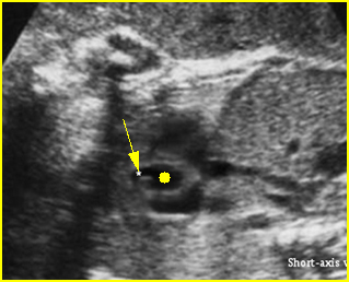
  • One great vessel with a single semilunar valve that overrides the VSD.
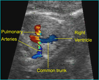
  • A direct continuity between one or two pulmonary arteries and the single arterial trunk.
  • Normal aortic root excluding this condition.
  • Abnormal truncal valve due to an abnormal number of cusps, thickening, stenosis, or insufficiency.
  • Color Doppler imaging can aid in the identification of pulmonary trunk, arising from the main trunk, as well as the assessment of the degree of truncal valve dysfunction.
  • Thymic hypoplasia or aplasia may be noted during echocardiography in the case of conotruncal defects related to deletion of chromosome 22q11.2.
  • Pitfalls:
    • Tetralogy of Fallot with pulmonary atresia or pulmonary atresia with ventricular septal defect are often confused with truncus arteriosus.
    • The absence of the pulmonary outflow tract and failure to image the pulmonary artery makes it difficult to differentiate truncus arteriosus from pulmonary atresia with VSD.
  • Usually diagnosed after 16-18 weeks.

Fig 1:  Ventricular septal defect (in case of truncus arteriosus)  Four-chamber view: interventricular septal defect (*)

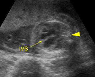
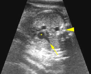
Fig 2:  Overriding aorta (common trunk) (in case of truncus arteriosus)   Long axis view: aortic root (solid circle) running from both ventricles (arrow = interventricular septum)

Fig 3:  Pulmonary artery arising from aorta   Short axis view: Pulmonary artery (arrow) arising from aorta (solid circle) in case of truncus arteriosus

Fig 4:  Truncus arteriosus:   Both pulmonary arteries arising from the enlarged common trunk

Video clips of Truncus Arteriosus

Truncus arteriosus : Common trunk (aortic root) overiding both ventricles; small pulmonary artery (arrow) originating from the common trunk (*), (arrowhead =spine)

Truncus arteriosus:  Both pulmonary arteries arising from the enlarged common trunk

Associations: Other intracardiac malformations, especially VSD and interrupted aortic arch, and extracardiac malformation (20-40%), often associated with chromosome 22q11 deletion (12-35%).

Management: Careful prenatal and postnatal search for associated anomalies and karyotype (including FISH for chromosome 22q11 deletion) are indicated. Delivery should be performed where immediate pediatric cardiac consultation is available.

Prognosis: Poor in most cases but better in cases of appropriate surgical correction. Repair of truncus arteriosus in the neonatal period can be performed routinely with excellent survival, even in patients with major associated abnormalities.

Read More

Tetralogy of Fallot (TOF)

Tetralogy of Fallot (TOF)

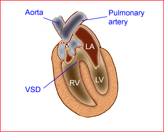
TOF consists of four anatomical malformations as follows: (1) ventricular septal defect; (2) pulmonary artery stenosis; (3) aortic valve that overrides the interventricular septum; and (4) hypertrophy of the right ventricle, which may not occur until the newborn period.

Fig 1

Incidence: 0.2-0.3 in 1000 live births, accounting for 7% of structural cardiac defects.

Sonographic findings:

 Fig 2, Fig 3, Fig 3, Fig5

  • Very large aortic root (the most common prenatal sonographic finding).
  • Normal aortic root excluding TOF.
  • Small pulmonary artery or stenosis of the right ventricular outflow tract.
  • Ventricular septal defect in the outlet portion of the septum and an overriding aorta, best seen in long-axis views.
  • Right ventricular hypertrophy.
  • Hydrops fetalis or polyhydramnios may be seen in the absence of the pulmonary valve.
  • Increased NT especially in fetuses with chromosomal 22q11 deletion.
  • Although Doppler studies show increased peak velocities in the pulmonary artery, suggestive of obstruction of blood flow in the right outflow tract, normal pulmonic velocity in TOF is commonly observed due to the parallel flow circulations created by the combination of ventricular septal defect and ductus arteriosus.
  • Reversal of flow in the arterial duct and failure of growth of the pulmonary trunk predict the need for early surgery to maintain pulmonary blood flow.
  • Pitfalls:
    • A frequent artifact resembles overriding of the aorta.
    • Overiding of the aorta can also be seen in double-outlet right ventricle, truncus arteriosus, or transposition of great arteries.
  • Usually diagnosed after 16-18 weeks, but possible after 13-15 weeks with transvaginal ultrasound in some cases.

Fig 1:  Schematic drawing of  Tetralogy of Fallot (LA = left atrium, LV = left ventricle, RV = right ventricle, VSD = ventricular septal defect)

Fig 2:  Overriding aorta (in case of TOF)  Long axis view of the heart: aortic root (solid circle) running from both ventricles (arrow = interventricular septum)

Fig 2:  Pulmonary stenosis (in case of TOF)  Long axis view of the heart: small pulmonary trunk (*) compared to aortic root (solid circle)

Fig 4:  Small Pulmonary trunk and ductal arch  Short axis view: Small ductal arch (arrow) giving no branch to the head (* = ascending aorta)

Fig 5:  Large aortic arch   Large aorta compared to ductal arch: aortic arch (*), giving the carotid branch (arrow) to the head

Video clips of Tetralogy of Fallot (TOF)

Overding aorta:  Longitudinal scan of the aoratic arch; * overiding aorta, solid circle = aortic arch, arrow = right ventricle

Pulmonary stenosis:  Short-axis view: very small pulmonary artery (*) compared to ascending aorta (solid circle), arrow = right ventricle, arrowhead = spine

 Associations: Other abnormalities, and chromosomal abnormalities found in 60% of cases. Chromosome 22q11 microdeletion may be seen in 11-34% of cases.

Management: Careful prenatal and postnatal search for associated anomalies and karyotype (including FISH for chromosome 22q11 deletion) are indicated. Delivery should be performed where immediate pediatric cardiac consultation is available.

Prognosis: Good prognosis in the case of a normal pulmonary valve, with surgical correction; poor prognosis if the pulmonary valve is absent which is related to hydrops fetalis.

Read More

รศ.นพ. กิตติภัต เจริญขวัญ หัวหน้าภาควิชาสูติศาสตร์และนรีเวชวิทยา

รศ.นพ. กิตติภัต เจริญขวัญ  หัวหน้าภาควิชาสูติศาสตร์และนรีเวชวิทยา :
ภาควิชาขอแสดงความยินดีกับ อ.ต้น (อาจารย์กิตติภัต) ที่ได้รับการสรรหาอย่างเป็นทางการ จากความทุ่มเท ใส่ใจ จริงจัง ต่อการพัฒนาภาควิชาตลอดเวลาที่ผ่านมา ทำให้รับประกันได้ว่าภาควิชากำลังเดินไปสู่ยุคแห่งครอบครัวที่มีความสุขและความเจริญในทุกด้าน

Read More

รศ.พญ.เกษมศรี ศรีสุพรรณดิฐ ผู้ช่วยคณบดี

รศ.พญ.เกษมศรี ศรีสุพรรณดิฐ  ผู้ช่วยคณบดี :
ภาควิชาขอแสดงความยินดีกับ อ.แป้ง ที่ได้รับการแต่งตั้งเป็นผู้ช่วยคณบดีด้านวิชาการ (กิจกรรมการศึกษาระดับคลินิก) ขอแสดงความยินดีกับรับผิดชอบด้านกิจกรรมการศึกษาระดับคลินิก ซึ่งเป็นงานท้าทายที่ชอบของอ.แป้ง จึงนับเป็นก้าวโชคดีของคณะอีกเช่นเดียวกัน

Read More