Nuchal Thickening
Redundant soft tissue in the back of the neck area has been known to be a feature of newborns and fetuses with Down’s syndrome. A sonographic sign of a thickened nuchal fold is used as a marker of an increased risk of fetal Down’s syndrome in the second trimester, typically after 15-20 weeks of gestation. This measurement has remained the most sensitive and specific single marker for the midtrimester detection of Down’s syndrome.
Sonographic examination:
Fig 1, Fig 2, Fig 3
- The measurement is done using a transverse section of the fetal head, angled posteriorly to include the cerebellum and the occipital bone. The following landmarks must be identified: cavum septi pellucidi, cerebral peduncles, cerebellar hemispheres and cisterna magma. The measurement is made from the outside of the occipital bone to the outer skin edge.
- A soft tissue thickening of 6 mm or greater between 15 and 20 weeks of gestation is considered abnormal. However, the 95th percentile of nuchal fold thickness measured by transvaginal sonography at 14-16 weeks is 3.0 mm.
- The observer variability for this measurement is only 1 mm among experienced practitioners.
- Combining second trimester serum testing and nuchal fold thickness is substantially more effective than either serum screening or ultrasound alone.
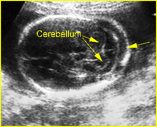
Fig 1: Nuchal edema Transcerebellar view of the skull: thickening of nuchal fold
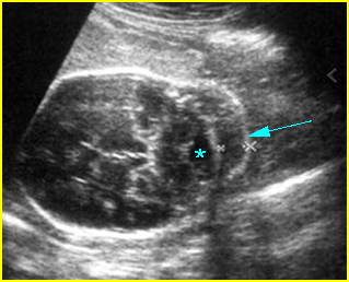
Fig 2: Nuchal edema Transcerebellar view of the skull: normal cisterna magna (*), thickening of nuchal fold (arrow)
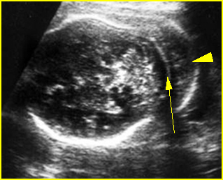
Fig 3: Nuchal edema Transcerebellar view of the skull: marked thickening of nuchal fold (arrowhead) (arrow = occipital bone)
Video clips of nuchal thickening
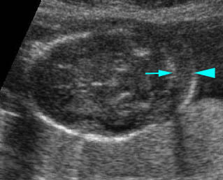
Nuchal thickening: Transcerebellar plane: thickened nuchal fold (arrowhead) (arrow = occipital bone)
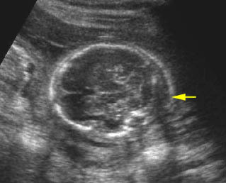
Nuchal thickening: Transcerebellar plane: thickened nuchal fold (arrow)
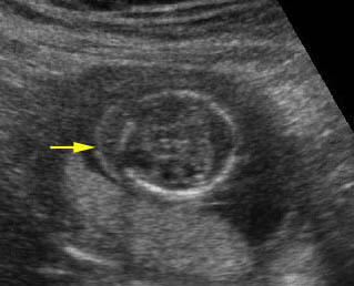
Nuchal thickening : Transcerebellar plane: thickened nuchal fold (arrow)
Nuchal thickness along with other sonomarkers during 15-20 weeks is incorporated into the sonographic scoring index for detection of Down’s syndrome in the second trimester as follows:
Sonographic index for Down’s syndrome in second trimester fetuses
Findings Score
Major anomaly 2
Nuchal fold >6 mm 2
Short femur 1
Short humerus 1
Pyelectasis 1
Hyperechoic bowel 1
Echogenic intracardiac focus 1
- This genetic sonogram scoring index was used to identify approximately 75% of fetuses with Down’s syndrome, with amniocentesis being recommended in 26.7% of a high-risk population.

