Other Neck Masses

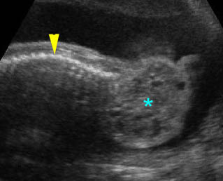
Cervical teratoma: Sagittal scan of the thorax and neck: solid-cystic mass (*) lateral to the neck (arrowhead = spine)

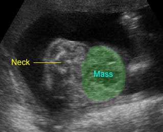
Fetal goiter : Large complex mass at the anterior aspect of the neck, postnatally proven to be goiter
Cervical Teratomas
Teratomas are neoplasms derived from pleuripotent cells and are composed of a diversity of tissues foreign to the anatomic site in which they arise.
Incidence: Rare, may be 1 in 20,000 to 1 in 40,000 live births. It most commonly occurs in the sacrococcygeal area and the neck accounts for about 5% of teratomas.
Sonographic findings:
- Typically, solid cystic, beginning with cystic masses and becoming larger and more complex with gestational age.
- Usually unilateral with an anterolateral location.
- Calcification seen in nearly half of the cases.
- Sometimes hyperextension secondary to a pressure effect of the large mass.
- Hydrops fetalis and hydramnios noted in 30% of cases.
- Three-dimensional ultrasound and color Doppler ultrasound showing intense arterial and venous flow with low resistance indices may be helpful.
- The main differential diagnoses include lymphangioma, hemangioma, or goiter.
Associations: Rarely related to other anomalies.
Management: Fetal resection or termination of pregnancy may be offered for a large mass with hydrops before viability. Cesarean section should be considered in the case of a large mass. The ex utero intrapartum treatment (EXIT) or operation on placenta support (OOPS) has been proposed in cases of potential airway obstruction.
Prognosis: Benign in more than 90% of cases but there is a high mortality rate mainly due to respiratory obstruction, prematurity and hydrops fetalis; definitive surgery may result in good outcome.
Recurrence risk: Rare.
Cervical meningomyelocele
(See spina bifida.)
Occipital Cephalocele
(see details in Part I)
Hemangioma/Lymphangioma
Hemangioma is a proliferation of vascular endothelium that may occur anywhere in the body including the face and neck, although this is relatively rare. Lymphangioma is a congenital malformation of lymphatic vessels. Hemangioma and lymphangioma show overlapping pathologic and sonographic features.
Incidence: Rare in fetal life, but rather common during the first year of life.
Sonographic findings :
- Variable depending on types of hemangiomas; capillary, arteriovenous, or venous angiomas.
- Hemangiomas: solid appearance due to numerous small vascular channels in most cases, but mixed solid and cystic masses are also seen.
- Small internal hypoechoic spaces are occasionally seen.
- Positive Doppler signal or demonstration of vascularization is helpful in the diagnosis of hemangioma.
- Lymphangiomas: cystic mass with multiloculated areas in most cases.
- High-out heart failure or hydrops fetalis may be noted in massive hemangioma.
Associations: Usually isolated abnormalities.
Management: In general, hemangioma/lymphangioma does not alter the standard obstetric management. Postnatal surgical correction is required for persistent cases.
Prognosis: Generally good, but prompt airway management is required in massive cases; postnatal spontaneous regression can be observed in 60% of hemangiomas.
Recurrence risk: Rare.
Goiter
Goiter is an enlarged thyroid gland, probably associated with maternal ingestion of iodides or antithyroid drugs or maternal Graves’ disease.
Incidence: Rare.
Sonographic findings:
- Typically solid and homogeneous but occasionally hypoechoic mass which is considered abnormal if it is 2 SD above normal.
- Symmetrical, bilobed, located at the anterior neck.
- Hyperextension of the fetal head may be noted in cases of large mass.
- Polyhydramnios presumably due to impaired swallowing may be observed.
- High-output cardiac failure with cardiomegaly and pleural effusion or hydrops fetalis may occasionally occur.
- MRI is helpful in identifying thyroid tissue.
- The main differential diagnoses include cervical teratoma and hemangioma.
- Usually diagnosed in the third trimester or late second trimester.
Associations: Usually isolated.
Management: If sonographically diagnosed, accurate diagnosis of fetal thyroid status should be obtained by fetal blood sampling and intrauterine therapy may be initiated, either by reduction of maternal antithyroid medication, or intra-amniotic injection of levothyroxin in hypothyroidism, and on administration of antithyroid drugs in hyperthyroidism.
Prognosis: Good with appropriate management, however, cretinism and mental retardation secondary to hypothyroidism can occur.
Recurrence risk: Rare.

