Cystic Hygroma
A congenital malformation of the lymphatic system, which most commonly develops from jugular lymph sac obstruction and enlargement at the posterior triangles of the neck. It is the most common mass of the neck.
Incidence: 1 in 1000 live births but 1 in 200 abortuses.
Natural history: Either an isolated finding or part of a syndrome, such as Turner syndrome, or trisomy 21, 18 and 13. Two-thirds of cases are related to chromosome abnormalities.
Fig 1,
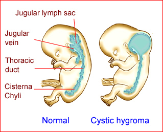
Fig 1: Schematic drawing: Normal lymphatic system compared to that of cystic hygroma
Sonographic findings:
Fig2, Fig3, Fig4
- Septated or non-septated cystic mass without a solid component in the mass.
- Typically, a symmetric or bilateral cystic structure with one or more septations, located in the posterior neck area.
- Dense midline septum, nuchal ligament.
- Normal cranial contents and skull as well as intact cervical spine, unlike meningocele.
- Diffuse hydrops and oligohydramnios in severe cases.
- The main differential diagnoses are cephalocele, cervical meningocele, cystic teratoma, and hemangioma.
- First diagnosable late in the first trimester, and usually early in the second trimester.
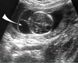
Fig 2: Cystic hygroma Oblique cross-sectional scan at the level of cerebellum: anechoic cyst (arrow) with central septum
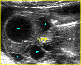
Fig 3: Cystic hygroma Oblique coronal scan at the neck: multiple anechoic cysts with thickened septum at the posterior aspect of the neck
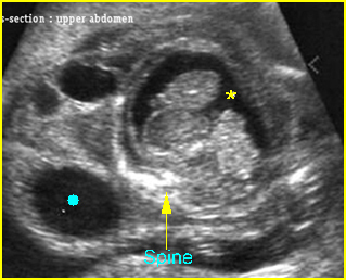
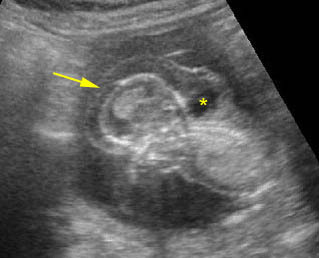
Fig 4: Cystic hygroma with hydropic changes Cross-section scan of the abdomen: anechoic cysts (solid circle) in subcutaneous edema (extending from the neck area) (* = ascites)
Video clips of cystic hygroma
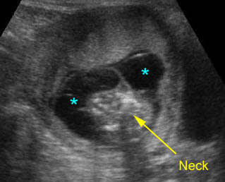
Cystic hygroma: Septate cystic mass (*) on the back of the neck
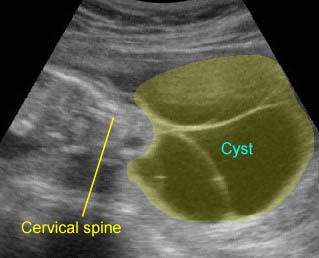
Large cystic hygroma: Large septate cyst on the back of the neck
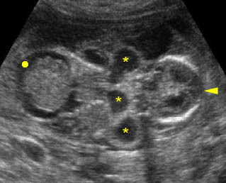
Cystic hygroma: Coronal scan of the fetal trunk: multiple cystic areas (*) around the neck as well as ascites (solid circle)
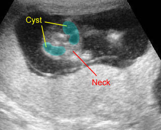
Early cystic hygroma: Small non-septate cyst on the back of the neck with early hydrops fetalis (subcutaneous edema)
Early hydrops fetalis: Sagittal scan of the fetus 13 weeks: generalized subcutaneous edema (arrow) with cystic fluid collection (on the back of the neck) (hydrops fetalis related to cystic hygroma)
Associations: About 60-70% of fetuses with cystic hygroma have aneuploidy, especially 45,XO, and trisomy 21, 18 and 13. The risk of aneuploidy is higher when the cyst is septated, and therefore large. Cystic hygroma in the first trimester is less likely to be associated with aneuploidy, and trisomy 21 is the most common. Compared to non-septated cystic hygromas, septated cystic hygromas are more likely to persist and be associated with aneuploidy, hydrops and other anomalies, and pregnancy loss. Turner syndrome, which is often related to cystic hygroma, may be associated with coarctation of aorta or other anomalies such as horse shoe kidneys, and widespread anatomic disruption due to hydrops.
The 45,X karyotype was found only in patients with septated hygromas and was present in 77% of cases. In contrast, trisomy 21 was the most common abnormal karyotype in non-septated lesions (31%).
Management: Careful search for associated anomalies and karyotyping should be performed. Termination of pregnancy should be considered if associated with chromosomal abnormalities or hydrops fetalis. Isolated cases need follow-up ultrasound study. Intrauterine injection of OK-432 was reported to be a safe and effective therapy in some cases.
Prognosis: Good in mild isolated cases in which spontaneous resolution can occur, but very poor if associated with chromosome abnormalities or hydrops fetalis. Sixty percent of affected fetuses develop signs of hydrops (e.g. ascites, pleural effusions, and edema).

