Proboscis
Proboscis, abnormal nose formation, is almost always related to hypotelorism and holoprosencephaly. The sonographic findings reveal an appendage with one or two openings which replaces the nose. It is located midline at the level of orbit, upper or lower, and is usually diagnosed in the second trimester but possibly in the first trimester. Associated abnormalities include holoprosencephaly, hypotelorism or cyclopia, cebocephaly, and abnormal chromosome, especially trisomy 13. The use of 3D prenatal US made additional diagnostic images possible.
Lateral nasal proboscis is a rare anomaly resulting in incomplete formation of one side of the nose and other variable abnormalities in the adjoining regions of the face, without associated brain malformations. With careful examination, it can also be diagnosed antenatally.
Fig 1, Fig 2, Fig 3, Fig 4, Fig 5, Fig 6
The main differential diagnoses are midline abnormal masses such as
- Anterior cephalocele
- Hemangioma
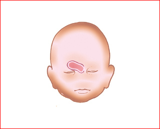
Fig 1: Schematic drawing: Facial abnormality related to holoprosencephaly: Ethmocephaly
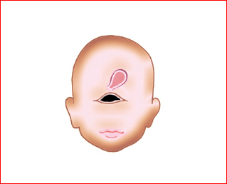
Fig 2: Schematic drawing: Facial abnormality related to holoprosen-cephaly: Cyclopia with proboscis
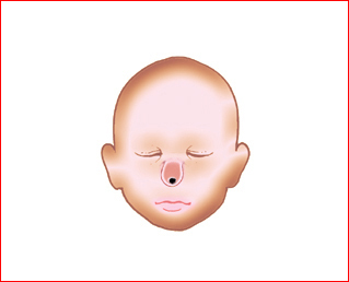
Fig 3: Schematic drawing: Facial abnormality related to holoprosencephaly: Cebocephaly
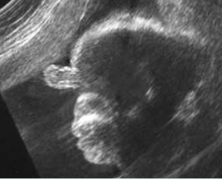
Fig 4: Proboscis Facial profile view: proboscis and absent nose
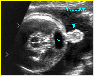
Fig 5: Proboscis and cyclopia Transverse scan of the skull at the level of orbits: fused orbit (*) and proboscis
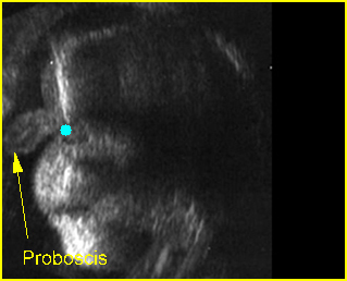
Fig 6: Proboscis Sagittal-coronal view: proboscis cyclopia (solid circle) and absent nose
Video clips of proboscis
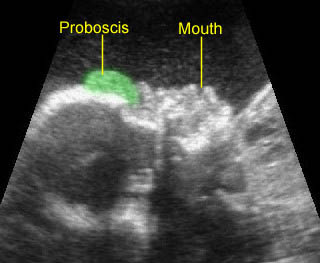
Proboscis: Mid-sagittal scan of the face showing a proboscis above the eye with absent nose
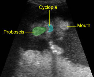
Proboscis: Coronal scan of the face showing a proboscis above the cyclopia
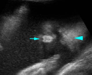
Proboscis: Coronal scan of the face: arrowhead = forehead, arrow = proboscis)
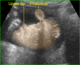
Proboscis: Facial profile view shows proboscis in a case of holoprosencephaly, note: absent nose

