Osteogenesis Imperfecta (OI)

Osteogenesis Imperfecta (OI)

OI is genetically a heterogeneous disorder consisting of both autosomal dominant and autosomal recessive entities caused by mutations in one or two structural genes for type I procollagen. The clinical heterogeneity is due to the different mutations in the genes: COL1A1 and COL1A2. OI is divided into four types as follows:

  • Type I (autosomal dominant) is characterized by bone fragility, blue sclera, hearing loss, and normal calvarium, with fractures ranging from none to multiple. DNA-based analysis is possible for early prenatal diagnosis.
  • Type II (new dominant mutations and autosomal recessive in <5%) is a perinatal lethal variety characterized by almost no ossification of the skull, beaded ribs, shortened crumpled long bones and multiple fractures in utero.
  • Type III (autosomal recessive, rare) is a non-lethal variety characterized by blue sclera (becoming white with time) and multiple fractures present at birth.
  • Type IV (autosomal dominant) is the mildest form (mild to moderate osseous fragility). Long bones and sclera are normal.

Incidence: 0.4 per 10,000 births for OI, and 0.2 per 10,000 births for OI type II.

Sonographic findings (OI type II) :

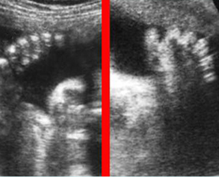
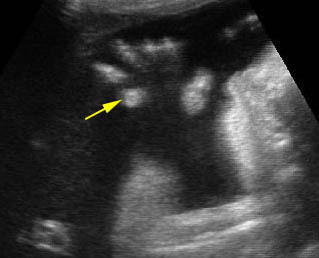
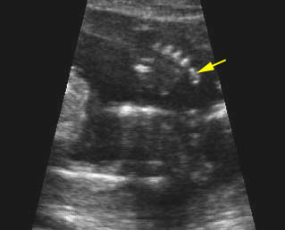
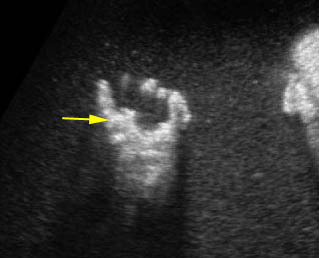
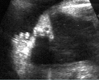
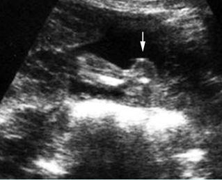
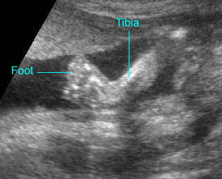
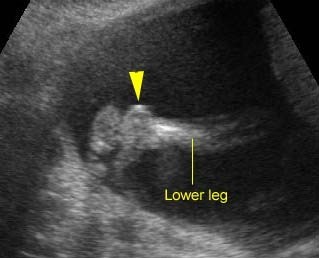
Fig 1, Fig 2, Fig 3, Fig 4, Fig 5, Fig 6

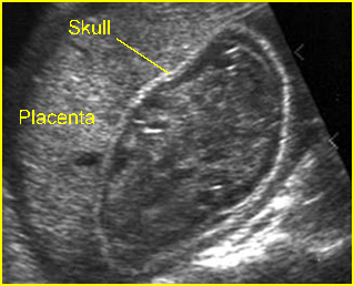
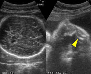
  • Variable hypomineralization, which may result in complete absence of acoustic shadow and may be so severe that the normally-difficult-to-visualize near-field brain is easily seen.
  • Micromelia (type IIB and IIC may be less severe).
  • Fractures of long bones.
  • Thickened long bones due to frequent fractures and secondary callus formation.
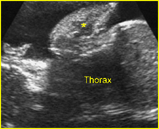
  • Small bell-shaped thorax with multiple rib fractures or beaded ribs.
  • Increased nuchal translucency thickness in the first trimester in some cases.
  • MRI complemented sonography may be helpful in further differentiating clinical and sonographic findings.
  • Sonographic features are variable among the three subtype as follows:
    • Type IIA: thick bones, multiple fractures, hypomineralization, beaded ribs
    • Type IIB: thick bones, multiple fractures, less beading of ribs; lower extremities are affected more than upper extremities
    • Type IIC: thin bones, multiple fractures, thin beaded ribs.
  • Pitfalls: Variable short limb lengths without fractures may be the only clue in the mild form.
  • Usually diagnosed in the second and third trimesters but possible in the late first trimester.
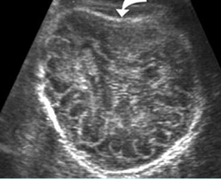
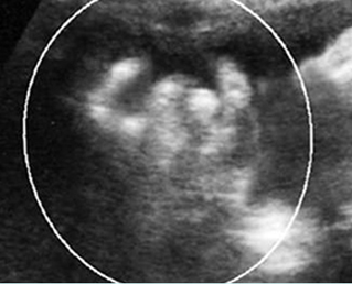
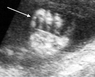
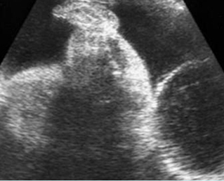
Fig 1:  Sonolucent and compressible skull   Poorly ossified and compressible cranium (arrow), cerebral sulci and gyri could easily be seen
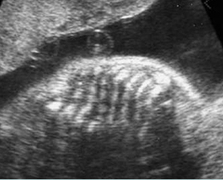
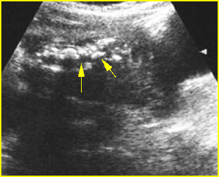
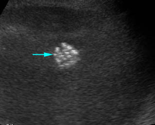
Fig 2:  Rib fractures   Multiple rib fractures with poor ossification in the fetus with osteogenesis imperfecta type IIA
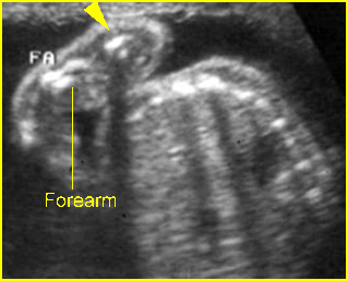
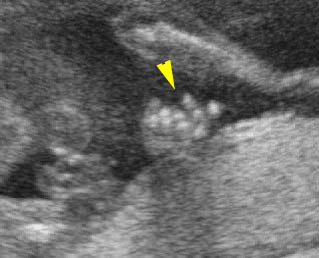
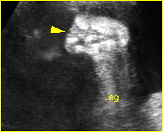
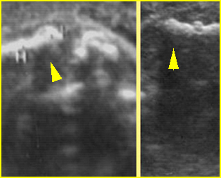
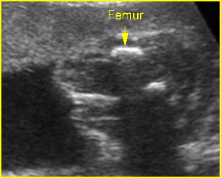
Fig 3:  Micromelia   Irregularity and severe shortening of long bone (arrowhead)
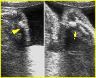
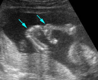
Fig 4:  Fracture in utero   Longitudinal scan of upper extremity: poorly ossified and fracture in osteogenesis imperfecta type IIA
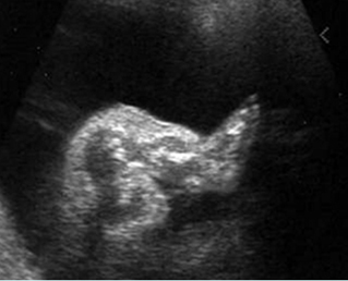
Fig 5:  Osteogenesis imperfecta type IIA   Longitudinal scan of lower extremity: shortened and irregular ossified long bones
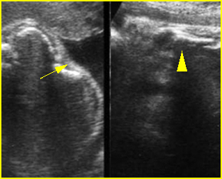
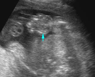
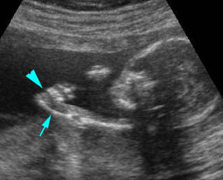
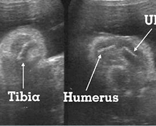
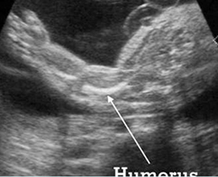
Fig 6:  Fracture of long bones   Longitudinal scan of rib and humerus: fracture rib (arrow) and humerus (arrowhead) with moderate ossification

Video clips of osteogenesis imperfecta (OI)
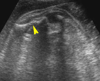
Osteogenesis imperfecta (IIA) :  Irregular long bone (arrowhead) with poorly ossified
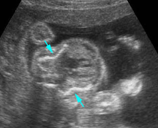
Osteogenesis imperfecta (IIA) :  Cross-sectional scan of the thorax: rib fractures (arrow)
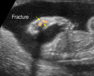
Osteogenesis imperfecta:  Long bone fracture of the forearm
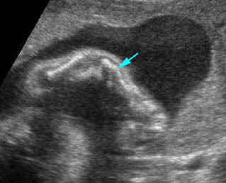
Fracture in utero :  Longitudinal scan of upper limb: callus formation (arrow) and irregularity of ulna secondary to previous fracture
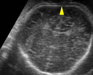
Sonolucent skull :  The thin skull is so poorly ossified that cerebral gyri and sulci could be seen easily

Associations: Rare, though concurrent anencephaly has been reported.

Management: Termination of pregnancy can be offered. For term pregnancy, cesarean delivery does not decrease fracture rates at birth in infants with non-lethal forms of OI.

Prognosis: OI type II is lethal.

Recurrence risk: Unlikely because OI type II is due to new mutations in most cases and there are only a few reports of autosomal recessive inheritance.

Read More

Spinal Dysraphism

Spinal Dysraphism

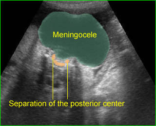
Spinal dysraphism, splaying of the posterior ossification centers of the spine, usually indicates spina bifida resulting from failure of closure of the posterior neuropore. However, spinal dysraphism can be due to other abnormalities as follows:
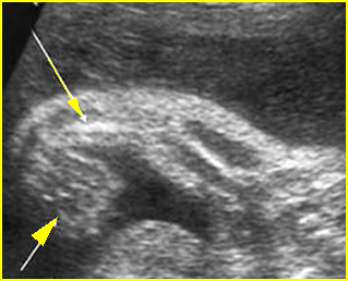
Fig 1, Fig 2, Fig 3

Major differential diagnoses

  • Spina bifida
    • meningocele
    • meningomyelocele
    • myeolochisis
  • Arnold-Chiari malformation type II
  • Pseudodysraphism: technical pitfalls (described previously)

Minor differential diagnoses

  • Diastematomyelia: a splitting of the spinal cord
  • Lipomyelomeningocele: asymmetric lesion of lipoma originating in the spinal cord or cauda equina
  • etc.
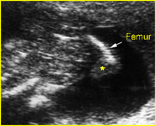
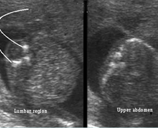
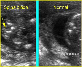
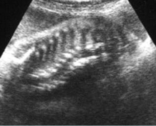
Fig 1:  Spina bifida   Cross-sectional scan of the lumbar and thoracic spine: separation of posterior ossification centers at the lumbar region with small protrusion of the overlying tissue

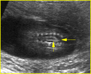
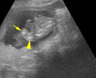
Fig 2:  Spina bifida   Coronal scan of the lumbosacral spine: separation of the ossification centers of the spine (arrow)

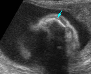
Fig 3:  Spina bifida  Cross-sectional scan of the lumbar and thoracic spine: separation of posterior ossification centers at the lumbar region with small protrusion of the overlying tissue (arrow)

Video clips of spinal dysraphism

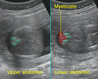
Spina bifida :  Cross-sectional scan of the normal spine at the level of upper abdomen, but wide separation of the posterior ossification centers at the lumbar spine with the protrusion of the spinal cord

Large meningocele :  Cross-sectional scan of the normal spine at the level of upper abdomen, but wide separation of the posterior ossification centers at the lumbar spine with the protrusion of the meningeal sac

Read More

Masses on the Back

Masses on the Back

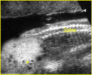
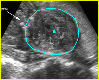
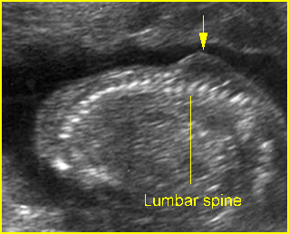
Lumbosacral masses can be caused by a number of specific anomalies, especially meningomyelocele, which is the most common cause. The differential diagnosis for lumbosacral mass can be summarized as follows:

Fig 1, Fig 2, Fig 3

Major differential diagnoses

Meningomyelocele (most common)

  • spinal dysraphism
  • solid-cystic mass located at the lumbosacral spine in most cases
  • associated cranial signs of spina bifida, such as lemon sign, banana sign, and ventriculomegaly

Sacrococcygeal teratoma (SCT) (uncommon)

  • solid-cystic, solid predominantly in most cases but entirely cystic in 15% of cases
  • high vascularization
  • intra-abdominal components in most cases with a displacement effect on internal structures
  •  usually located in the sacrococcygeal area

Limb-body wall complex (uncommon)

  • solid-cystic asymmetric mass
  • abnormal spinal curvature
  • no specific location
  • severe abdominal wall defects
  •  limb defects
  • no or very short umbilical cord

Amniotic band syndrome (uncommon)

  • solid-cystic asymmetric mass
  • no specific location
  • associated limb reduction/constriction defects
  • amniotic band in the amniotic cavity.

Minor differential diagnoses

  • Artifacts: extrafetal mass, such as chorioangioma attached to the fetal back
  • Rare tumors: lipomas, lipomyelomeningocele, and large hemangioma.

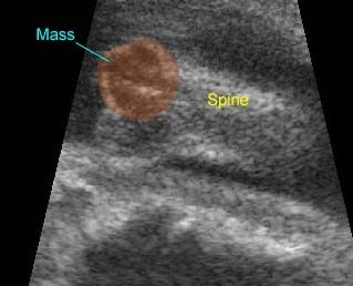
Fig 1:  Sacrococcygeal teratoma   Sagittal scan of the spine: abnormal complex solid mass (*) beneath the sacral spine

Fig 2:  Large meningomyelocele   Scan of lower spine: large solid mass with heterogeneous echodensity (solid circle) (arrow = sacrum)

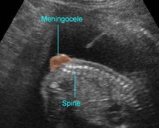
Fig 3:  Lumbar meningocele   Sagittal scan of the spine: small solid mass protruding from lumbar region

Video clips of masses on the back

Sacrococcygeal Teratoma :  Mass on the back: the complex cyst at the caudal end of the spine

Spina bifida :  Sagittal scan of the fetal spine showing sacral meningocele

Read More

Abnormal Spinal Curvature

Abnormal Spinal Curvature

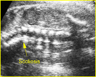
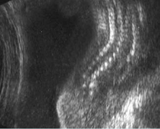
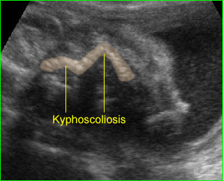
These abnormalities include kyphoscoliosis, hemivertebrae, angulations or disorganized vertebral structures. Scoliosis, an abnormal lateral deflection of the spinal column, is the most common in this category.

Fig 1, Fig 2, Fig 3, Fig 4

Sonographic differential diagnosis:

  • Normal
  • Meningomyelocele (common)
  • LBWC (uncommon)
  • Amniotic band syndrome (uncommon)
  • Skeletal dysplasia
  • VATER or VACTERL associations (rare)
  • Isolated hemivertebrae
  • Iniencephaly (very rare)
  • Caudal regression syndrome

Note:

  • Hemivertebrae are an example of congenital scoliosis resulting from failure of segmentation, which is mostly associated with other anomalies.
  • The normal flexible skeleton can be subject to strong deformation forces in utero resulting in transient scoliosis.
  • Gross skeletal defects, including absent ribs or hemivertebrae, associated with lateral spinal deflection probably represent truly pathologic scoliosis.

Fig 1:  Scoliosis   Coronal scan of the spine: scoliosis of spine at the thoracic region (arrow)

Fig 2:  Angulation of spine   Coronal scan of the spine: angulation of spine at the thoraco-lumbar region (arrow)

Fig 3:  VATER associations   Coronal scan of the spine: scoliosis

Fig 4:  Disorganized spine :   Coronal scan of the spine: scoliosis and disorganized

Video clips of abnormal spinal curvature

Limb-body wall complex:  Coronal scan of the fetal spine: scoliosis associated with limb body wall complex

Severe kyphoscoliosis :  Severe kyphoscoliosis in case of iniencephaly

Read More

Abnormal Hands and Feet

Abnormal Hands and Feet

Hands and feet should be examined to exclude polydactyly, brachydactyly, abnormal hand posture, sandal gap, clinodactyly, Rocker-bottom foot, clubfoot, etc. These abnormalities are commonly part of several syndromes.

Polydactyly Fig 1

Polydactyly is the presence of an additional digit, which can be postaxial (on the ulnar or fibular side) or preaxial (on the radial or tibial side) and with a central form. This extra digit may range from a fleshy nubbin to a complete digit. Postaxial polydactyly is the most common form and is inherited autosomal dominantly as an isolated finding in most cases. However, it is significantly related to some syndromes, especially trisomy 13 or short-rib polydactyly syndrome. Preaxial polydactyly, especially a triphalangeal thumb, is most likely to be part of a syndrome.

The major differential diagnoses for polydactyly include

  • Isolated polydactyly
  • Chromosome abnormalities, especially trisomy 13
  • Meckel Gruber syndrome
  • Asphyxiating thoracic dystrophy
  • Short-rib polydactyly syndrome
  • Achondroectodermal dysplasia (Ellis-Van Creveld syndrome)
  • Smith-Lemli-Opitz syndrome.

Fig 1:  Polydactyly

Post-axial polydactyly

Polydactyly :  Post-axial polydactyly (fleshy finger)

Pre-axial polydactyly

Clubfoot Fig 2

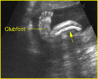
A clubfoot is one of the most common congenital anomalies, occurring in 1:250 to 1 in 1000 births. In 95% of cases, the sole is turned medially (talipes equinovarus). This can be caused by external compression in the case of oligohydramnios or by internal factors including abnormal bone formation, spina bifida, muscular defects or genetic causes (15% have a family history of clubfoot). Most clubfeet are found in otherwise normal infants. However, about 10% of cases are associated with several syndromes including Pena-Shokier phenotype, trisomy 18 and 13, etc.

Rocker bottom foot Fig 3

Vertical talus, or eversion of the planar arch, produces a rocker-bottom (convex outward) appearance of the bottom of the foot. This is most often associated with chromosomal abnormalities, particularly trisomy 13 and 18, although it may also be seen as an isolated anomaly, with caudal dysplasia sequence, neural tube defects, and neuromuscular disorders, and as part of the Potter sequence

Fig 2:  Clubfoot  Longitudinal scan of the lower leg: plantar view of the foot seen in the same plane of longitudinal view of tibia and fibula

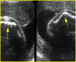
Fig 3:  Rocker bottom foot   Prominent calcaneous bone (arrow) associated with trisomy 13

Clubfoot :  The plantar view of the foot can be demonstrated on the same plane of the longitudinal view of the tibia

Rocker-bottom foot :  Sagittal scan of the fetal foot showing the prominent heel

Abnormal hand posture Fig 4, Fig 5, Fig 6

Fetal hand malformation may be isolated or associated with chromosomal abnormalities, limb reduction defect, a neuromuscular disorder, or skeletal dysplasia. Ultrasound evaluation of the fetal hand requires assessment of the number and configuration of the fingers, the presence and position of the thumb, and the relationship of the thumb and fingers to each other and to the hand and wrist. The phalanges should be evaluated in the long-axis rather than the short-axis view. Short-axis views may represent the metacarpals rather than the phalanges. The normal fetal hand is most often in a resting position with loosely curled fingers, which the fetus periodically opens. Sonographic assessment requires skill on the part of the examiner in following the movement of the hand and documenting the extended fingers and thumb should the hand open during the examination.

Clenched hands Fig 7, Fig 8

Clenched hands or abnormal hand postures are often related to other abnormalities and more than half of cases have aneuploidy, predominantly trisomy 18 (88% of aneuploid fetuses).

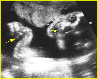
Fig 4:  Pena-Shokier phenotype  Fixed flexion of both elbow and wrist joint (arrow) as well as persistent mouth opening (*)

Fig 5:  VATER associations  Longitudinal scan of forearm: absent radius with abnormal hand posture

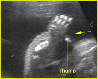
Fig 6:  Hitchhiker thumbs   Abnormal posture and wide separation of the thumb (arrow) in the fetus with diastrophic dysplasia (25 weeks)

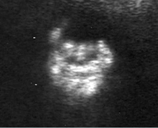
Fig 7:  Clenched hand   Overlapping fingers of the fetus with trisomy 18

Fig 8: Clenched hand   Overlapping fingers of the fetus with trisomy 18

Overlaping fingers :  Clenched hand (arrowhead) in trisomy 18

Fixed flexion:  Fixed flexion of the wrist joint in association with trisomy 18

Edematous hand

Clinodactyly Fig 9, Fig 10

Clinodactyly is the permanent curvature or deflection of one or more fingers. It is frequently associated with abnormal chromosomes especially trisomy 21.

Syndactyly

Syndactyly is the fusion of digits and may consist of bony or soft tissue fusion. This may also be difficult to detect sonographically, especially those cases consisting of soft tissue fusion. Syndactyly is associated with a number of syndromes. It may be seen in the SRPD syndromes with the characteristics of polydactyly, and it may be seen in triploidy or Down’s syndrome.

Fig 9: Clinodactyly   Curved-in position of the fifth finger (arrow) of the fetus with trisomy 21

Fig 10:  Clinodactyly  Scan of the hand: disproportionately small middle phalange of the fifth finger (arrow)

Clinodactyly :  Clinodactyly with hypoplasia of the middle phalanx of the fifth finger

Read More

Limb Reduction Defects

Limb Reduction Defects

Limb reduction deformities, presenting alone or as part of a specific syndrome, occur in approximately 0.5 per 10,000 births. Half of them are simple transverse reduction deficiencies of one forearm or hand without associated anomalies and the remaining half have additional anomalies.

Fig 1, Fig 2, Fig 3

Isolated limb reduction:

  • isolated limb deficiency more commonly occurs in the upper extremities rather than the leg, which generally occurs within the context of a syndrome, as do bilateral amputations or reduction of all limbs
  • sporadic occurrence in most cases with negligible risk of recurrence

Limb reduction associated amniotic band:

  • secondary to entanglement with mesodermic bands derived from the chorionic surface of the amnion after the latter has separated from the chorion
  • isolated or multiple limb reductions, with or without other defects

Phocomelia:

  • typically hands and feet are present, but the intervening arms and legs are absent
  • main differential diagnoses include Robert’s syndrome, variants of TAR syndrome, and Grebe’s syndrome
  • can be caused by exposure to thalidomide but this is only of historical interest

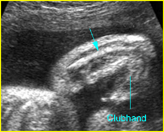
Radial ray defects: radial clubhand may be part of

  • TAR syndrome (thrombocytopenia with absent radius)
  • Fanconi’s syndrome: an autosomal recessive disorder consisting of pancytopenia anemia, leukopenia and thrombocytopenia and skeletal anomalies, especially radial clubhand
  • Aase’s syndrome: an autosomal recessive disorder characterized by hypoplastic anemia and radial clubhand with bilateral triphalangeal thumb and a hypoplastic radius
  • Holt-Oram syndrome: an autosomal dominant disorder characterized by congenital heart defects, radial hypoplasia and triphalangeal or absent thumb
  • VATER association: a sporadic disorder resulting from defective mesodermal development during embryogenesis consisting of vertebral segmentation (70%), anal atresia (80%), tracheoesophageal fistula (70%), esophageal atresia, and radial (65%) and renal defects (53%)
  • Goldenhar’s syndrome: characterized by hemifacial microsomia, hypoplasia of the malar and maxillary or mandibular region, vertebral anomalies, and radial defects
  • Klipple-Feil syndrome
  • Chromosomal abnormalities, particularly trisomy 18 and 21.

Fig 1:  Transverse limb reduction  Scan of the foot: absent distal end of foot

Fig 2:  Absent radius syndrome  Longitudinal scan of upper limb (15 weeks): short ulna (arrow) without radius with fixed flexion of wrist joint (arrowhead)

Fig 1:  Limb reduction   Longitudinal scan of lower limb: absent tibia and fibula, abnormal foot (*) arising from femur (arrow) in the fetus with VATER association

Video clips of limb reduction defects

Radial ray defects :  Longitudinal scan of the upper extremity: absent radius (arrow) with fixed flexion of the wrist joint (arrowhead)

Radial ray defects :  Longitudinal scan of the upper extremity at 13 weeks: absent radius (with fixed flexion of the wrist joint (arrow)

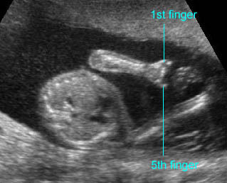
Transverse limb defect :  Absent fingers; only the 1st and 5th fingers remained

Transverse limb defect :  Absent hand with shortenings of all long bones (14 weeks) (arrow = severely shortened forearm, arrow = humerus)

Read More

Long Bone Bowing

Long Bone Bowing

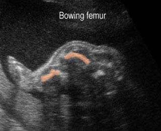
The degree of long-bone curvature should be examined. Campomelic (bent-bow) dysplasia is characterized by ventral bowing of the long bones. Thanatophoric dysplasia and osteogenesis imperfecta also have bowed extremities.

Fig 1, Fig  2

The major differential diagnoses are as follows:

  • Campomelic dysplasia (bowing but no shortening)
  • Osteogenesis imperfecta type II (related to fracture and callus formation)
  • Thanatophoric dysplasia (severe shortened long bones)
  • Achondrogenesis
  • Hypophosphatasia

Fig 1:  Campomelic dysplasia   Longitudinal scan of tibia: anterior bowing of well ossified tibia

Fig 2:  Osteogenesis imperfecta type IIB  Irregular and angular fracture of long bones (arrow) but rather well-ossified

Video clips of fracture in utero

Long bone shortening :  Severe long bone shortening with bowing and normal echogenicity in case of thanatophoric dysplasia

Campomelia :  Anterior bowing of the tibia (arrow)

Read More

Fracture In Utero

Fracture In Utero

The detection of rib or long bone fractures in association with severe micromelia suggests a diagnosis of osteogenesis imperfecta. Fractures may be subtle or may lead to angulation.

Fig 1, Fig2, Fig 3

The major differential diagnoses for fractures in utero are as follows:

  • Osteogenesis imperfecta type II (most common)
  • Hypophosphatasia (rare)
  • Campomelic dysplasia (not a true fracture but bowing).

Fig 1:  Fracture in utero  Longitudinal scan of upper extremity: poorly ossified and fracture in osteogenesis imperfecta type IIA

Fig 2:  Callus formation of long bone  Longitudinal scan of humerus and femur: irregularity due to callus (arrowhead) in osteogenesis imperfecta type IIA

Fig 3:  Rib fractures  Multiple rib fractures with poor ossification in the fetus with osteogenesis imperfecta type IIA

Video clips of fracture in utero

Fracture in utero :  Longitudinal scan of upper limb: callus formation (arrow) and irregularity of ulna secondary to previous fracture

Osteogenesis imperfecta :  Cross-sectional scan of the thorax: rib fractures (arrow)

Read More

Sonolucent Bones

Sonolucent Bones

Diffuse demineralization of the skull and long bones almost always occurs with fetal skeletal dysplasia syndromes. In the case of severe demineralization of the bony calvarium, the cranium is thin without an acoustic shadow and so poorly ossified that the intracranial structure can easily be seen. This increased visualization of the intracranial structures may be confused with such abnormalities as exencephaly due to acrania or acalvaria. Unlike exencephaly, however, there is an intact but poorly ossified skull. Careful scanning reveals concomitant limb anomalies.

Fig 1, Fig 2, Fig 3, Fig 4

The main differential diagnoses of the sonolucent skull are as follows:

  • Osteogenesis imperfecta (most common)
  • Hypophosphatasia (rare)
  • Achondrogenesis type I (rare).

Fig 1:  Hypophosphatasia  Longitudinal scan of long bones: shortened and poorly ossified bones

Fig 2:  Poorly ossified and compressible skull   Cross-sectional scan of skull: deformable skull with sonolucency in osteogenesis imperfecta type IIA

Fig 3:  Micromelia in Thanatophoric dysplasia   Severe shortenings of long bones (arrow) but normal ossification

Fig 4:  Sonolucent and shortened arm   Longitudinal scan of the arm: Sonolucent and shortened humerus (*) in case of achondrogenesis

Video clips of sonolucent bones

Sonolucent skull : The thin skull is so poorly ossified that cerebral gyri and sulci could be seen easily

Achondrogenesis:  Mid-sagittal view of the spine: extremely poor ossification of the spine (arrow)

Read More

Bone Shortening

Bone Shortening

All long bones should be measured in each extremity, although subjective evaluation may be sufficient in most cases. The severity of bone shortening varies from extreme to mild.

Fig 1, Fig 2, Fig 3

The major differential diagnoses of bone shortenings are as follows:

Severe shortening

  • thanatophoric dysplasia (micromelia) (common)
  • osteogenesis imperfecta type II (micromelia) (common)
  • achondrogenesis (micromelia) (common)
  • short-rib polydactyly syndrome (uncommon)
  • diastrophic dysplasia (uncommon)
  • homozygous achondroplasia (uncommon)
  • mesomelic dysplasia (uncommon)
  • chondrodysplasia punctata, rhizomelic type

Mild-to-moderate shortening

  • osteogenesis imperfecta type II (common)
  • heterozygous achondroplasia (common)
  • asphyxiating thoracic dysplasia (common)
  • campomelic dysplasia (uncommon)
  • hypophosphatasia (uncommon)
  • chondroectodermal dysplasia (uncommon).

Fig 1:  Long bone shortening   Longitudinal scan of the thigh and leg: shortening of thigh and leg, compared with foot (achondrogenesis)

Fig 2:  Short humerus  Longitudinal scan of long bones: shortened but well ossified humerus of the fetus with Juene syndrome

Fig 3:  Micromelia in Thanatophoric dysplasia   Severe shortenings of long bones (arrow) but normal ossification

Video clips of bone shortening

Thanatophoric displasia:  Severe shortenings of the long bone (arrowhead) compared to the skull

Long bone shortening : Severe long bone shortening with bowing and normal echogenicity in case of thanatophoric dysplasia

Read More