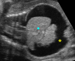
Duodenal Atresia

Duodenal Atresia

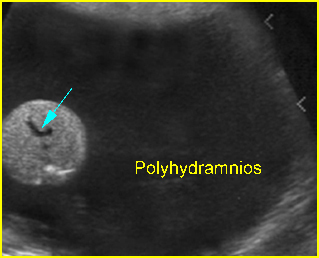
Duodenal atresia is the most common type of congenital small bowel obstruction resulting from failure to recanalize the duodenal lumen during the 11th week of gestation.

     Fig 1

Incidence: 1 in 5000-10,000 births. One-third have Down’s syndrome.

Sonographic findings:

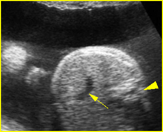
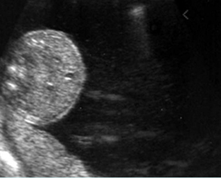
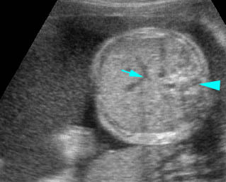
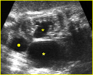
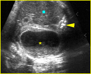
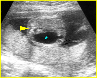
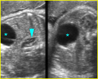
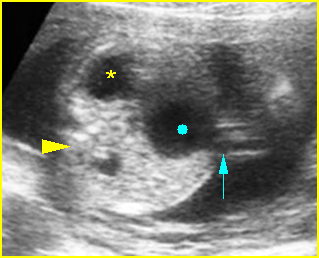
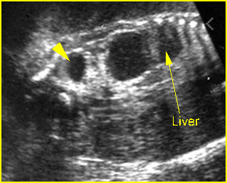
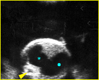
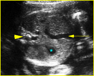
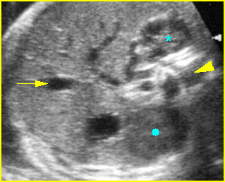
  • Double bubble sign and a narrow channel connecting the stomach and duodenum (the second bubble will be in the location of the duodenal bulb, which is typically in the center of the abdomen, just to the right of the midline). Fig 2, Fig 3, Fig 4
  • The continuity between the two fluid-filled structures must be demonstrated, and if the connection cannot be shown, a number of other possibilities should be considered, such as choledocal cyst (most common) and the less common renal cyst, hepatic cyst, omental cyst, bowel duplication or ovarian cyst.
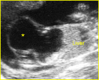
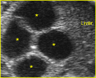
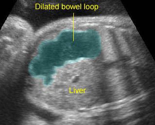
  • A more prominent appearance with a C loop of dilated bowel may occur when duodenal atresia is associated with esophageal atresia. Fig4, Fig5
  • Pitfalls:
    • A prominent normal stomach with a visible incisura angularis may be confused with a double bubble sign.
    • Bisection of the normal stomach in an oblique scan plane, giving the spurious appearance of a double bubble. This misinterpretation can be corrected by scanning the abdomen in a true transverse plane, so as to demonstrate the typical tapering configuration of the gastric antrum.
    • Any right upper quadrant mass such as a choledocal cyst, bile-filled gallbladder, or hepatic cyst may be misinterpreted as a distended duodenum.
  • Polyhydramnios occurs in nearly all cases diagnosed in the third trimester.
  • Perforation secondary to obstruction can occur, and ascites or meconium peritonitis may be found.
  • Usually first diagnosable in the second trimester, although it can occasionally be diagnosed before 20 weeks or even in the first trimester.
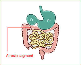
Fig 1:  Schematic drawing of duodenal atresia, The fluid-filled stomach (St) and duodenum (D) cause double bubble sign on ultrasound examination
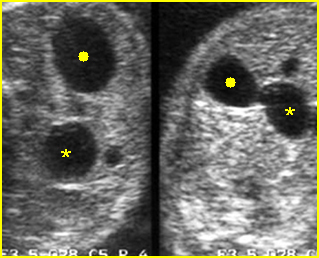
Fig 2:  Duodenal atresia  Cross-sectional scan of the abdomen: Double bubble sign: two cystic masses (* = proximal duodenum, solid circle = stomach) with connection together in the upper abdomen

Fig 3:  Duodenal atresia   Cross-sectional scan of the abdomen: Double bubble sign: two cystic masses (* = stomach, solid circle = proximal duodenum ) in the upper abdomen
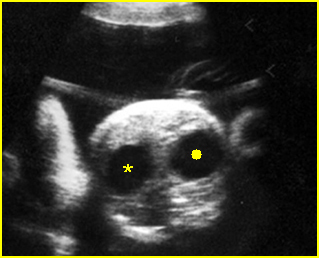
Fig 4:  Duodenal atresia   Cross-sectional scan of the abdomen: Double bubble sign: two cystic masses (* = proximal duodenum, solid circle = stomach) in the upper abdomen

Video clips of duodenal atresia
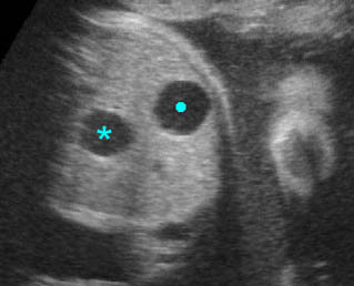
Double bubble sign:  Cross-sectional scan of the abdomen: two connecting cystic masses (*) in the upper abdomen with polyhydramnios
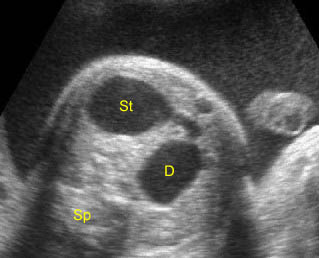
Duodenal atresia:  Double bubble sign with continuation representing stomach (St) and duodenum (D) (Sp = spine)

Duodenal atresia:  C-loop of duodenum and stomach secondary to duodenal and esophageal atresia

Associations: Gastrointestinal, genitourinary and cardiovascular anomalies occur in more than 50% of cases and trisomy 21 is found in 30% of cases (but only 5% of fetuses with trisomy 21 show duodenal atresia). Furthermore, duodenal obstruction also increases the risk of prenatal asphyxia and death, probably caused by bradycardia/asystole following vagal overactivity due to distension of the upper gastrointestinal tract.

Management: A careful search for associated anomalies and karyotyping are indicated. The delivery should occur where appropriate personnel are available for surgical correction.

Prognosis: A high mortality rate of about 20-40% due to associated abnormalities or preterm delivery. Uncomplicated cases have a good prognosis.

Recurrence risk: Sporadic (with rare familial reports).

Read More

Esophageal Atresia

Esophageal Atresia

This sporadic malformation results in an abrupt termination of the esophagus as a blind pouch. In approximately 90% of cases, esophageal atresia is associated with a tracheoesophageal fistula, which most often connects the trachea to the distal esophagus. Additional abnormalities occur in a high percentage of fetuses with esophageal atresia, and these include cardiac, chromosomal, gastrointestinal, genitourinary, and central nervous system lesions.

Incidence: About 1 in 3500 births, and nearly two-thirds are male.

Sonographic findings:

            Fig 1, Fig 2, Fig 3

  • Polyhydramnios: massive polyhydramnios (amniotic fluid index (AFI)>40) is commonly seen in the third trimester.
  • Absent fluid-filled stomach in its normal location in the left upper quadrant.
  • The absent visible stomach can be identified in about 42% of fetuses. Conversely, the combination of polyhydramnios and gastric non-visualization is only seen in less than half of fetuses with a tracheoesophageal fistula.
  • Proximal esophageal pouch (pouch sign): a fluid-filled pouch corresponding to the atretic segment of the esophagus may be seen in the neck or distal esophageal segment.
  • Fetal growth restriction: about 40% of infants with esophageal atresia are associated with growth restriction.
  • Usually first diagnosable in the second trimester, mostly after 24 weeks.
  • Among fetuses with ultrasonographic features suggestive of esophageal atresia, 50% have the disorder confirmed postnatally.
  • In case of suspicion, MRI may be helpful.
  • Pitfalls:
    • Non-visualization of the stomach may be a transient normal finding.
    • Several cases of tracheoesophageal fistula with a connection to the stomach through the lungs have a visible stomach.
    • When combined with duodenal atresia, it may be very difficult to detect due to masking from polyhydramnios and double bubble sign.

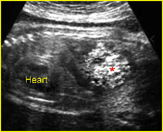
Fig 1:  Esophageal atresia  Cross-sectional scan of the abdomen: absent stomach at the level of umbilical vein complex (arrow) with marked polyhydramnios

Fig 2:  Esophageal atresia  Cross-sectional scan of the abdomen: absent stomach at the level of umbilical vein complex (arrow) with polyhydramnios (arrowhead = spine)

Fig 3:  Esophageal atresia   Cross-sectional scan of the abdomen: absent stomach with marked polyhydramnios

Video clips of esophageal atresia

Esophageal atresia:  Cross-sectional scan of the abdomen: persistently absent stomach (arrow = umbilical vein, arrowhead = spine)

Associations: Associated malformations are seen in at least 50% of cases of tracheoesophageal fistula, including gastrointestinal, genitourinary, and cardiovascular anomalies, trisomy 21 and 18.

Management: A careful search for associated anomalies is indicated. The pregnancy with fetal isolated esophageal atresia should be closely followed-up and monitored for preterm delivery. The delivery should occur where appropriate personnel are available for surgical correction.

Prognosis: Poor overall (due to associated anomalies and prematurity), but the prognosis is much improved with isolated esophageal atresia.

Recurrence risk: Sporadic.

Read More

Masses at the Anterior Body Wall

Masses at the Anterior Body Wall

Differential diagnoses of the masses at the anterior body wall include

Fig 1, Fig 2, Fig 3, Fig 4, Fig 5, Fig 6

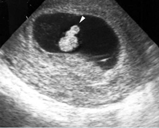
  • Physiologic omphalocele (8-12 weeks); size of <7 mm, no liver content
  • Omphalocele
  • Gastroschisis
  • Ectopia cordis
  • Limb-body wall defect
  • Bladder exstrophy
  • Cloacal exstrophy
  • Stack of umbilical cord (positive Doppler signal or color flow)
  • Cord edema or localized Wharton’s jelly near the umbilicus
  • Cord cysts (omphalomesenteric cyst or allantoic cyst)
  • Placental mass (chorioangioma) close to the anterior wall
  • Artifacts: pseudo-omphalocele secondary to oblique plane

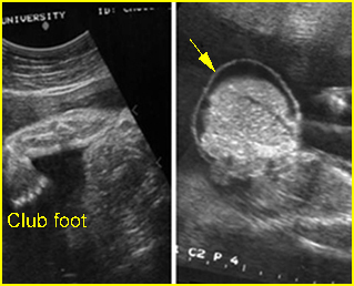
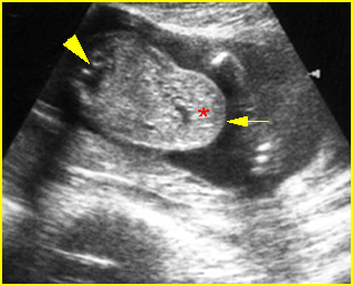
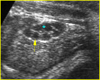
Fig 1:  Omphalocele  Clubfoot and free floating sac containing liver with covering membrane (arrow)

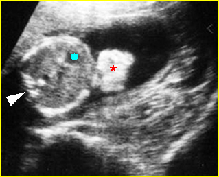
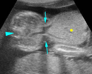
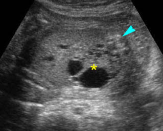
Fig 2:  Gastroschisis  Free floating echogenic bowel (*) in the amniotic fluid (arrowhead = spine, solid circle = intra-abdominal stomach)

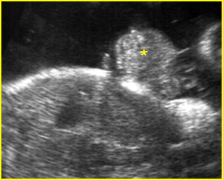
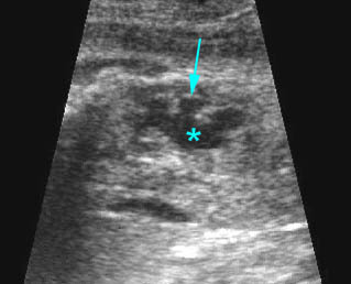
Fig 3:  Bladder extrosphy  Sagittal view of the fetal abdomen: complex extra-abdominal mass (*) below the umbilicus, finally proven to be bladder extrosphy

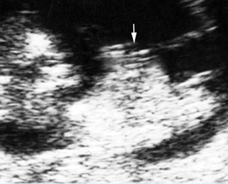
Fig 4:  Physiologic omphalocele  Sagittal scan of the fetus (10 weeks): bowel contents protruding into the proximal umbilical cord (arrow)

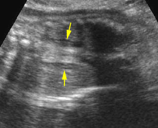
Fig 5:  Pseudo-omphalocele  Oblique cross-sectional scan of the fetus: the liver (*) covered by normal abdominal wall (arrow) protrudes anteriorly, may be mistaken for omphalocele (arrowhead = spine)

Fig 6:  Physiologic omphalocele  Cross-sectional scan of the embryo (8 weeks): bowel contents protruding into the proximal umbilical cord (arrowhead)

Video clips of masses at the anterior body wall

Omphalocele:  Cross-sectional scan: large omphalocele with liver content (solid circle) (arrow = the defect, arrowhead = spine)

Umbilical cord cyst:  Focus on the proximal cord: cord cyst with coarse particles (*) may be due to liquidfaction of the Wharton’s jelly

Gastroschisis:   Free floating bowel mass anterior to abdominal wall

True anterior wall defect

  • Omphalocele
  • Gastroschisis
  • Limb-body wall complex
  • Amniotic band syndrome
  • Bladder exstrophy
  • Cloacal exstrophy
  • Pentralogy of Cantrel

Cystic mass close to the umbilicus

  • Allantoic cyst (urachal cyst)
  • Omphalomesenteric cyst
  • Pseudocyst (liquefaction of Wharton’s jelly)
  • Umbilical vein varix
  • Omphalocele

The approach of the anterior wall defects

  •  Relationship of the cord insertion to the defect: various sites of the defect suggest the nature of pathology as follows:
    • above the umbilicus: pentalogy of Cantrell
    • at the umbilicus: omphalocele
    • paraumbilical area: gastroschisis
    • below the umbilicus: bladder/cloacal exstrophy
    • defects throughout the abdomen: limb-body wall complex
    • severe and asymmetric defect: amniotic band syndrome
  • Characteristics of herniated organs: a herniated organ can suggest the nature of pathology as follows:
    • bowel: either a gastroschisis, omphalocele, or LBWC
    • liver only: highly suggestive of omphalocele (but most omphaloceles also include portions of bowel), very unlikely to be gastroschisis
    • bowel only (in omphalocele): more often related to chromosomal abnormalities
    • solid mass at the lower abdomen: bladder or cloacal exstrophy
  • Presence of covering membranes:
    • presence: omphaloceles (be careful, the covering membrane may not always be seen or may be ruptured), limb-body wall complex (not always)
    • absence: gastroschisis
    • the presence of a herniated liver in a ventral wall defect without a covering membrane is more suggestive of a ruptured omphalocele
  • Associated anomalies:
    • multiple anomalies: omphalocele, LBWC
    • extra-, intra-abdominal bowel obstruction: gastroschisis
    • scoliosis: LBWC
    • non-visualization of bladder: bladder or cloacal exstrophy
    • amputation defects: amniotic band syndrome
Read More

Renal Cysts

Renal Cysts

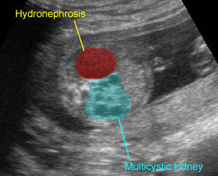
Sonographic differential diagnosis of common renal cystic conditions

Fig 1, Fig 2, Fig 3, Fig 4

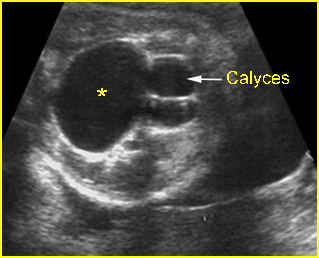
Fig 1:  Hydronephrosis  Cross-sectional scan of the abdomen: markedly dilated renal pelvis (*) with calyces (arrow)

Fig 2:  Multicystic kidney  Sagittal scan of the abdomen: multiple cysts varying in size in the kidney

Fig 3:  Hydronephrosis  Coronal scan of the abdomen: bilateral dilated renal pelvis (*) (solid circle = bladder)

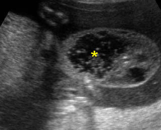
Fig 4:  Normal kidney   The kidneys consisting of multiple normal pyramids (arrow), looking like multicystic kidney (* = renal pelvis)

Video clips of renal cysts

Renal cystic dysplasia:  Cross-sectional scan of the abdomen: muliticystic kidney (*) with oligohydramnios and absent contralateral kidney

Severe hydronephrosis:

  • visible renal parenchyma
  • peripheral oval cysts communicating with each other and the renal pelvis
  • often ureteral dilatation
  • underlying cause in some cases such as bladder outlet obstruction
  • abnormalities in the contralateral kidney (10-40%)

Autosomal recessive polycystic kidneys

  • bilateral enlarged echogenic kidneys with reniform shape
  • oligohydramnios

Multicystic kidney disease

  • multiple round non-communicating cysts of variable size
  • multiple cysts with grape-like appearance in many cases
  • unilateral (77%)
  • abnormalities in the contralateral kidney (39%)

Renal cystic dysplasia

  • echogenic parenchyma and subcapsular or cortical cyst
  • evidence of hydronephrosis in some cases
  • evidence of an underlying cause such as bladder outlet obstruction in some cases
Read More

No Visible Bladder

No Visible Bladder

Non-visualization of the bladder with oligohydramnios

  • Severe intrauterine growth restriction (common)
  • Bilateral renal agenesis (common)
  • Bilateral ureteral pelvic junction obstruction
  • Bilateral multicystic dysplastic kidney
  •  Infantile polycystic kidney disease
  • Cloacal exstrophy (very rare)
  • Donor twin in twin-to-twin transfusion syndrome

Non-visualization of one fetal kidney

  • Shadowing from the fetal spine
  • Pelvic kidney
  • Unilateral renal agenesis
  • Crossed renal ectopia

Bilateral renal agenesis:  Coronal scan of the abdomen, no visible kidney, oligohydramnios, elongated adrenal glands (arrow) seen in the renal fossa instead

Read More

Abdominal Cysts

Abdominal Cysts

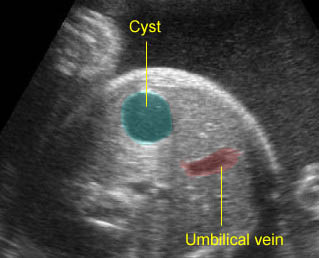
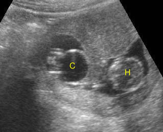
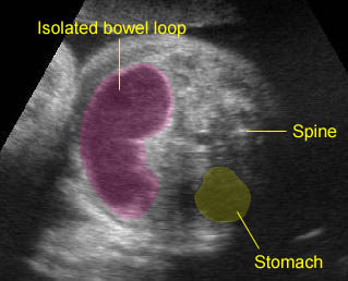
Sonographic differential diagnosis of abdominal cyst is often necessary to obtain the following critical information to narrow the diagnosis:

Fig 1, Fig 2, Fig 3, Fig 4, Fig 5, Fig 6, Fig 7, Fig 8, Fig 9

  •  Location
    • close to the spine: usually related to renal abnormalities, etc.
    • right upper quadrant: typically hepatic cyst or choledochal cyst
    • close to the umbilicus: mesenteric cyst, umbilical vein varix, meconium pseudocyst, ovarian cyst
    • lower abdomen: urachal cyst, ovarian cyst, hydrometrocolpos, megacystis
  • Size and shape of the lesion
    • unilocular, round: ovarian cyst
    • multiple loops: bowel obstruction
    • tubular: dilated ureter
  • Fetal gender
    • always female: ovarian cyst, hydrometrocolpos
    • usually female: hepatic cyst, choledochal cyst
    • usually male: bladder outlet obstruction, ureterocele, hydronephrosis
  • The change in appearance over time
    • dilated bowel
    • partial obstruction of the urinary tract
  • Other information is often helpful: the wall and contents of the cyst, the relationship to other abdominal organs, peristalsis activity, etc.

Fig 1:  Fetal dermoid cyst  Cross-section at the upper abdomen: cystic mass (*) containing echogenic debris or sebum (arrowhead = spine, circle = liver)

Fig 2:  Fetal ovarian cyst  Cross-section of the fetal upper abdomen: simple anechoic cyst (*) (arrowhead = spine)

Fig 3:  Fetal ovarian cyst  Cross-sectional scan of the abdomen: Simple anechoic cyst (*) in the rightside of the abdomen, note normal kidney and adrenal gland (arrowhead)

Fig 4:  Large urachal cyst  Sagittal scan of the fetal trunk: large cystic mass (*) connecting with the bladder, externally protruding from the lower abdomen

Fig 5:  Large umbilical vein aneurysm  Cross-sectional scan of the abdomen: cystic mass (solid circle) connecting with umbilical vein (arrow) with Doppler signal on flow study (* = stomach, arrowhead = spine)

Fig 6:  Choledocal duct cyst  Coronal scan of the abdomen: anechoic cystic mass (*) located at the right upper abdomen with pressure effect on the liver (arrowhead = renal cystic dysplasia)

Fig 7:  Duodenal atresia  Cross-sectional scan of the abdomen: Double bubble sign: two cystic masses (* = proximal duodenum, solid circle = stomach) in the upper abdomen

Fig 8:  Proximal jejunal atresia  Cross-sectional scan of the abdomen: marked dilatation of the bowel loop with continuation of stomach (solid circle), duodenum (*) and proximal jejunal

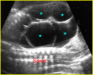
Fig 9:  Small bowel obstruction  Cross-sectional scan of the abdomen: Multiple loop of small bowel loops with marked dilatation (*)

Video clips of abdominal cysts

Hepatic cyst:  Isolated cystic mass located at right upper abdomen

Bladder outlet obstruction:  Coronal scan: Megacystis (C) at 11 weeks of gestation (H = head)

Renal cysts:  Multicystic kidney and marked hydronephrosis of the contralateral kidney

Bowel duplication:  Sausage cystic mass secondary to bowel duplication; isolated bowel loop with no other visible bowel loops

Duodenal atrestia:  Double bubble sign with continuation representing stomach (St) and duodenum (D) (Sp = spine)

Proximal jejunal atresia:  Dilated bowel loop at the upper abdomen representing the proximal jejunal loop separated from the stomach and large bowel

Renal cystic dysplasia:  Cross-sectional scan of the abdomen: muliticystic kidney (*) with oligohydramnios and absent contralateral kidney

The main sonographic differential diagnoses include

Choledochal cyst/hepatic cyst:

  • anechoic single cyst in liver, choledochal cyst located adjacent to hepatic ducts
  • more common in females
  • not associated with other anomalies

Multicystic kidneys:

  • anechoic multicysts of variable size which are non-communicating
  • located posteriorly
  • unilateral or bilateral
  • male or female

Hydronephrosis:

  • anechoic single or often multiple communicating cysts within renal fossa
  • more common in males
  • probably associated with other GU anomalies

Hydroureter:

  • anechoic tubular cyst in nature
  • communicating with renal pelvis and bladder
  • located posteriorly
  • more common in males
  • may be associated with megacystis

Meconium pseudocyst:

  • echogenic cyst with calcified wall at mid-abdomen
  • associated with bowel obstruction
  • male or female

Mesenteric/omental cyst:

  • isolated anechoic cyst, unilocular or less commonly septated
  • often mobile
  • located centrally
  • male or female

Urachal cyst:

  • isolated anechoic simple
  • communicates with the bladder and also commonly with the umbilicus
  • located anteriorly
  • male or female

Ovarian cyst:

  • isolated unilocular, sometimes septated, or daughter cyst
  • usually unilateral
  • located lower or laterally
  • only in females

Duodenal atresia:

  • double bubble sign in upper abdomen
  • connection between the two bubbles
  • polyhydramnios
  • male or female
  • commonly related to trisomy 21

Small bowel obstruction:

  • multiple bowel loops with echogenic contents
  • tubular in nature
  • located centrally or throughout the abdomen
  • peristalsis
  • male or female

Hydrometrocolpos:

  • anechoic to echogenic cyst
  • located retrovesically
  • only in females
Read More

Hepatosplenomegaly

Hepatosplenomegaly

Enlargement of the liver or spleen is usually related to systemic abnormalities such as fetal hydrops, however it may occur secondary to an isolated finding such as a liver tumor (hemangioendothelioma).
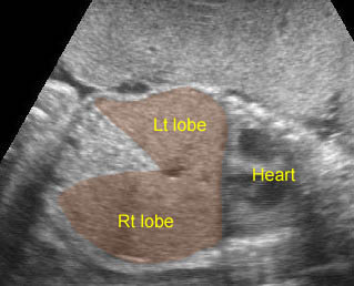
Sonographic findings:

Fig 1, Fig 2

  • Fetal liver measurements are usually obtained in a longitudinal plane, from the dome of the right hemidiaphragm to the tip of the right lobe.
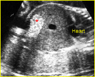
  • Commonly associated with ascites or other signs of hydrops fetalis.
  • Tables of normal values are available for analyzing both fetal liver and spleen measurements.
  • Hepatosplenomegaly could be seen in more than 90% of cases of hydrops fetalis from hemoglobin Bart’s disease.
  • Splenic circumference is an excellent predictor of severe anemia.
  • Hepatomegaly is constantly demonstrated in Beckwith-Wiedemann syndrome.

Differential diagnosis:

  • Hydrops fetalis due to various causes, especially hemoglobin Bart’s disease.
  • Severe anemia or myeloproliferative disorder.
  • Congenital infection; for example, cytomegalovirus infection and syphilis.
  • Beckwith-Wiedemann syndrome.
  • Down’s syndrome.

Associations: Hydrops fetalis due to various causes.

Prognosis: Depends on underlying causes.

Fig 1:  Splenomegaly  Cross-sectional scan of the abdomen: enlarged spleen behind the stomach (*) (arrow= the umbilical vein, arrowhead = spine)

Fig 2:  Splenomegaly  
Cross-sectional scan of the upper abdomen: The enlarged spleen (solid circle) behind the stomach, Note adrenal gland (*) close to the spine (arrowhead) (arrow = umbilical vein)

Video clips of hepatosplenomegaly

Hepatomegaly:  Coronal scan of the upper abdomen shows the enlarged liver, both left and right lobe

Read More

Echogenic Bowel

Echogenic Bowel

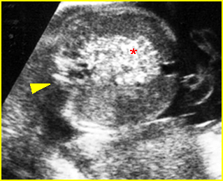
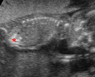
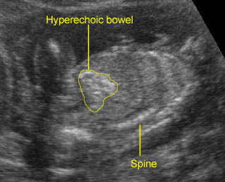
An increased echogenicity of bowels is associated with an increased prevalence of a variety of unfavorable outcomes including cystic fibrosis, chromosomal abnormalities such as trisomy 21, 18, 13 and triploidy, cytomegalovirus infection, intestinal obstruction, and severe fetal growth restriction. Finally, this is also commonly normal variant.

The incidence of intrauterine fetal demise in fetuses with unexplained echogenic bowel is also increased. However, there is no evidence of any serious long-term bowel pathology associated with isolated fetal echogenic bowel.

Sonographic findings:

  • The bowels display high echogenicity, typically occurring in the third trimester but in some cases in the second trimester.

      Fig 1, Fig 2, Fig 3

  • Echogenic bowels should be diagnosed only when a brightness equivalent to that of the surrounding pelvic bones is achieved.
  • Bowel echodensity correlates better with liver density than with bone density. Thus, comparison of bowel to liver echogenicity may be a more effective clinical tool in the evaluation of fetal intra-abdominal echodensities.
  • Differential diagnosis of echogenic bowel including:
    • normal variant (0.5% of normal fetuses)
    • high gain settings
    • bloody amniotic fluid
    • meconium ileus (cystic fibrosis)
    • Down’s syndrome.

Note: The use of special image processing techniques, particularly harmonic and compound-imaging techniques, can artificially enhance the apparent level of echogenicity of the bowel. Additionally, the echogenicity of normal bowel increases with transducer frequency, especially when the 8 MHz transducer is used, although this effect is uniform whereas true hyperechoic bowel tends to be focal.

Fig 1:  Hyperechoic bowel  Coronal scan of the abdomen: hyperechogenic bowel (*)

Fig 2:  Hyperechoic bowel  Sagittal scan of the abdomen: hyperechoic bowel (*)

Fig 3:  Hyperechoic bowel   Cross-sectional scan of the abdomen: hyperechoic bowel (*) (arrowhead = spine)

Video clips of echogenic bowel

Hyperechoic bowels:  Sagittal scan of the fetal trunk: * = bright echogenic bowel, comparable with bone echodensity

Hyperechoic bowel

Read More

Ascites

Ascites

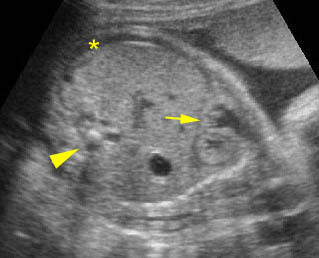
Ascites, fluid collection within the fetal abdomen, is always abnormal. In severe cases, ascites can be readily appreciated, however, mild ascites, often seen in early hydrops fetalis, may be confused with pseudoascites (described previously). Therefore, first of all, one must distinguish between pseudoascites (a symmetric hypoechoic rim of the anterior abdominal wall) and true ascites. The following findings are often related to true ascites but not to pseudoascites:

  • Fluid between bowel loops or outlines the visceral structures (liver, omentum, umbilical vein, falciform ligament)
  • Other signs of hydropic changes
  • Polyhydramnios.

The major causes of ascites are as follows:

  • Hydrops fetalis secondary to various causes (most common)
  • Intrauterine infection
  • GI or urinary tract obstruction with perforation
  • Abdominal tumors (rare)
  • Perforation of the common bile duct (very rare)
  • Chyloperitoneum (very rare).

Note: Ascites associated with hydrops fetalis is usually accompanied by fluid collection in other spaces such as subcutaneous edema, pleural effusion or pericardial effusion. Isolated ascites is most often secondary to intra-abdominal disorders.

Mild ascites:  Oblique cross-sectional scan: * = mild ascites, arrow = diaphragm, arrowhead = spine

Marked ascites:  Coronal scan of the fetal trunk: marked fluid collection in the abdomen (*) (solid circle = liver)

Read More