Chest Masses
A sonographic appearance of chest masses usually reveals the change in echogenicity (cystic, solid or mixed) and mediastinal shift. Sonographic characteristics of the masses can often help to narrow the diagnostic considerations. For example, nearly all chest masses are unilateral, and the bilateral solid masses are likely to be laryngeal or tracheal atresia. A completely solid mass may be a congenital cystic adenomatoid malformation (CCAM), sequestration, or obstructed lung. A solid and left lower lung mass has a high chance of being a pulmonary sequestration. A right lung solid-cystic mass is probably CCAM. A cystic mass with peristalsis is specific to a diaphragmatic hernia.
Fig 1, Fig 2, Fig 3
The main differential diagnoses of chest masses are as follows:
Cystic
- CDH (occasional peristalsis, absent stomach in the abdomen)
- CCAM
- Foregut duplication cyst (neurenteric, bronchogenic, enteric cyst)
- Loculated fluid collection
- Mediastinal meningocele
Cystic and Solid
- CDH (occasional peristalsis, absent stomach in the abdomen)
- CCAM
- Foregut duplication cyst (neurenteric, bronchogenic, enteric cyst)
- Teratoma (pericardial)
Solid
- Sequestration (more common at lower lobe, vascular supply from aorta)
- CDH (occasional peristalsis, absent stomach in the abdomen)
- CCAM
- Bronchial atresia
- Bronchogenic with bronchial compression
- Pericardial teratoma.
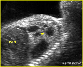
Fig 1: Cystic adenomatoid malformation Oblique sagittal scan of the fetal trunk: solid-cystic mass (*), mostly cystic, in the left chest (CCAM type I)
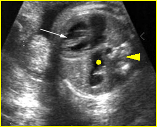
Fig 2: Diaphragmatic hernia Cross-sectional scan of thorax at the level of four-chamber view: cystic structure of bowel loop (solid circle) located in the chest with cardiac displacement to the right (arrowhead = spine)
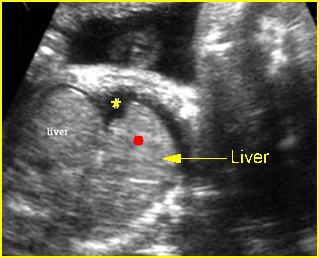
Fig 3: Right diaphragmatic hernia Sagittal scan of the chest: part of liver (solid circle) upward displacement to the chest (* = pleural effusion)
Video clips of chest masses
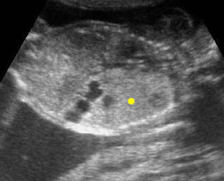
CCAM (Congenital cystic adenomatoid malformation: CCAM) Coronal scan of the chest: large echogenic mass (solid circle) in the left thorax with some cystic changes
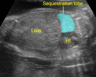
Lung sequestration: Extralobar sequestration: Echogenic lung mass with lobar in shape

Diaphragmatic hernia: Cross-sectional scan: The stomach is herniated into the chest with cardiac displacement

