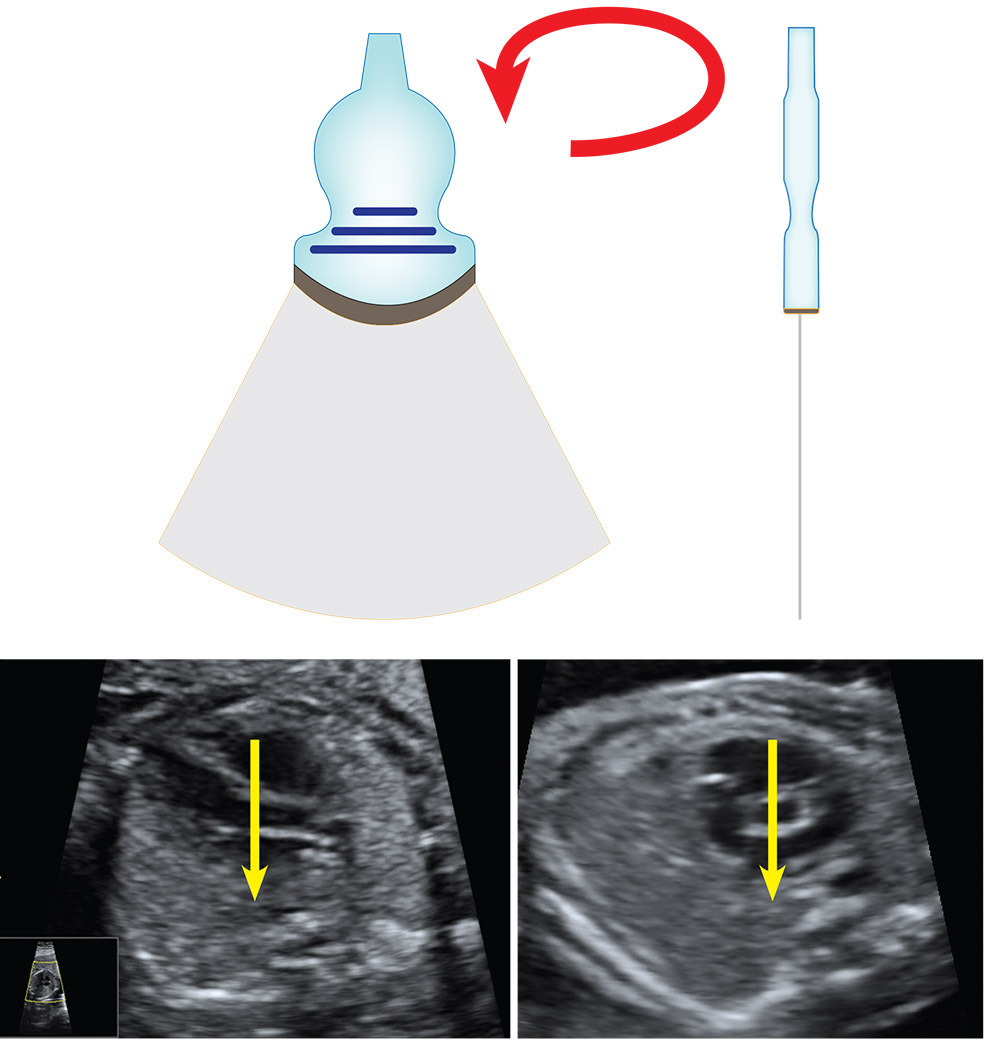
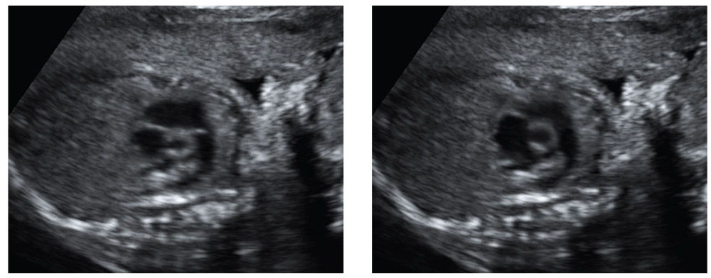
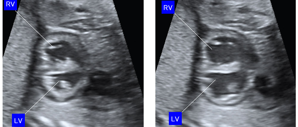
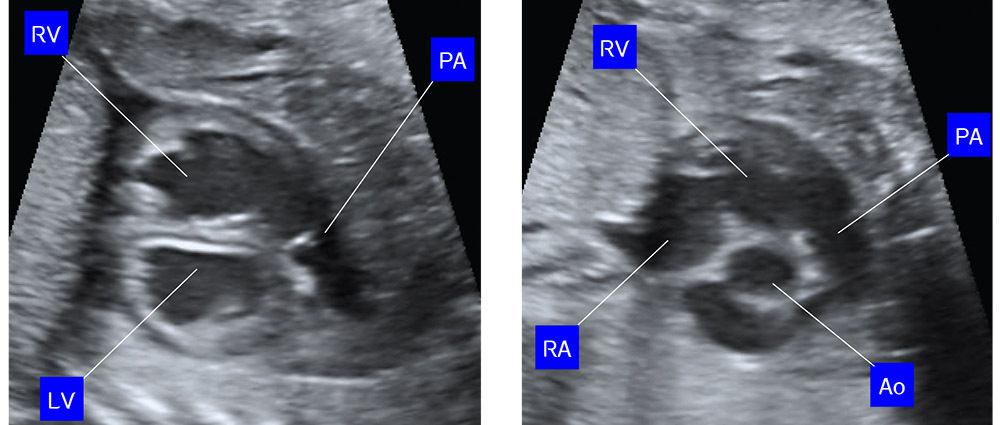
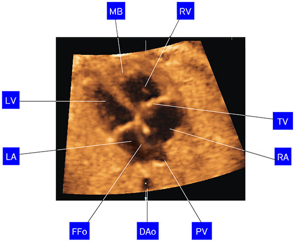
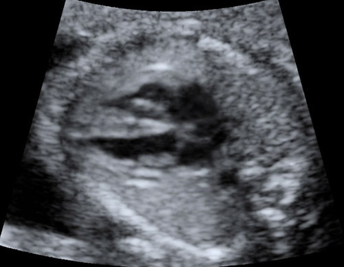
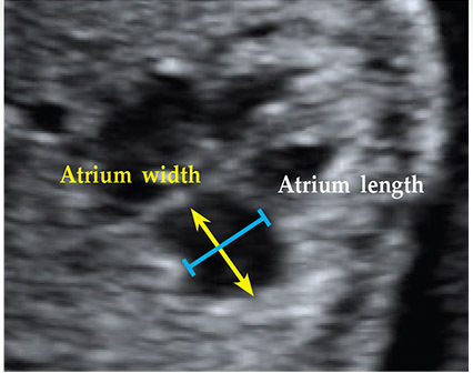
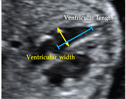
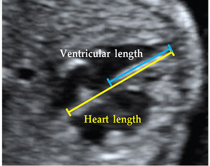
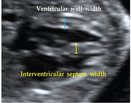
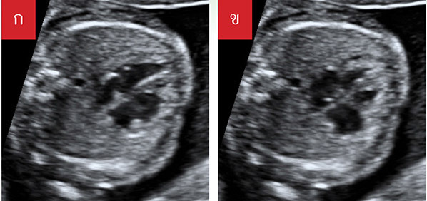
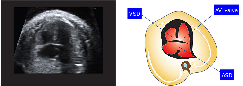
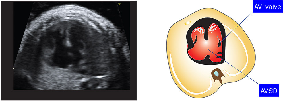
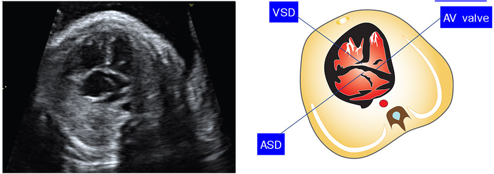
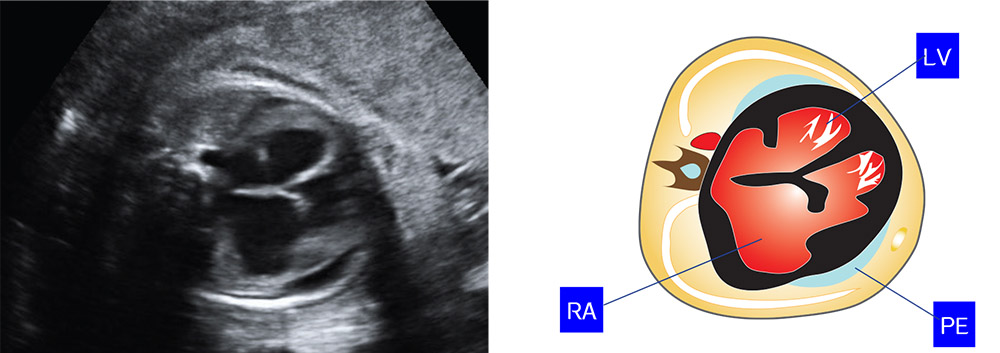
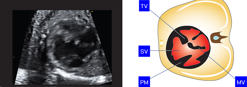
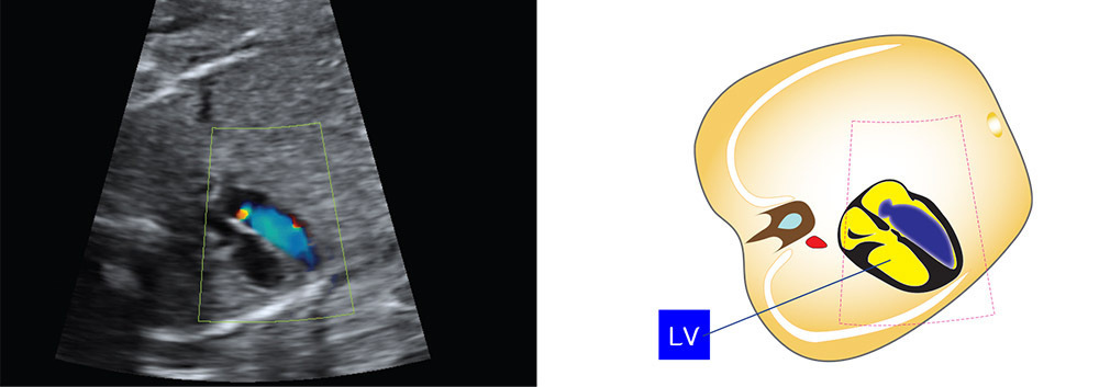
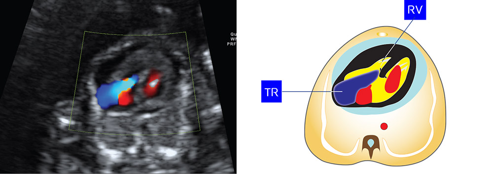
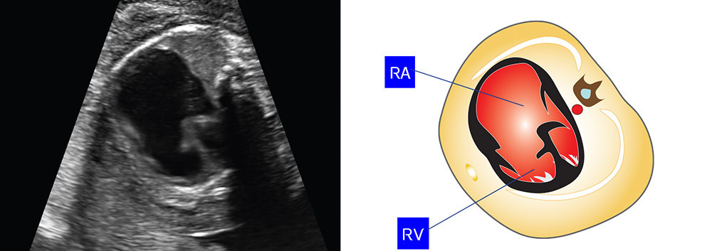
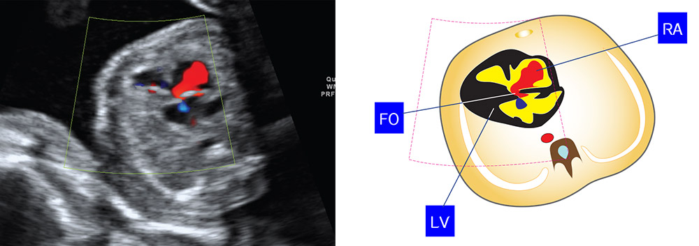
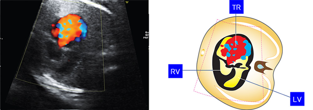
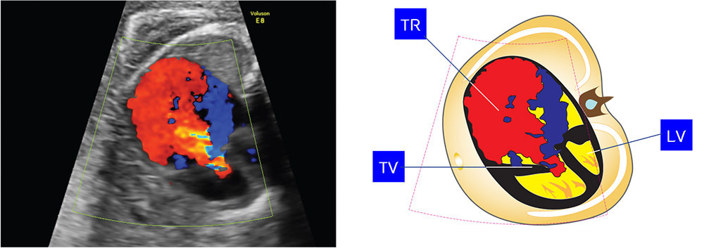
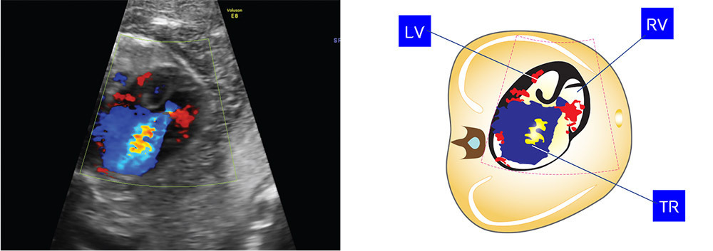
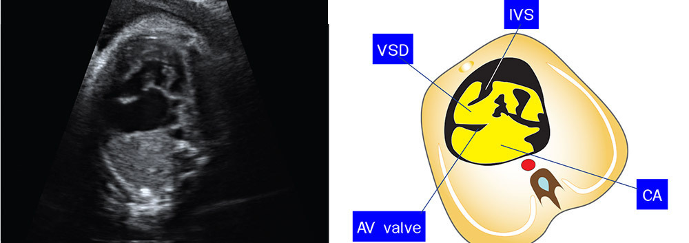
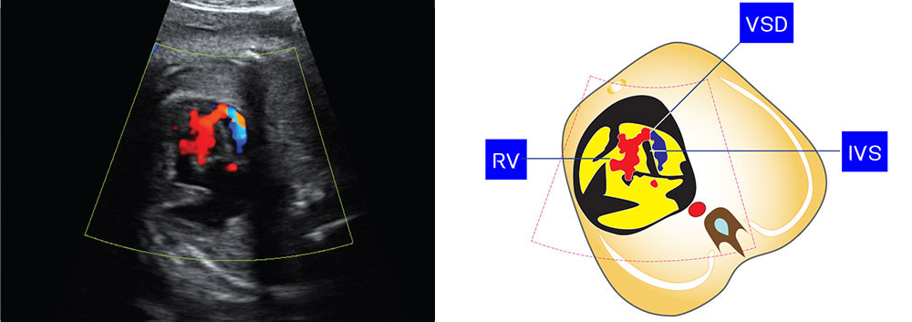
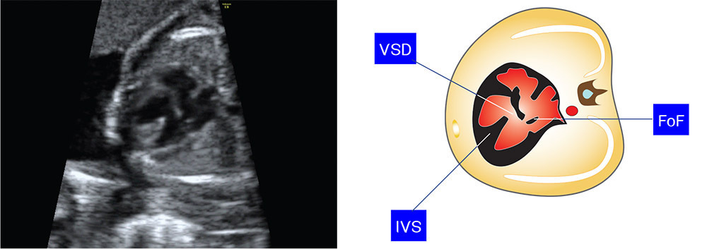
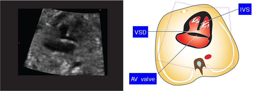
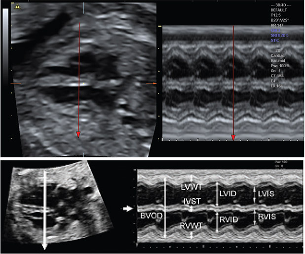
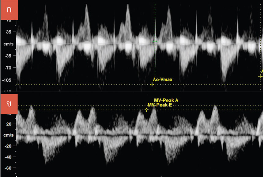
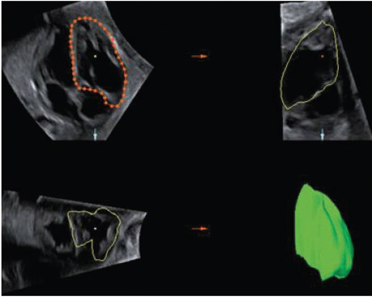
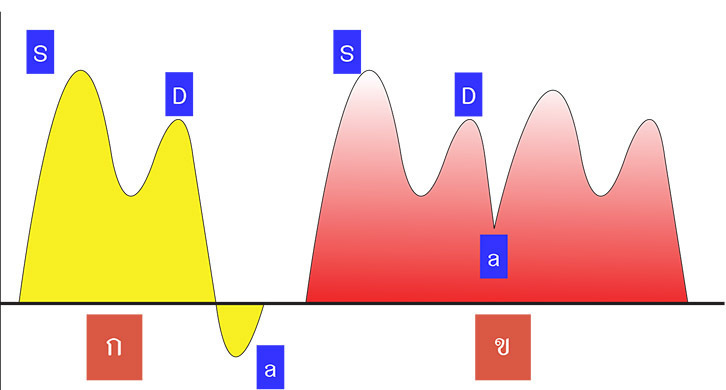
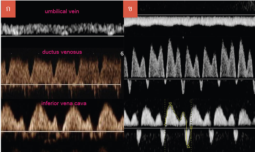
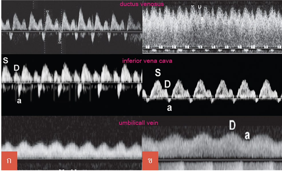
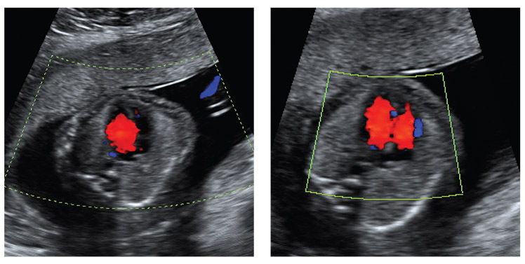
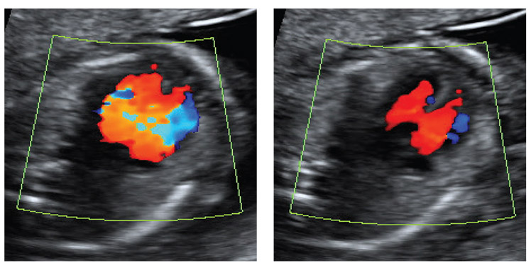
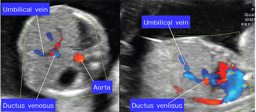
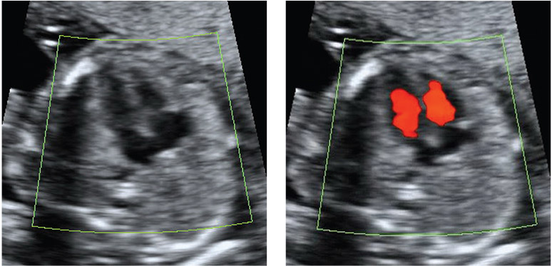
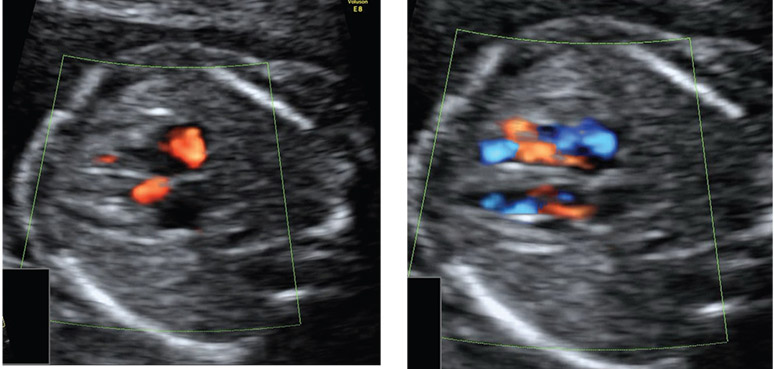
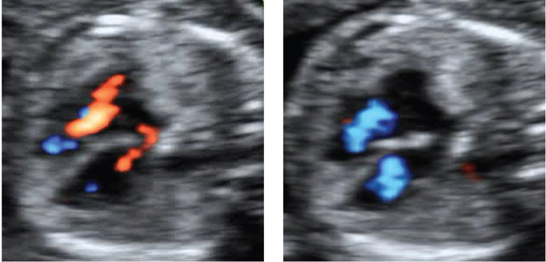
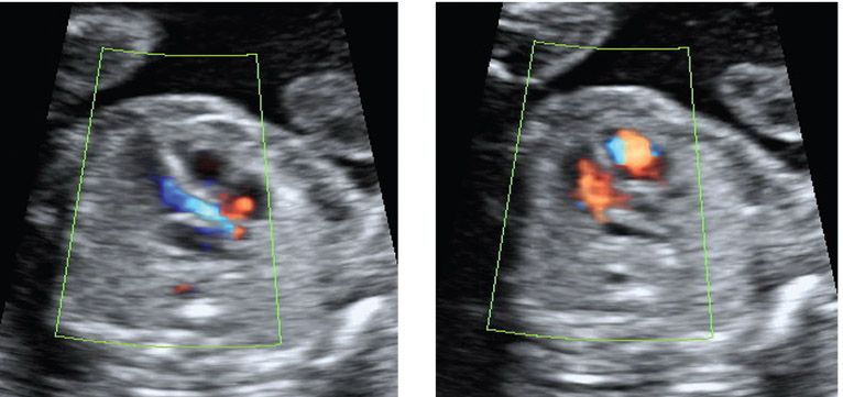
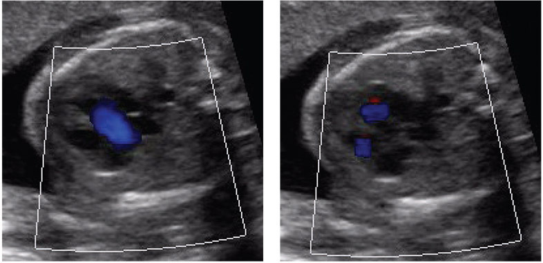
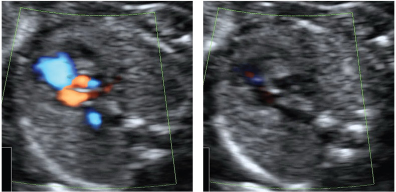
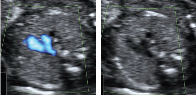
ต่อมาในปี 2011 ได้ศึกษาการทำงานของหัวใจด้วย shortening fraction(SF) จาก STIC-M ใน hydrops ที่เกิดจากทั้งสองสาเหตุ โดยที่กลุ่ม high output พบว่าค่า SF ไม่แตกต่างจากทารกปกติหรืออาจลดลงเล็กน้อย แต่ต่างจากกลุ่มที่เป็น low output ที่ค่า SF จะลดลงมากอย่างมีนัยสำคัญทางสถิติ (p<0.001)(36)
จากหลาย ๆ การศึกษา ต่างให้ข้อมูลในทางเดียวกันว่าการทำงานของหัวใจในทารกบวมน้ำในกลุ่ม high output นั้น แม้ว่ามีลักษณะบวมน้ำปรากฏแล้วก็ตาม แต่การทำงานของหัวใจยังดีอยู่ เนื่องจากเป็นกระบวนการ ตอบสนองเพื่อให้อยู่รอดของทารกในครรภ์ ซึ่งบ่งชี้ว่าการพยากรณ์โรคยังค่อนข้างดี ต่างจากกลุ่ม low output ที่จะพบว่าการทำงานของหัวใจแย่ลงมากแล้ว บอกถึงพยากรณ์โรคที่ไม่ดีของทารก
- มารดาที่เป็นเบาหวาน
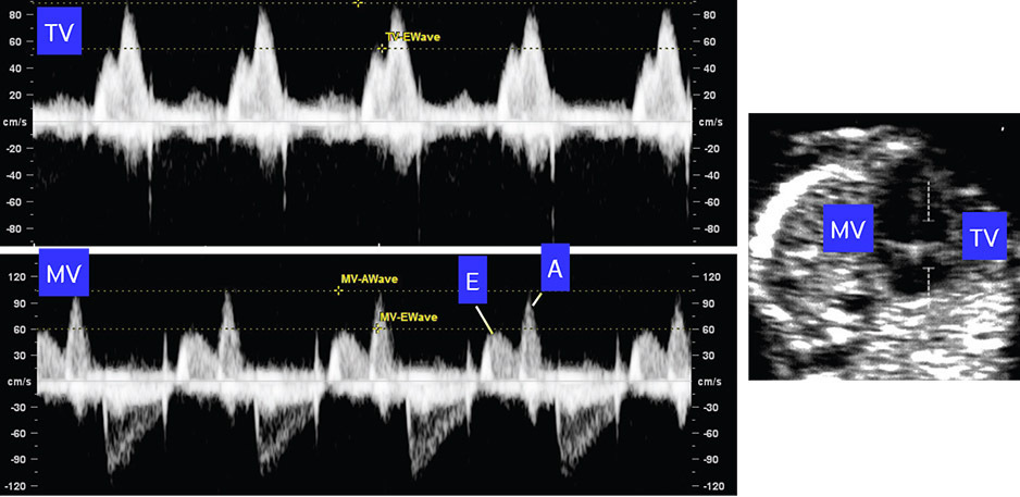
สตรีตั้งครรภ์ที่มีระดับน้ำตาลในร่างกายสูง(Hyperglycemia) จะเพิ่มความเสี่ยงต่อระบบหัวใจและหลอดเลือดของทารกในครรภ์ ทั้งที่ทำให้เกิดหัวใจพิการแต่กำเนิดและกล้ามเนื้อหัวใจโต เบาหวานในสตรีตั้งครรภ์เป็นสาเหตุสำคัญที่ทำให้เกิด fetal hypertrophic cardiomyopathy ซึ่งมีลักษณะสำคัญคือ หัวใจโต ค่า SF เพิ่มขึ้น ความหนาของ interventricular septum เพิ่มขึ้น และมี outflow tract obstruction โดยทารกของสตรีตั้งครรภ์ที่เป็นเบาหวาน มักตรวจพบลักษณะดังนี้ คือ มี diastolic dysfunction จาก ค่า Pulsatility index ของ precordial vein เพิ่มสูงขึ้น ค่า E/A ratio ลดต่ำลง ค่า IRT และ annular peak velocity เพิ่มขึ้น ถ้าตรวจ marker อื่น ๆ เช่น cord blood ดู atrial และ B-type natriuretic peptide (ANP/BNP) และ troponin จะพบว่าค่าสูงขึ้นด้วย ซึ่งmarker เหล่านี้อาจพบได้แม้ว่าจะควบคุมระดับน้ำตาลได้ดี(1)
- ภาวะรกเสื่อม(placental insufficiency)
รกเสื่อมสภาพจะมีผลต่อการเจริญเติบโตของทารกในครรภ์ เกิดภาวะทารกโตช้าในครรภ์(fetal growth restriction,IUGR) ได้ถึงร้อยละ 10 และเป็นสาเหตุสำคัญทีทำให้เกิดทารกตาบปริกำเนิดหรือมีภาวะทุพพลภาพ หัวใจของทารกมีการปรับสภาพต่อผลกระทบจากภาวะรกเสื่อม ซึ่งอาจเห็นได้ตั้งแต่แรกหรือเมื่อมีอาการรุนแรงมากแล้ว
Early-Onset FGR
กรณีที่เกิดรกเสื่อมสภาพเร็วหรือก่อนอายุครรภ์ 34 สัปดาห์ ส่วนใหญ่มักมีภาวะแทรกซ้อนอย่างอื่นร่วมด้วยหรือเกิดในมารดาที่มีโรคทางอายุรกรรม การศึกษาในช่วงแรก ๆ จะตรวจพบแต่ systolic dysfunction ในรายที่รุนแรง ส่วน EF นั้นจะเปลี่ยนแปลงก็ต่อเมื่อเป็นระยะสุดท้าย ซึ่งทำให้วินิจฉัยได้ค่อนข้างช้า ไม่สามารถลดภาวะแทรกซ้อนต่าง ๆ ลงได้ การศึกษาในปัจจุบัน ได้ใช้ค่าอื่น ๆ เข้ามาช่วยในการประเมินการทำงาน เช่น การตรวจ systolic annular peak velocities ซึ่งจะลดลงตั้งแต่ระยะแรก ๆ ของ FGR นานก่อนที่จะมีการเปลี่ยนแปลงของเส้นเลือดอื่น ๆ(37) และยังพบว่าจะมีปัญหาในจังหวะคลายตัว ตั้งแต่เริ่มมี FGR ซึ่งจะตรวจเจอ ค่า PIV ของ precordial vein เริ่มสูงขึ้น E/A ratio และ IRT ก็เพิ่มขึ้นเช่นกัน การเปลี่ยนแปลงที่เกิดขึ้นเป็นกระบวนการปรับตัวต่อภาวะขาดออกซิเจนเรื้อรัง และ ภาวะน้ำเกิน แต่ถ้าความรุนแรงมากขึ้นจนเกิดภาวะ academia ซึ่งสัมพันธ์กับผลลัพธ์การตั้งครรภ์ที่ไม่ดี จะตรวจพบ ค่า MPI สูงขึ้น และ systolic annular peak velocities ลดลงมาก โดยที่ abnormal wave form ของ DV จะเป็นค่าที่ใช้พยากรณ์ความรุนแรงได้ดีที่สุด มีความไวร้อยละ 40-60
Late-Onset FGR
ในรายที่เป็น FGR แต่เกิดหลังอายุครรภ์ 34 สัปดาห์ หลายคนอาจคิดว่าคงไม่น่ารุนแรง แต่ในปัจจุบันมีรายงานที่บ่งชี้ว่า รายที่เป็น late FGR จะมี placental insufficiency ที่รุนแรงไม่มาก ความบกพร่องของพัฒนาการทางสมองและสติปัญญา รวมถึงความเสี่ยงในการเกิดโรคหัวใจและหลอดเลือดในอนาคต การตรวจวินิจฉัยส่วนมาก Doppler ของ umbilical artery จะปกติ แต่สามารถตรวจเจอว่า ค่า MPI จะเริ่มสูงขึ้น ร่วมกับ systolic annular peak velocities ที่ลดลง
- Twin-to-Twin Transfusion Syndrome
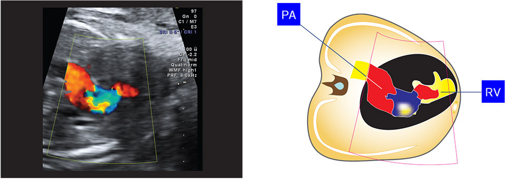
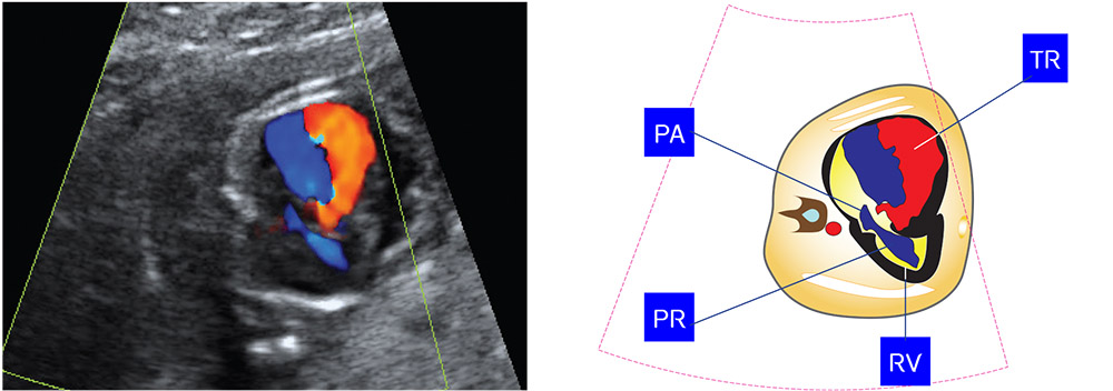
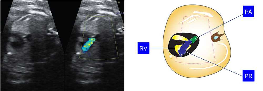
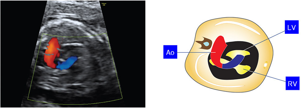
เป็นภาวะแทรกซ้อนที่พบได้บ่อยใน Monozygotic twins เกิดจากการเชื่อมกันของเส้นเลือดบริเวณรก ทำให้มีการถ่ายเทเลือดจากแฝดคนหนึ่งไปยังอีกคนหนึ่ง จะแบ่งเป็นแฝดให้และแฝดรับ โดยที่แฝดให้จะตัวเล็กกว่า มี FGR ในขณะที่แฝดรับจะตัวโตกว่ามีภาวะบวมน้ำ ส่วนใหญ่ ในอดีตมีการแบ่งความรุนแรงของโรคโดยการใช้ Quintero staging ซึ่งมีการประเมินการทำงานของหัวใจโดยในระยะที่ 3 ที่ใช้ doppler waveform ของ Umbilical artery หรือ umbilical vein ในระยะรุนแรง หัวใจของทารกจะเริ่มผิดปกติ โดยจะพบมี หัวใจโต systolic dysfunction right outflow tract obstruction แต่ในระยะเริ่มต้นพบว่าร้อยละ 20-60 อาจมีการทำงานของหัวใจที่ผิดปกติแล้วได้เช่นกัน(38)
แต่ในปัจจุบันมีรายงานการศึกษา โดยกำหนดการให้คะแนนเพื่อใช้ ในการประเมินใหม่ที่เน้นแต่การทำงานของหัวใจด้วย การตรวจคลื่นเสียงหัวใจทารกและ คลื่นเสียงดอพเลอร์ เรียกว่า CHOP (Children’s Hospital of Philadelphia) score(15) เน้นการประเมินที่แฝดรับเป็นหลัก และพบว่าวิธีนี้ผลที่ได้สัมพันธ์กับกับ Quintero stagingโดยมีวิธีการประเมินดังตาราง 5
ตารางที่ 5 แสดงการประเมินTTTS ด้วย CHOP (Children’s Hospital of Philadelphia) score
| Cardiovascular parameter score |
Description |
| Donor
0
1
2 |
Positive
Absent
Negative |
| Recipient
0
1
0
1
2
- Ventricular systolic function
0
1
2
0
1
2
0
1
2
0
1
0
1
0
1
2
0
1
0
1
- Right ventricular outflow tract (RVOT)
0
1
2
3 |
<2SD
>2SD
<1/3
>1/3 และ < 0.5
> 0.5
>30%
<30% and >20%
<20%
- Color flow area of regurgitant jet
Absent
<25% atria
>25% atria
- Color flow area of regurgitant jet
Absent
<25% atria
>25% atria
- Merging of E- and A-waves
Absent
Present
- Merging of E- and A-waves
Absent
Present
Positive
Absent
Negative
No
Yes
No
Yes
PA > Ao
PA = Ao
Ao > PA
RVOT obstruction |
หมายเหตุ EA, E- and A-waves on pulsed Doppler interrogation; MV, mitral valve; TV, tricuspid valve
จาก CHOP score สามารถแบ่งความรุนแรงของ TTTS ตามคะแนนดังนี้ คะแนน 0-5 (cardiovascular grade 1); 6-10 (cardiovascular grade 2); 11-15 (cardiovascular grade 3) และ 16-20 (cardiovascular grade 4)ซึ่งจัดเป็นขั้นที่รุนแรงที่สุด จากการใช้ score นี้สามารถบอกพยากรณ์โรคได้ แต่ในรายที่ใช้ก่อนทำการรักษาด้วย laser coagulation ในสถานที่สามารถทำได้และเป็นการรักษาหลัก การประเมินด้วยวิธีนี้อาจไม่มีประโยชน์ในการทำนายผลการรักษา แต่ช่วยบอกว่าควรได้รับการรักษาอย่างเหมาะสมเมื่อไหร่หรือการพิจารณาให้คลอด แต่ที่มีประโยชน์ คือ การประเมินผลการรักษาหลังทำ laser coagulation ซึ่งพบว่า ค่า PI ของ DV, MPI จะลดลง การมี tricuspid regurgitation และ การรวมกันของ EA wave ในแฝดรับ ส่วนแฝดให้ ปริมาณสารถน้ำในกายจะเพิ่มขึ้น เลือดที่ไหลไปทาง UV เพิ่มขึ้นรวมถึง ค่า DV-PI การมี tricuspid regurgitation ซึ่งหลังการรักษาจะดีขึ้นใน 4 สัปดาห์
- ความผิดปกติอื่น ๆ
สาเหตุอื่น ๆ เช่น ทารกมีความผิดปติของโครโมโซมคู่ที่ 21 ในช่วงไตรมาสแรก จะพบ tricuspid regurgitation, reversed a wave ของ DV, E/A ration ของหัวใจซีกขวาเพิ่มขึ้น รวมถึง ค่า MPI ที่เพิ่มขึ้นตามด้วย และจะตรวจพบได้จนถึงช่วงไตรมาสที่สองและสาม
เอกสารอ้างอิง
(1) Crispi F, Gratacos E. Fetal cardiac function: technical considerations and potential research and clinical applications. Fetal Diagn Ther 2012;32(1-2):47-64.
(2) Mielke G, Benda N. Blood flow velocity waveforms of the fetal pulmonary artery and the ductus arteriosus: reference ranges from 13 weeks to term. Ultrasound Obstet Gynecol 2000 Mar;15(3):213-8.
(3) Pombo JF, Troy BL, Russell RO, Jr. Left ventricular volumes and ejection fraction by echocardiography. Circulation 1971 Apr;43(4):480-90.
(4) Carvalho JS, O’Sullivan C, Shinebourne EA, Henein MY. Right and left ventricular long-axis function in the fetus using angular M-mode. Ultrasound Obstet Gynecol 2001 Dec;18(6):619-22.
(5) Gardiner HM, Pasquini L, Wolfenden J, Barlow A, Li W, Kulinskaya E, et al. Myocardial tissue Doppler and long axis function in the fetal heart. Int J Cardiol 2006 Oct 26;113(1):39-47.
(6) Ho SY, Nihoyannopoulos P. Anatomy, echocardiography, and normal right ventricular dimensions. Heart 2006 Apr;92 Suppl 1:i2-13.
(7) Abuhamad AZ, Chaoui R. A Practical Guide to Fetal Echocardiography: Normal and Abnormal Hearts. Lippincott Williams & Wilkins; 2009.
(8) Fernandez PL, Tamariz-Martel MA, Maitre Azcarate MJ, Lopez ZM, Rico GF, Cazzaniga BM, et al. Contribution of Doppler atrioventricular flow waves to ventricular filling in the human fetus. Pediatr Cardiol 2000 Sep;21(5):422-8.
(9) Reed KL, Sahn DJ, Scagnelli S, Anderson CF, Shenker L. Doppler echocardiographic studies of diastolic function in the human fetal heart: changes during gestation. J Am Coll Cardiol 1986 Aug;8(2):391-5.
(10) Xie GY, Berk MR, Smith MD, Gurley JC, DeMaria AN. Prognostic value of Doppler transmitral flow patterns in patients with congestive heart failure. J Am Coll Cardiol 1994 Jul;24(1):132-9.
(11) Stirnemann JJ, Mougeot M, Proulx F, Nasr B, Essaoui M, Fouron JC, et al. Profiling fetal cardiac function in twin-twin transfusion syndrome. Ultrasound Obstet Gynecol 2010 Jan;35(1):19-27.
(12) Mahle WT, Rychik J, Tian ZY, Cohen MS, Howell LJ, Crombleholme TM, et al. Echocardiographic evaluation of the fetus with congenital cystic adenomatoid malformation. Ultrasound Obstet Gynecol 2000 Dec;16(7):620-4.
(13) Crispi F, Hernandez-Andrade E, Pelsers MM, Plasencia W, Benavides-Serralde JA, Eixarch E, et al. Cardiac dysfunction and cell damage across clinical stages of severity in growth-restricted fetuses. Am J Obstet Gynecol 2008 Sep;199(3):254-8.
(14) Makikallio K, McElhinney DB, Levine JC, Marx GR, Colan SD, Marshall AC, et al. Fetal aortic valve stenosis and the evolution of hypoplastic left heart syndrome: patient selection for fetal intervention. Circulation 2006 Mar 21;113(11):1401-5.
(15) Rychik J, Tian Z, Bebbington M, Xu F, McCann M, Mann S, et al. The twin-twin transfusion syndrome: spectrum of cardiovascular abnormality and development of a cardiovascular score to assess severity of disease. Am J Obstet Gynecol 2007 Oct;197(4):392-8.
(16) Makikallio K, Rasanen J, Makikallio T, Vuolteenaho O, Huhta JC. Human fetal cardiovascular profile score and neonatal outcome in intrauterine growth restriction. Ultrasound Obstet Gynecol 2008 Jan;31(1):48-54.
(17) Mori Y, Rice MJ, McDonald RW, Reller MD, Wanitkun S, Harada K, et al. Evaluation of systolic and diastolic ventricular performance of the right ventricle in fetuses with ductal constriction using the Doppler Tei index. Am J Cardiol 2001 Nov 15;88(10):1173-8.
(18) Tei C, Ling LH, Hodge DO, Bailey KR, Oh JK, Rodeheffer RJ, et al. New index of combined systolic and diastolic myocardial performance: a simple and reproducible measure of cardiac function–a study in normals and dilated cardiomyopathy. J Cardiol 1995 Dec;26(6):357-66.
(19) Tsutsumi T, Ishii M, Eto G, Hota M, Kato H. Serial evaluation for myocardial performance in fetuses and neonates using a new Doppler index. Pediatr Int 1999 Dec;41(6):722-7.
(20) Hernandez-Andrade E, Lopez-Tenorio J, Figueroa-Diesel H, Sanin-Blair J, Carreras E, Cabero L, et al. A modified myocardial performance (Tei) index based on the use of valve clicks improves reproducibility of fetal left cardiac function assessment. Ultrasound Obstet Gynecol 2005 Sep;26(3):227-32.
(21) Hernandez-Andrade E, Figueroa-Diesel H, Kottman C, Illanes S, Arraztoa J, Acosta-Rojas R, et al. Gestational-age-adjusted reference values for the modified myocardial performance index for evaluation of fetal left cardiac function. Ultrasound Obstet Gynecol 2007 Mar;29(3):321-5.
(22) van Splunder IP, Wladimiroff JW. Cardiac functional changes in the human fetus in the late first and early second trimesters. Ultrasound Obstet Gynecol 1996 Jun;7(6):411-5.
(23) Van MT, Gucciardo L, Lewi P, Lewi L, Van SD, Devlieger R, et al. Validation of the fetal myocardial performance index in the second and third trimesters of gestation. Ultrasound Obstet Gynecol 2009 Jan;33(1):58-63.
(24) Raboisson MJ, Fouron JC, Lamoureux J, Leduc L, Grignon A, Proulx F, et al. Early intertwin differences in myocardial performance during the twin-to-twin transfusion syndrome. Circulation 2004 Nov 9;110(19):3043-8.
(25) Letti Muller AL, Barrios PM, Kliemann LM, Valerio EG, Gasnier R, Magalhaes JA. Tei index to assess fetal cardiac performance in fetuses at risk for fetal inflammatory response syndrome. Ultrasound Obstet Gynecol 2010 Jul;36(1):26-31.
(26) Ichizuka K, Matsuoka R, Hasegawa J, Shirato N, Jimbo M, Otsuki K, et al. The Tei index for evaluation of fetal myocardial performance in sick fetuses. Early Hum Dev 2005 Mar;81(3):273-9.
(27) Duan Y, Harada K, Wu W, Ishii H, Takada G. Correlation between right ventricular Tei index by tissue Doppler imaging and pulsed Doppler imaging in fetuses. Pediatr Cardiol 2008 Jul;29(4):739-43.
(28) Yagel S, Cohen SM, Shapiro I, Valsky DV. 3D and 4D ultrasound in fetal cardiac scanning: a new look at the fetal heart. Ultrasound Obstet Gynecol 2007 Jan;29(1):81-95.
(29) Quintero RA, Morales WJ, Allen MH, Bornick PW, Johnson PK, Kruger M. Staging of twin-twin transfusion syndrome. J Perinatol 1999 Dec;19(8 Pt 1):550-5.
(30) Turan OM, Turan S, Gungor S, Berg C, Moyano D, Gembruch U, et al. Progression of Doppler abnormalities in intrauterine growth restriction. Ultrasound Obstet Gynecol 2008 Aug;32(2):160-7.
(31) Wieczorek A, Hernandez-Robles J, Ewing L, Leshko J, Luther S, Huhta J. Prediction of outcome of fetal congenital heart disease using a cardiovascular profile score. Ultrasound Obstet Gynecol 2008 Mar;31(3):284-8.
(32) Pereira S, Ganapathy R, Syngelaki A, Maiz N, Nicolaides KH. Contribution of fetal tricuspid regurgitation in first-trimester screening for major cardiac defects. Obstet Gynecol 2011 Jun;117(6):1384-91.
(33) Pedra SR, Smallhorn JF, Ryan G, Chitayat D, Taylor GP, Khan R, et al. Fetal cardiomyopathies: pathogenic mechanisms, hemodynamic findings, and clinical outcome. Circulation 2002 Jul 30;106(5):585-91.
(34) Pedra SR, Hornberger LK, Leal SM, Taylor GP, Smallhorn JF. Cardiac function assessment in patients with family history of nonhypertrophic cardiomyopathy: a prenatal and postnatal study. Pediatr Cardiol 2005 Sep;26(5):543-52.
(35) Tongsong T, Tongprasert F, Srisupundit K, Luewan S. Venous Doppler studies in low-output and high-output hydrops fetalis. Am J Obstet Gynecol 2010 Nov;203(5):488-6.
(36) Tongsong T, Wanapirak C, Piyamongkol W, Sirichotiyakul S, Tongprasert F, Srisupundit K, et al. Fetal ventricular shortening fraction in hydrops fetalis. Obstet Gynecol 2011 Jan;117(1):84-91.
(37) Comas M, Crispi F, Cruz-Martinez R, Martinez JM, Figueras F, Gratacos E. Usefulness of myocardial tissue Doppler vs conventional echocardiography in the evaluation of cardiac dysfunction in early-onset intrauterine growth restriction. Am J Obstet Gynecol 2010 Jul;203(1):45-7.
(38) Michelfelder E, Gottliebson W, Border W, Kinsel M, Polzin W, Livingston J, et al. Early manifestations and spectrum of recipient twin cardiomyopathy in twin-twin transfusion syndrome: relation to Quintero stage. Ultrasound Obstet Gynecol 2007 Dec;30(7):965-71.