Hydronephrosis

Hydronephrosis

Hydronephrosis is the dilatation of the fetal renal collecting system. It is the most common fetal abnormality detected by antenatal ultrasound. It can result from ureteropelvic junction (UPJ, 41%), ureterovesical junction (UVJ, 23%), duplication of the collecting system (13%), and bladder outlet (10%). Renal dysplasia can occur in some fetuses with severe or early-onset obstruction.

UPJ obstruction is a functional obstruction and is bilateral in 30% of cases. It affects males more than twice as often as females. Most cases do not become dysplastic.

Vesicoureteral reflux is much more common among males and is often bilateral. It often resolves within the first years of life. Reflux that does not resolve spontaneously or is very severe at birth can be corrected surgically.

Incidence: 1-5 per 1000 births (ureteropelvic junction obstruction: male/female, 4:1, ureterovesicular junction: male>female).

Sonographic findings:

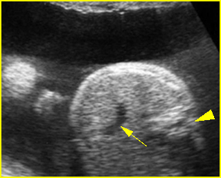
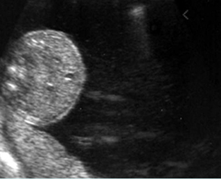
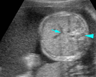
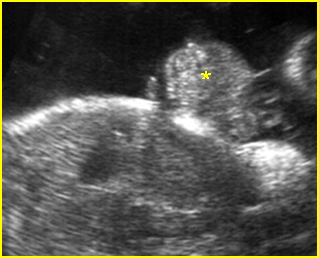
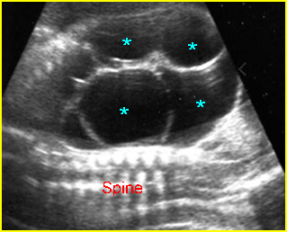
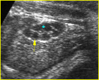
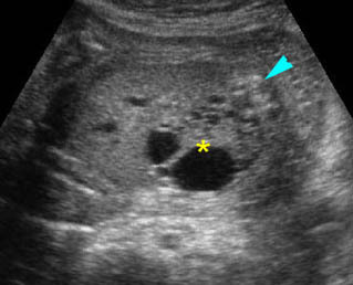
  • A diagnosis of hydronephrosis should be made when there is calicectasis or the antero-posterior diameter of the renal pelvis, measured on a transverse view through the kidney, is at least 8 mm at 16-20 weeks or at least 10 mm after 20 weeks of gestation. Fig 1, Fig 2, Fig 3, Fig 4, Fig 5 Fig 6, Fig 7
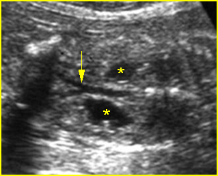
Fig 1:  Hydronephrosis  Coronal scan of the posterior abdomen: mild dilated renal pelvis (*) (arrow = bifurcation of aorta)
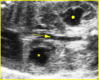
Fig 2:  Hydronephrosis  Coronal scan of the abdomen: bilateral markedly dilated renal pelvis (*) (arrow = aorta)
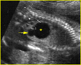
Fig 3:  Hydronephrosis  Sagittal scan of the abdomen: markedly dilated renal pelvis (*) and calyx (arrow)
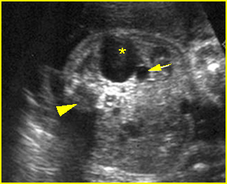
Fig 4:  Hydronephrosis   Cross-sectional scan of the abdomen: markedly dilated renal pelvis (*) and calyx (arrow) (arrowhead = spine)
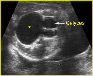
Fig 5:  Hydronephrosis   Cross-sectional scan of the abdomen: markedly dilated renal pelvis (*) with calyces (arrow)
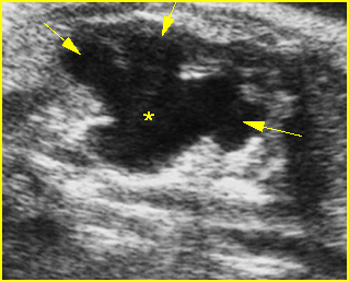
Fig 6:  Hydronephrosis  Parasagittal scan of the abdomen: markedly enlarged renal pelvis (*) and calyces (arrow)
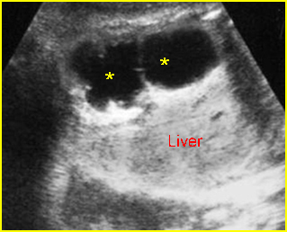
Fig 7:  Severe hydronephrosis  Sagittal scan of the abdomen: markedly enlarged renal pelvis (*) with echogenic thin parenchyma

Video clips of large hydronephrosis
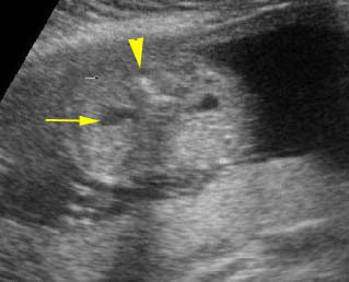
Mild pyelectasis:   Cross-sectional scan of the abdomen: bilateral mild dilatation of renal pelvis (arrow) (arrowhead = spine)
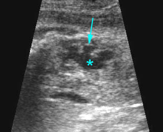
Hydronephrosis:  Sagittal scan of the fetal kidney: dilatated renal pelvis (*), and calyces (arrow)

  • The Society for Fetal Urology recommends a scoring system to grade dilation of the upper urinary tract as follows:
    • Grade 0: no dilation Fig 8
    • Grade I: renal pelvic dilation with or without infundibula visible Fig 9
    • Grade II: renal pelvic dilation with calices visible Fig 10, Fig 11
    • Grade III: renal pelvis and calyces dilated Fig 12
    • Grade IV: features of grade III with parenchymal thinning Fig 13
    • Ultrasound grading of hydronephrosis correlates with the severity of cortical damage or the decrease in renal function on postnatal renal scan.
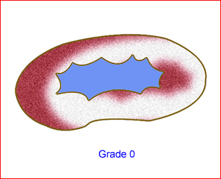
Fig 8:  Schematic drawing of grade 0: no dilation
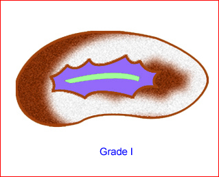
Fig 9: Schematic drawing of grade I: renal pelvic dilation without calices visible
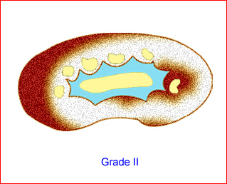
Fig 10:  Schematic drawing of grade II: renal pelvic dilation with calices visible
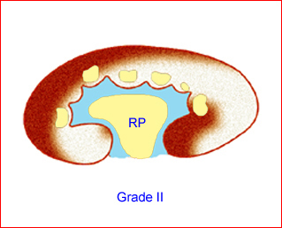
Fig 11: Schematic drawing of grade II: renal pelvic dilation with calices visible
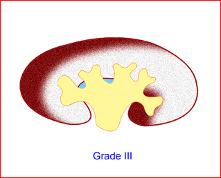
Fig 12:  Schematic drawing of grade III: renal pelvis and calices dilated
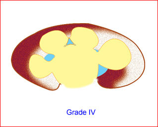
Fig 13: Schematic drawing of grade IV: renal pelvis and calices dilated as well as parenchymal thining

  • Proposed criteria for the diagnosis of fetal hydronephrosis in different studies:
    • Arger (1985): AP diameter >10 mm or ratio of AP diameter of pelvis/kidney >0.5
    • Corteville (1991: AP diameter >4 mm at <33 weeks or >7 mm at >33 weeks, or ratio of AP diameter of pelvis/kidney >0.28
    • Mandell (1991): AP diameter >5 mm at 20 weeks, or >8 mm at 20-30 weeks, or >10 mm at >30 weeks
    • Adra (1995): AP diameter >8 mm from 28 weeks to term
    • Ouzounian (1996): AP diameter >5 mm from 16 weeks to term
    • Dudley (1997): AP diameter >5 mm from 16 weeks to term.
  • UPJ obstruction: The ureters are not dilated and the amniotic fluid volume is usually normal.
  • Vesicoureteral reflux: the hydroureter will be visualized. In severe cases, the ureter may be markedly dilated and tortuous. Care must be taken not to mistake such a ureter for a dilated bowel loop. This error can be avoided by following the dilated ureter proximally to the renal pelvis and distally to the bladder.
  • Dysplastic kidney, which possibly occurs with UPJ obstruction, can be diagnosed when the parenchyma becomes abnormally echogenic or cystic.
  • The contralateral kidney and related obstructions, such as ureteral dilatation, bladder distension, urethral dilatation and amniotic fluid, must be carefully evaluated.

Associations: A renal pelvis dilated to >4 mm increases the risk of trisomy 21, 18, 13.

Management: Serial sonographic monitoring is necessary to assess the degree of severity. In cases of severe obstruction, early delivery may be beneficial and the delivery should occur in a tertiary center since neonatal evaluation and treatment are necessary.

Prognosis: Good for mild forms of isolated cases, but poor in cases of dysplastic kidney especially secondary to prolonged bladder outlet obstruction. Mild fetal hydronephrosis (>4 mm) at mid-trimester appears to be associated with an excellent prognosis, probably representing physiological renal pelvic dilatation. Most cases resolve before delivery. Moderate/severe (>7 mm) fetal hydronephrosis is associated with a poorer outcome.

Recurrence risk: Sporadic in the absence of a recognizable syndrome.

Read More

Bilateral Renal Agenesis

Bilateral Renal Agenesis

Renal agenesis is the absence of one or both kidneys, resulting from unilateral or bilateral failure of development of the ureteric bud secondary to multicystic dysplastic kidney. Bilateral renal agenesis combined with fetal deformities secondary to oligohydramnios (pulmonary hypoplasia, abnormal facies, and abnormal limb postures) is called Potter’s syndrome.
Incidence: Unilateral renal agenesis occurs in 1 per 1000 births (male/female; 1:1) whereas bilateral agenesis is a lethal anomaly, sporadically occurring in 1-4 per 10,000 births with a male to female ratio of 2-3:1.

Sonographic findings:

  • Non-visualization of kidneys and bladder.
  • Oligohydramnios in the second trimester, however, amniotic fluid can be relatively normal at up to 16 weeks of gestation.
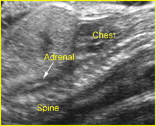
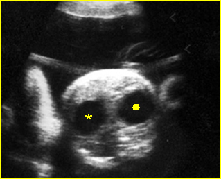
  • Adrenal glands filling the renal fossae, producing the lying down adrenal sign. Fig 1, Fig 2
  • Absence of both renal arteries using color-flow Doppler imaging.
  • Compression effects such as dolichocephaly and small thorax. Fig 2
  • Careful scanning is required before making the diagnosis because the absence of amniotic fluid degrades the sonographic image.
  • Potential pitfalls in the diagnosis include
    • There is an inability to image the kidneys and bladder due to the poor image quality secondary to severe oligohydramnios; amnioinfusion may be helpful in this case. Color flow is also helpful in demonstrating renal vasculature to confirm the presence of kidney.
    • In the absence of kidney, the ipsilateral adrenal is often flattened or lying down and is mistaken for a kidney. Note that the adrenal gland has no central sinus echoes and no reniform shape in the longitudinal view.
    • Pelvic kidney: It is important to scan the fetal pelvis to be sure that the kidney is truly absent rather than in an ectopic location.
    • Crossed renal ectopia may be mistaken for unilateral renal agenesis. In this case, the united kidney is bilobed, enlarged and often with findings of obstruction.
  • Usually first diagnosable in the early second trimester, at 15-18 weeks.
  • In case of inconclusive findings, MRI is usually helpful.

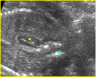
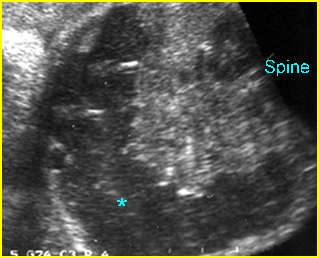
Fig 1:  Bilateral renal agenesis  Coronal scan of the abdomen: absent kidneys with oligohydramnios and adrenal gland replacement (*) (solid circle = spine)

Fig 2:  Bilateral renal agenesis   Sagittal scan of the abdomen: absent kidneys with oligohydramnios and adrenal gland replacement (arrow)

Video clips of large bilateral renal agenesis

Bilateral renal agenesis:  Coronal scan of the abdomen, no visible kidney, oligohydramnios, elongated adrenal glands (arrow) seen in the renal fossa instead

Associations: About half of the cases may have anomalies of other organ systems; all systems can be involved.

Management: Termination of pregnancy can be offered. Vaginal delivery without electronic fetal monitoring in labor is appropriate.

Prognosis: Fatal for bilateral agenesis but excellent for unilateral agenesis.

Recurrence risk: Sporadic with rare autosomal dominant inheritance.

Read More

Large Bowel Obstruction

Large Bowel Obstruction

Large bowel obstruction is generally more difficult to diagnose than small bowel obstruction because there is considerable variability in the diameter of the normal fetal colon. The colon diameters of normal fetuses can overlap with those of fetuses with abnormally distended large bowel. During the latter part of the third trimester, the normal fetal colon can be particularly prominent and striking and could be mistaken as abnormal.

Incidence: About 1 in 2000 births.

Sonographic findings:

  • Dilated colon with or without dilated small bowel. Fig1
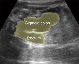
  • Dilated V- or U-shaped segment of bowel in the fetal pelvis suggestive of anorectal atresia. Fig2
  • Calcified intraluminal meconium. Fig3
  • The sensitivity of ultrasound for the diagnosis is rather low.
  • Congenital duplication of the cecum or colon shows peristaltic cyst with haustra at the lower abdomen, which may mimic colonic obstruction.

Fig 1:  Meconium ileus  Cross-sectional scan of the abdomen: Markedly dilated transverse colon (*) with meconium contents

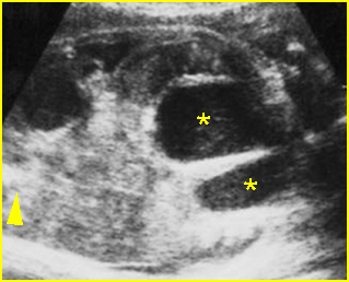
Fig 2:  Anorectal atresia   Oblique cross-sectional scan of the abdomen: U-shaped cystic mass located at the lower abdomen (*), separated from the bladder (arrowhead=spine)

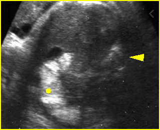
Fig 3:  Hyperechoic colon  Cross-sectional scan of the abdomen: hyperechoic colon (solid circle) (arrowhead = spine)

Video clips of large bowel obstruction

Distal bowel obstruction:  Cross-sectional scan of the lower abdomen: dilated rectum (*), the lesions became proven to be Hirschprung disease in neonatal life (this may be temporary normal variant) (solid circle = bladder)

Anorectal obstruction:  Dilated rectum and sigmoid colon resulting in V-shape appearance secondary to anorectal atresia

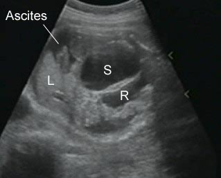
Anorectal atresia:  Anorectal atresia with ruptured large bowel: Oblique cross-sectional scan of the abdomen shows ascites with dilated rectum (R) and sigmoid colon

Associations: Related to other malformations in most cases including VACTERL syndrome, multiple congenital anomalies, caudal regression syndrome, and abnormal chromosome (15%).

Management: A careful search for associated anomalies is indicated. In general the disorder does not alter the standard obstetric care, however, the pregnancy should be managed in a tertiary center.

Prognosis: Poor overall (due to associated anomalies and prematurity), but good in isolated cases with surgical correction.

Recurrence risk: Sporadic with rare familial cases.

Read More

Meconium Peritonitis

Meconium Peritonitis

Meconium peritonitis is a condition resulting from a small bowel perforation, leading to an inflammatory reaction of the peritoneum and intra-abdominal calcifications.

Incidence: About 1 in 2000 births.

 Sonographic findings:

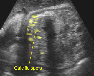
  • Calcifications (bright echogenic area) in the abdomen, often with ascites.
  • The calcifications can occur anywhere within the peritoneal cavity but must be distinguished from intraluminal or intrahepatic calcifications. Fig1, Fig2
  • Typically the calcifications are linear in nature, sometimes rimming a hypoechoic mass or pseudocyst. Fig3
  • Pseudocyst, a hypoechoic mass representing extraluminal meconium, resulting from a contained bowel perforation.
  • Usually associated with ascites and polyhydramnios.
  • Typically diagnosed in the third trimester.
  • Persistent ascites, pseudocyst or dilated bowel loop are most sensitive for predicting postnatal surgery.

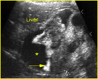
Fig 1:  Meconium peritonitis  Coronal scan of the abdomen: ascites (*) and calcification (arrow) outlining the visceral structures

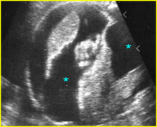
Fig 2:  Meconium peritonitis  Cross-sectional scan of the abdomen: ascites (*) and calcification outlining the visceral structures

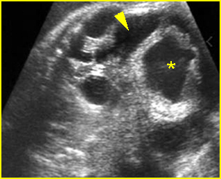
Fig 3:  Meconium peritonitis  Oblique coronal scan of the abdomen: dilated large bowel loops (*) with thickened wall and ascites (arrowhead)

Video clips of meconium peritonitis

Intraperitoneal calcifications:  Intraperitoneal calcifications; postnatally proven to be due to meconium peritonitis

Anorectal atresia:  Anorectal atresia with ruptured large bowel: Oblique cross-sectional scan of the abdomen shows ascites with dilated rectum (R) and sigmoid colon

Associations: Cystic fibrosis is found in 15% of cases with meconium peritonitis in neonatal series but is rarely found in prenatal series. Some cases are related to pavovirus B 19.

Management: Sonographic monitoring may be beneficial in assessing fluid volume, fetal growth and the degree of the severity. Early delivery may be appropriate if there is marked bowel dilatation. The pregnancy should be managed in a tertiary center.

Prognosis: Rather good in isolated cases, with most cysts disappearing spontaneously during pregnancy.

Recurrence risk: Rare for isolated cases, but may be as high as 25% when associated with cystic fibrosis.

Read More

Small Bowel Obstruction

Small Bowel Obstruction

Small bowel obstructions are non-duodenal bowel atresias with congenital obliteration of the lumen of segments of the large or small intestine, usually caused by in utero vascular accident. There are several types of atresias, the most common of which results in blind ends of bowel separated from each other by a gap.

Incidence: About 1 in 3000-5000 births.

Sonographic findings:

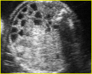
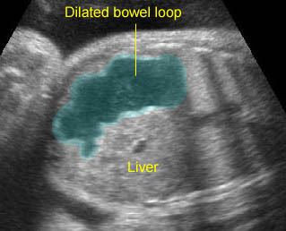
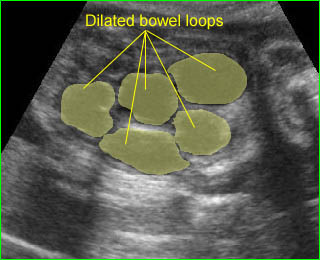
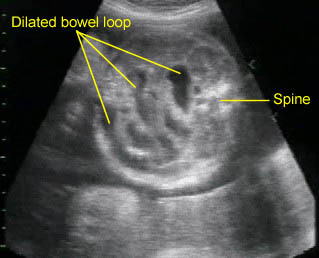
  • Multiple dilated loops of small bowel (more than 7 mm in diameter or 15 mm in length), often with increased peristalsis.

Fig 1, Fig 2, Fig 3, Fig 4, Fig 5, Fig 6, Fig 7, Fig 8

  • Polyhydramnios usually appears in the third trimester and more likely with a proximal atresia.
  • The abdominal circumference may be large.
  • Differentiating small bowel from large bowel obstruction is usually possible by the location of the loops, the absence of haustra in small bowel, and the presence of polyhydramnios. The dilated ureter can easily be differentiated from bowel, as urine is echogenic-free, whereas the bowel contains low-level echoes.
  • It is usually difficult to differentiate jejunal atresia from ileal atresia or volvulus, although the extent of bowel dilatation is often a clue (more loops are present with ileal abnormalities).
  • MRI is helpful in determining the site of obstruction.
  • Pitfalls:
    • Cystic masses, such as bowel duplication, mesenteric cyst or urinary tract cyst including hydronephrosis as well as multicystic kidneys can also be confused with fluid-filled dilated bowel. These anomalies, however, are rarely related to polyhydramnios.
    • Occasionally, small bowel atresia can present as a cyst-like mass.
  • Usually detected after 24 weeks

Fig 1:  Small bowel obstruction   Coronal scan of the abdomen: Multiple dilated loops of small bowel

Fig 2:  Small bowel obstruction   Cross-sectional scan of the abdomen: Multiple loops of small bowel with marked dilatation (arrowhead = spine)

Fig 3:  Small bowel obstruction   Coronal scan of the abdomen: Multiple loop of small bowel loops with marked dilatation (*) (arrow = diaphragm)

Fig 4:  Small bowel obstruction  Coronal scan of the abdomen: Multiple dilated loops of small bowel (*)

Fig 5:  Small bowel obstruction   Coronal scan of the fetal abdomen: markedly dilated loops of the small bowels

Fig 6:  Small bowel obstruction   Cross-sectional scan of the abdomen: multiple dilated small bowel loops (*) (arrow = spine)

Fig 7:  Small bowel obstruction  Cross-sectional scan of the abdomen: Markedly dilated loops of small bowel (solid circle) (arrowhead = spine)

Fig 8:  Small bowel obstruction   Cross-sectional scan of the abdomen: Multiple dilated loops of small bowel (arrowhead = spine)

Video clips of small bowel obstruction

Proximal jejunal atresia:  Dilated bowel loop at the upper abdomen representing the proximal jejunal loop separated from the stomach and large bowel

Small bowel obstruction:  Multiple markedly dilated bowel loops of small bowels

Small bowel obstruction:  Multiple dilated bowel loops of small bowels

Associations: Unlike duodenal atresia, small bowel atresia is associated with a low rate of extragastrointestinal tract abnormalities and chromosomal abnormalities. However, additional GI anomalies, such as volvulus, malrotation, and duplication, are relatively common. Other complication such as intrauterine hemorrhage or bowel perforation may rarely be associated with intestinal atresia. Cystic fibrosis is commonly related to jejunal and ileal atresia.

Management: Delivery should be at term where appropriate personnel are available for surgical correction, however, early delivery might be beneficial in cases with massive bowel dilatation.

Prognosis: Depends on associated anomalies, the health of the remaining bowel and whether perforation or necrosis has occurred. The prognosis is good if it occurs as an isolated abnormality.

Recurrence risk: Sporadic (with rare autosomal recessive inheritance).

Read More

Duodenal Atresia

Duodenal Atresia

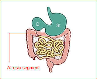
Duodenal atresia is the most common type of congenital small bowel obstruction resulting from failure to recanalize the duodenal lumen during the 11th week of gestation.

     Fig 1

Incidence: 1 in 5000-10,000 births. One-third have Down’s syndrome.
Sonographic findings:

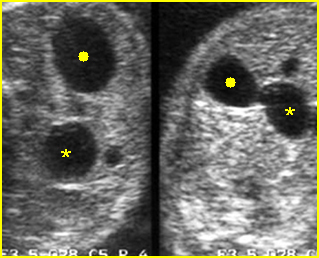
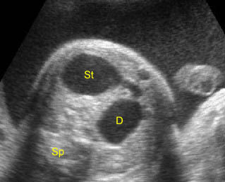
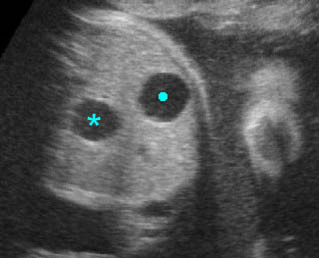
  • Double bubble sign and a narrow channel connecting the stomach and duodenum (the second bubble will be in the location of the duodenal bulb, which is typically in the center of the abdomen, just to the right of the midline). Fig 2, Fig 3, Fig 4
  • The continuity between the two fluid-filled structures must be demonstrated, and if the connection cannot be shown, a number of other possibilities should be considered, such as choledocal cyst (most common) and the less common renal cyst, hepatic cyst, omental cyst, bowel duplication or ovarian cyst.
  • A more prominent appearance with a C loop of dilated bowel may occur when duodenal atresia is associated with esophageal atresia. Fig4, Fig5
  • Pitfalls:
    • A prominent normal stomach with a visible incisura angularis may be confused with a double bubble sign.
    • Bisection of the normal stomach in an oblique scan plane, giving the spurious appearance of a double bubble. This misinterpretation can be corrected by scanning the abdomen in a true transverse plane, so as to demonstrate the typical tapering configuration of the gastric antrum.
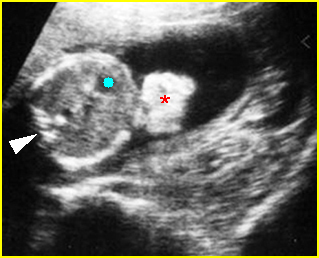
    • Any right upper quadrant mass such as a choledocal cyst, bile-filled gallbladder, or hepatic cyst may be misinterpreted as a distended duodenum.
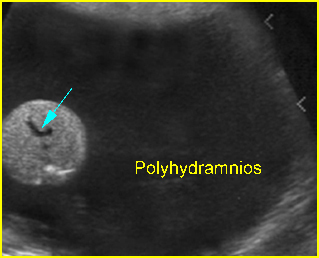
  • Polyhydramnios occurs in nearly all cases diagnosed in the third trimester.
  • Perforation secondary to obstruction can occur, and ascites or meconium peritonitis may be found.
  • Usually first diagnosable in the second trimester, although it can occasionally be diagnosed before 20 weeks or even in the first trimester.

Fig 1:  Schematic drawing of duodenal atresia, The fluid-filled stomach (St) and duodenum (D) cause double bubble sign on ultrasound examination

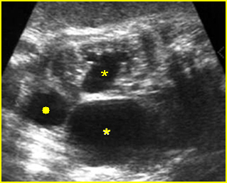
Fig 2:  Duodenal atresia  Cross-sectional scan of the abdomen: Double bubble sign: two cystic masses (* = proximal duodenum, solid circle = stomach) with connection together in the upper abdomen

Fig 3:  Duodenal atresia   Cross-sectional scan of the abdomen: Double bubble sign: two cystic masses (* = stomach, solid circle = proximal duodenum ) in the upper abdomen

Fig 4:  Duodenal atresia   Cross-sectional scan of the abdomen: Double bubble sign: two cystic masses (* = proximal duodenum, solid circle = stomach) in the upper abdomen

Video clips of duodenal atresia

Double bubble sign:  Cross-sectional scan of the abdomen: two connecting cystic masses (*) in the upper abdomen with polyhydramnios

Duodenal atresia:  Double bubble sign with continuation representing stomach (St) and duodenum (D) (Sp = spine)

Duodenal atresia:  C-loop of duodenum and stomach secondary to duodenal and esophageal atresia

Associations: Gastrointestinal, genitourinary and cardiovascular anomalies occur in more than 50% of cases and trisomy 21 is found in 30% of cases (but only 5% of fetuses with trisomy 21 show duodenal atresia). Furthermore, duodenal obstruction also increases the risk of prenatal asphyxia and death, probably caused by bradycardia/asystole following vagal overactivity due to distension of the upper gastrointestinal tract.

Management: A careful search for associated anomalies and karyotyping are indicated. The delivery should occur where appropriate personnel are available for surgical correction.

Prognosis: A high mortality rate of about 20-40% due to associated abnormalities or preterm delivery. Uncomplicated cases have a good prognosis.

Recurrence risk: Sporadic (with rare familial reports).

Read More

Esophageal Atresia

Esophageal Atresia

This sporadic malformation results in an abrupt termination of the esophagus as a blind pouch. In approximately 90% of cases, esophageal atresia is associated with a tracheoesophageal fistula, which most often connects the trachea to the distal esophagus. Additional abnormalities occur in a high percentage of fetuses with esophageal atresia, and these include cardiac, chromosomal, gastrointestinal, genitourinary, and central nervous system lesions.

Incidence: About 1 in 3500 births, and nearly two-thirds are male.

Sonographic findings:

            Fig 1, Fig 2, Fig 3

  • Polyhydramnios: massive polyhydramnios (amniotic fluid index (AFI)>40) is commonly seen in the third trimester.
  • Absent fluid-filled stomach in its normal location in the left upper quadrant.
  • The absent visible stomach can be identified in about 42% of fetuses. Conversely, the combination of polyhydramnios and gastric non-visualization is only seen in less than half of fetuses with a tracheoesophageal fistula.
  • Proximal esophageal pouch (pouch sign): a fluid-filled pouch corresponding to the atretic segment of the esophagus may be seen in the neck or distal esophageal segment.
  • Fetal growth restriction: about 40% of infants with esophageal atresia are associated with growth restriction.
  • Usually first diagnosable in the second trimester, mostly after 24 weeks.
  • Among fetuses with ultrasonographic features suggestive of esophageal atresia, 50% have the disorder confirmed postnatally.
  • In case of suspicion, MRI may be helpful.
  • Pitfalls:
    • Non-visualization of the stomach may be a transient normal finding.
    • Several cases of tracheoesophageal fistula with a connection to the stomach through the lungs have a visible stomach.
    • When combined with duodenal atresia, it may be very difficult to detect due to masking from polyhydramnios and double bubble sign.

Fig 1:  Esophageal atresia  Cross-sectional scan of the abdomen: absent stomach at the level of umbilical vein complex (arrow) with marked polyhydramnios

Fig 2:  Esophageal atresia  Cross-sectional scan of the abdomen: absent stomach at the level of umbilical vein complex (arrow) with polyhydramnios (arrowhead = spine)

Fig 3:  Esophageal atresia   Cross-sectional scan of the abdomen: absent stomach with marked polyhydramnios

Video clips of esophageal atresia

Esophageal atresia:  Cross-sectional scan of the abdomen: persistently absent stomach (arrow = umbilical vein, arrowhead = spine)

Associations: Associated malformations are seen in at least 50% of cases of tracheoesophageal fistula, including gastrointestinal, genitourinary, and cardiovascular anomalies, trisomy 21 and 18.

Management: A careful search for associated anomalies is indicated. The pregnancy with fetal isolated esophageal atresia should be closely followed-up and monitored for preterm delivery. The delivery should occur where appropriate personnel are available for surgical correction.

Prognosis: Poor overall (due to associated anomalies and prematurity), but the prognosis is much improved with isolated esophageal atresia.

Recurrence risk: Sporadic.

Read More

Masses at the Anterior Body Wall

Masses at the Anterior Body Wall

Differential diagnoses of the masses at the anterior body wall include

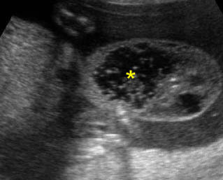
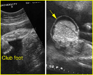
Fig 1, Fig 2, Fig 3, Fig 4, Fig 5, Fig 6

  • Physiologic omphalocele (8-12 weeks); size of <7 mm, no liver content
  • Omphalocele
  • Gastroschisis
  • Ectopia cordis
  • Limb-body wall defect
  • Bladder exstrophy
  • Cloacal exstrophy
  • Stack of umbilical cord (positive Doppler signal or color flow)
  • Cord edema or localized Wharton’s jelly near the umbilicus
  • Cord cysts (omphalomesenteric cyst or allantoic cyst)
  • Placental mass (chorioangioma) close to the anterior wall
  • Artifacts: pseudo-omphalocele secondary to oblique plane

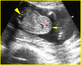
Fig 1:  Omphalocele  Clubfoot and free floating sac containing liver with covering membrane (arrow)

Fig 2:  Gastroschisis  Free floating echogenic bowel (*) in the amniotic fluid (arrowhead = spine, solid circle = intra-abdominal stomach)

Fig 3:  Bladder extrosphy  Sagittal view of the fetal abdomen: complex extra-abdominal mass (*) below the umbilicus, finally proven to be bladder extrosphy

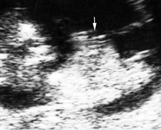
Fig 4:  Physiologic omphalocele  Sagittal scan of the fetus (10 weeks): bowel contents protruding into the proximal umbilical cord (arrow)

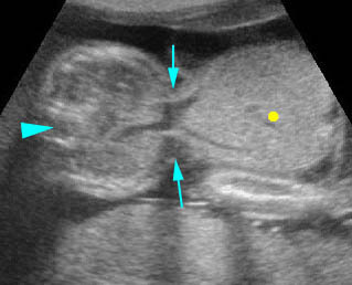
Fig 5:  Pseudo-omphalocele  Oblique cross-sectional scan of the fetus: the liver (*) covered by normal abdominal wall (arrow) protrudes anteriorly, may be mistaken for omphalocele (arrowhead = spine)

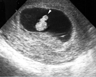
Fig 6:  Physiologic omphalocele  Cross-sectional scan of the embryo (8 weeks): bowel contents protruding into the proximal umbilical cord (arrowhead)

Video clips of masses at the anterior body wall

Omphalocele:  Cross-sectional scan: large omphalocele with liver content (solid circle) (arrow = the defect, arrowhead = spine)

Umbilical cord cyst:  Focus on the proximal cord: cord cyst with coarse particles (*) may be due to liquidfaction of the Wharton’s jelly

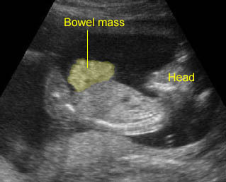
Gastroschisis:   Free floating bowel mass anterior to abdominal wall

True anterior wall defect

  • Omphalocele
  • Gastroschisis
  • Limb-body wall complex
  • Amniotic band syndrome
  • Bladder exstrophy
  • Cloacal exstrophy
  • Pentralogy of Cantrel

Cystic mass close to the umbilicus

  • Allantoic cyst (urachal cyst)
  • Omphalomesenteric cyst
  • Pseudocyst (liquefaction of Wharton’s jelly)
  • Umbilical vein varix
  • Omphalocele

The approach of the anterior wall defects

  •  Relationship of the cord insertion to the defect: various sites of the defect suggest the nature of pathology as follows:
    • above the umbilicus: pentalogy of Cantrell
    • at the umbilicus: omphalocele
    • paraumbilical area: gastroschisis
    • below the umbilicus: bladder/cloacal exstrophy
    • defects throughout the abdomen: limb-body wall complex
    • severe and asymmetric defect: amniotic band syndrome
  • Characteristics of herniated organs: a herniated organ can suggest the nature of pathology as follows:
    • bowel: either a gastroschisis, omphalocele, or LBWC
    • liver only: highly suggestive of omphalocele (but most omphaloceles also include portions of bowel), very unlikely to be gastroschisis
    • bowel only (in omphalocele): more often related to chromosomal abnormalities
    • solid mass at the lower abdomen: bladder or cloacal exstrophy
  • Presence of covering membranes:
    • presence: omphaloceles (be careful, the covering membrane may not always be seen or may be ruptured), limb-body wall complex (not always)
    • absence: gastroschisis
    • the presence of a herniated liver in a ventral wall defect without a covering membrane is more suggestive of a ruptured omphalocele
  • Associated anomalies:
    • multiple anomalies: omphalocele, LBWC
    • extra-, intra-abdominal bowel obstruction: gastroschisis
    • scoliosis: LBWC
    • non-visualization of bladder: bladder or cloacal exstrophy
    • amputation defects: amniotic band syndrome
Read More

Renal Cysts

Renal Cysts

Sonographic differential diagnosis of common renal cystic conditions

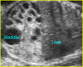
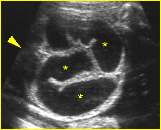
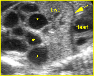
Fig 1, Fig 2, Fig 3, Fig 4

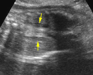
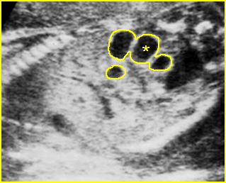
Fig 1:  Hydronephrosis  Cross-sectional scan of the abdomen: markedly dilated renal pelvis (*) with calyces (arrow)

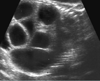
Fig 2:  Multicystic kidney  Sagittal scan of the abdomen: multiple cysts varying in size in the kidney

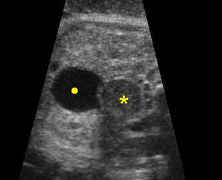
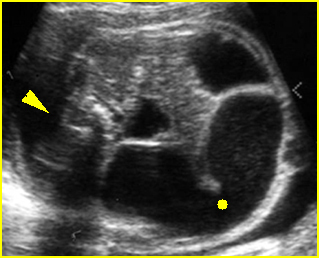
Fig 3:  Hydronephrosis  Coronal scan of the abdomen: bilateral dilated renal pelvis (*) (solid circle = bladder)

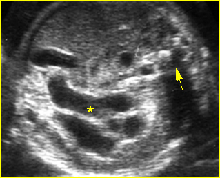
Fig 4:  Normal kidney   The kidneys consisting of multiple normal pyramids (arrow), looking like multicystic kidney (* = renal pelvis)

Video clips of renal cysts

Renal cystic dysplasia:  Cross-sectional scan of the abdomen: muliticystic kidney (*) with oligohydramnios and absent contralateral kidney

Severe hydronephrosis:

  • visible renal parenchyma
  • peripheral oval cysts communicating with each other and the renal pelvis
  • often ureteral dilatation
  • underlying cause in some cases such as bladder outlet obstruction
  • abnormalities in the contralateral kidney (10-40%)

Autosomal recessive polycystic kidneys

  • bilateral enlarged echogenic kidneys with reniform shape
  • oligohydramnios

Multicystic kidney disease

  • multiple round non-communicating cysts of variable size
  • multiple cysts with grape-like appearance in many cases
  • unilateral (77%)
  • abnormalities in the contralateral kidney (39%)

Renal cystic dysplasia

  • echogenic parenchyma and subcapsular or cortical cyst
  • evidence of hydronephrosis in some cases
  • evidence of an underlying cause such as bladder outlet obstruction in some cases
Read More