01 / PREPARATION
Biological sample preparation
Support for the preparation steps needed for electron microscopy, with a workflow selected for the specimen and imaging goal.


Electron microscopy and biological sample preparation support for research. Discuss your sample and research question with our team before choosing a TEM or SEM workflow.

Select an instrument to see how it supports different imaging questions.
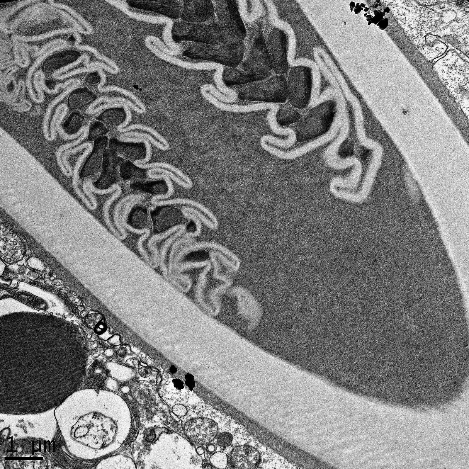
JEM-2200FS supports examination of fine internal structures in appropriately prepared thin biological sections.
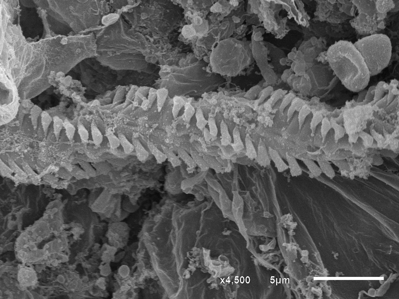
JSM-6610LV supports examination of surface morphology and microstructure in suitably prepared samples.
Representative images supplied by the EM Unit. The original scale bars are retained in each image.
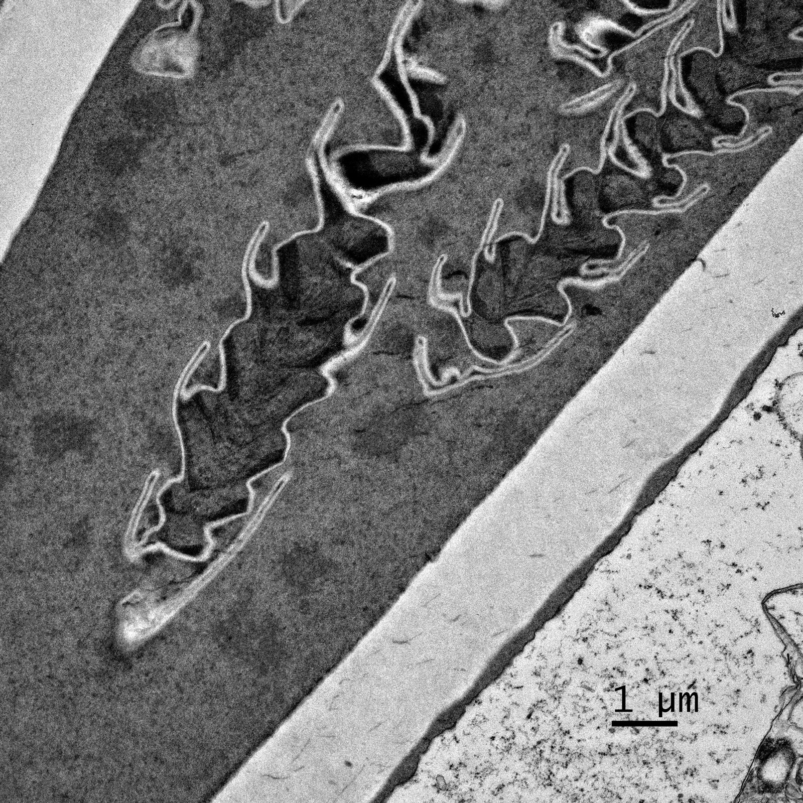
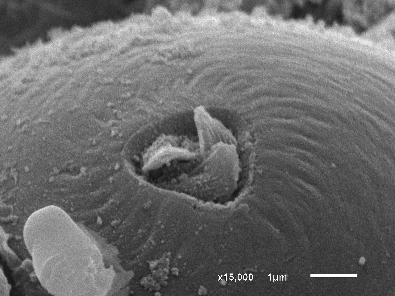
Support for the preparation steps needed for electron microscopy, with a workflow selected for the specimen and imaging goal.
Microscope access and technical assistance to observe internal ultrastructure or surface features.
Discuss specimen collection, preparation and imaging strategy when developing a protocol for biological samples.
Rates in Thai baht (THB), according to Faculty of Medicine announcement ร 11/2569, Appendix 1, page 5. Select a service group below to view all rates.
Swipe horizontally to view all four rate categories.
| Service | Unit | Faculty of Medicine personnel | CMU personnel | Public sector | Private sector |
|---|---|---|---|---|---|
| 10.1 Biological sample preparation — whole process | sample | 1,000 | 1,200 | 2,000 | 2,500 |
| 10.2 Osmium tetroxide treatment only (up to 5 samples) | sample | 500 | 700 | 900 | 1,100 |
| 10.3 Dehydration process | sample | 100 | 200 | 400 | 600 |
| 10.4 Critical point drying | run | 300 | 600 | 800 | 1,000 |
| 10.5 Mounting on stub | stub | 20 | 40 | 80 | 100 |
| 10.6 Gold coating (not more than 6 stubs per run) | run | 200 | 400 | 600 | 800 |
| 10.7 SEM instrument use | hour | 200 | 400 | 800 | 1,200 |
| 10.8 SEM image | image | 20 | 30 | 40 | 50 |
| Service | Unit | Faculty of Medicine personnel | CMU personnel | Public sector | Private sector |
|---|---|---|---|---|---|
| 11.1 Biological sample preparation with OsO₄ | sample | 2,000 | 2,500 | 3,000 | 3,500 |
| 11.2 Biological sample preparation without OsO₄ | sample | 1,000 | 1,500 | 2,000 | 2,500 |
| 11.3 Sectioning (up to 2 grids per block) | sample | 400 | 550 | 750 | 1,000 |
| 11.4 Negative staining — user supplies grid | grid | 150 | 250 | 500 | 750 |
| 11.5 Negative staining — Formvar-carbon grid included | grid | 300 | 500 | 700 | 900 |
| 11.6 TEM instrument use | hour | 800 | 1,200 | 1,500 | 2,500 |
| 11.7 TEM image | image | 20 | 30 | 40 | 50 |
Reference: Faculty of Medicine, Chiang Mai University, announcement ร 11/2569, Appendix 1, page 5. The full official announcement takes precedence.
Select your user category, services and quantities to estimate the cost before submitting your samples.
Choose each required service and enter its quantity. Instrument hours are rounded up; other quantities must be whole numbers.
Begin with a consultation so the preparation plan fits the specimen and the result you need.
Share your sample type and research or imaging objectives.
Agree on a suitable preparation protocol and sample requirements.
Coordinate sample submission and instrument time with the unit.
Complete the agreed imaging work and receive image files.
Discuss your sample and plan your electron microscopy study with our team.