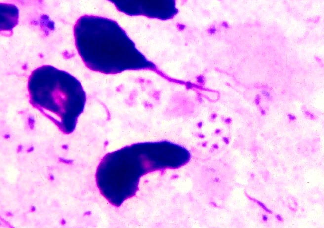
Pneumocystis carinii
- Impression smear, mouse lung, Giemsa-stained
- Cyst appear as “halo”, about 5 micrometer in diameter
- Intracystic bodies are small, 1-2 micrometer, each has reddish nucleus and blue cytoplasm
- Trophozoites can be seen as minute bodies distributed all over the smear
- Recently classified as a fungus
Facebook Comments Box
Contact