หน่วยจอประสาทตาและน้ำวุ้นตา (Retina and Vitreous Unit)
หน่วยจอประสาทตาและน้ำวุ้นตา (Retina and Vitreous Unit) ให้บริการตรวจและรักษาเกี่ยวกับโรคของจอประสาทตาและน้ำวุ้นตา อาทิเช่น เบาหวานขึ้นตา (Diabetic retinopathy), จอตาเสื่อมจากอายุ (Age-related macular degeneration), โรคหลอดเลือดโป่งพองในชั้นคอรอยด์ (Polypoidal choroidal vasculopathy), เส้นเลือดดำอุดตัน (Retinal vein occlusion), โรคเส้นเลือดแดงจอตาอุดตัน (Retinal artery occlusion), โรคจอรับภาพผิดปกติในผู้ป่วยสายตาสั้น (Myopic maculopathy), จอตาหลุดลอก (Retinal detachment), จุดรับภาพเป็นรู (Macular hole), พังผืดที่จุดรับภาพ (Epimacular membrane), โรคการอักเสบของจอตาจากการติดเชื้อ (Infectious retinitis) หรือโรคจอตาติดเชื้อไวรัสในผู้ป่วยเอดส์ (Cytomegalovirus retinitis) เป็นต้น นอกจากนี้ยังให้บริการตรวจโรคทางจอตาและต้อกระจกที่มีความซับซ้อน เช่น โรคจอประสาทตาผิดปกติจากกรรมพันธุ์ (Congenital retinal dystrophy), อุบัติเหตุทางตา (Ocular trauma), อุบัติเหตุทางตาที่มีลูกตาแตกชนิดรุนแรง (Severe penetrating ocular injury), สิ่งแปลกปลอมฝังในลูกตาจากอุบัติเหตุ (Intraocular foreign body), ต้อกระจกชนิดที่ต้องการเครื่องมือหรืออุปกรณ์พิเศษในการผ่าตัด (Complicated cataract) และโรคทางจอประสาทตาที่ซับซ้อนอื่นๆ (Complex retinal diseases) เป็นต้น
In the treatment, the Retinal and Vitreous Unit provides a variety of treatment services according to the suitability of the disease and the patient, such as laser photocoagulation, photodynamic therapy, cryotherapy, and injections to reduce the growth of blood vessels into the vitreous. (Intravitreal anti-vascular endothelial growth factor), etc. In addition, the Retina and Vitreous Unit also provides surgical services for retinal diseases. Vitreous and cataracts with expert faculty, nursing team, surgical cameras and modern equipment. And provides surgery without hospitalization (One day surgery) in patients with many diseases.
The retinal and vitreous units support the referral of patients from various hospitals. In the northern region for 40 years, there are about 22,000 outpatients per year, about 1500 laser operations per year, about 2000 retinal and vitreous surgery patients per year, and about 6000 intraocular injection patients per year.
ทั้งนี้ ทางหน่วยจอประสาทตาและน้ำวุ้นตามีการฝึกอบรมแพทย์ประจำบ้านต่อยอด (หลักสูตร 2 ปี) ปีละ 2 ตำแหน่ง ตั้งแต่ปี พ.ศ. 2551
The retinal and vitreous clinics consist of specialized teachers with expertise. by checking out at the retinal examination room, 6th floor, Sriphat Building according to the date and time as follows:
| Check-up Day | Check-up Time | Teacher's name | Special Clinic |
| Monday | 9.00-16.00 น. | Patikulsila, Direk | Associate Professor | Retina Clinic |
| Chaikitmongkol, Voraporn | Associate Professor | Retina Clinic | ||
| Tuesday | 9.00-16.00 น. | Choovuthayakorn, Janejit | Associate Professor | Retina Clinic |
| Wednesday | 9.00-16.00 น. | Nanegrungsunk, Onnisa | Instructor | Retina Clinic |
| Thursday | 9.00-12.00 น. | Ausayakhun, Somsanguan | Professor | CMV Retinitis Clinic |
| Apivatthakakul, Atitaya | Instructor | |||
| Wiwatwongwana, Atchareeya | Assistant Professor | ROP Clinic | ||
| Friday | 9.00-16.00 น. | Kunavisarut, Paradee | Professor | Retina Clinic |
| Apivatthakakul, Atitaya | Instructor | Uveitis & Retina Clinic |
Retina and vitreous examination services
– Diabetic retinopathy
– โรคจอตาเสื่อมจากอายุ (Age-related macular degeneration)
– โรคหลอดเลือดโป่งพองในชั้นคอรอยด์ (Polypoidal choroidal vasculopathy)
– โรคเส้นเลือดดำจอตาอุดตัน (Retinal vein occlusion)
– โรคเส้นเลือดแดงจอตาอุดตัน (Retinal artery occlusion)
– โรคจอรับภาพผิดปกติในผู้ป่วยสายตาสั้น (Myopic maculopathy)
– โรคจอตาหลุดลอก (Retinal detachment)
– โรคจุดรับภาพเป็นรู (Macular hole)
– โรคพังผืดที่จุดรับภาพ (Epimacular membrane)
– โรคการอักเสบของจอตาจากการติดเชื้อ (Infectious retinitis) หรือโรคจอตาติดเชื้อไวรัสในผู้ป่วยเอดส์ (Cytomegalovirus retinitis)
– Congenital retinal dystrophy
– Ocular trauma
– อุบัติเหตุทางตาที่มีลูกตาแตกชนิดรุนแรง (Severe penetrating ocular injury)
– สิ่งแปลกปลอมฝังในลูกตาจากอุบัติเหตุ (Intraocular foreign body)
– โรคต้อกระจกชนิดที่ต้องการเครื่องมือหรืออุปกรณ์พิเศษในการผ่าตัด (Complicated cataract)
– Complex retinal diseases
Special equipment and imaging equipment provided for the examination and treatment of retinal and vitreous diseases.
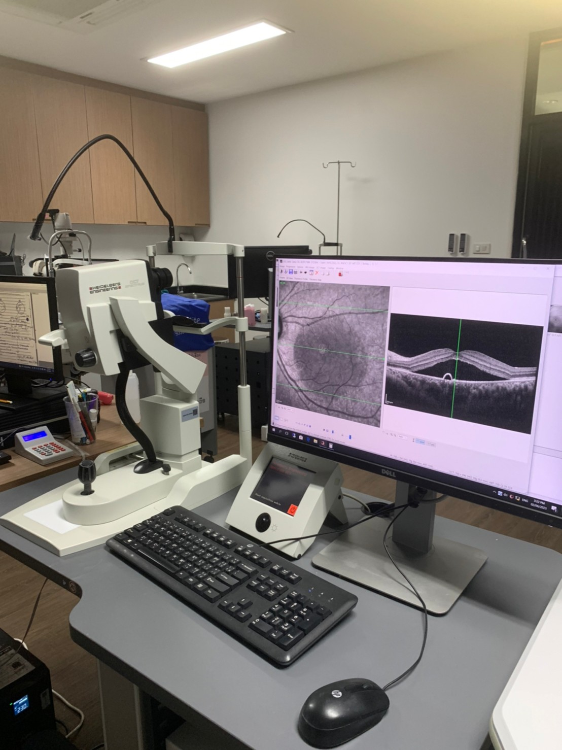
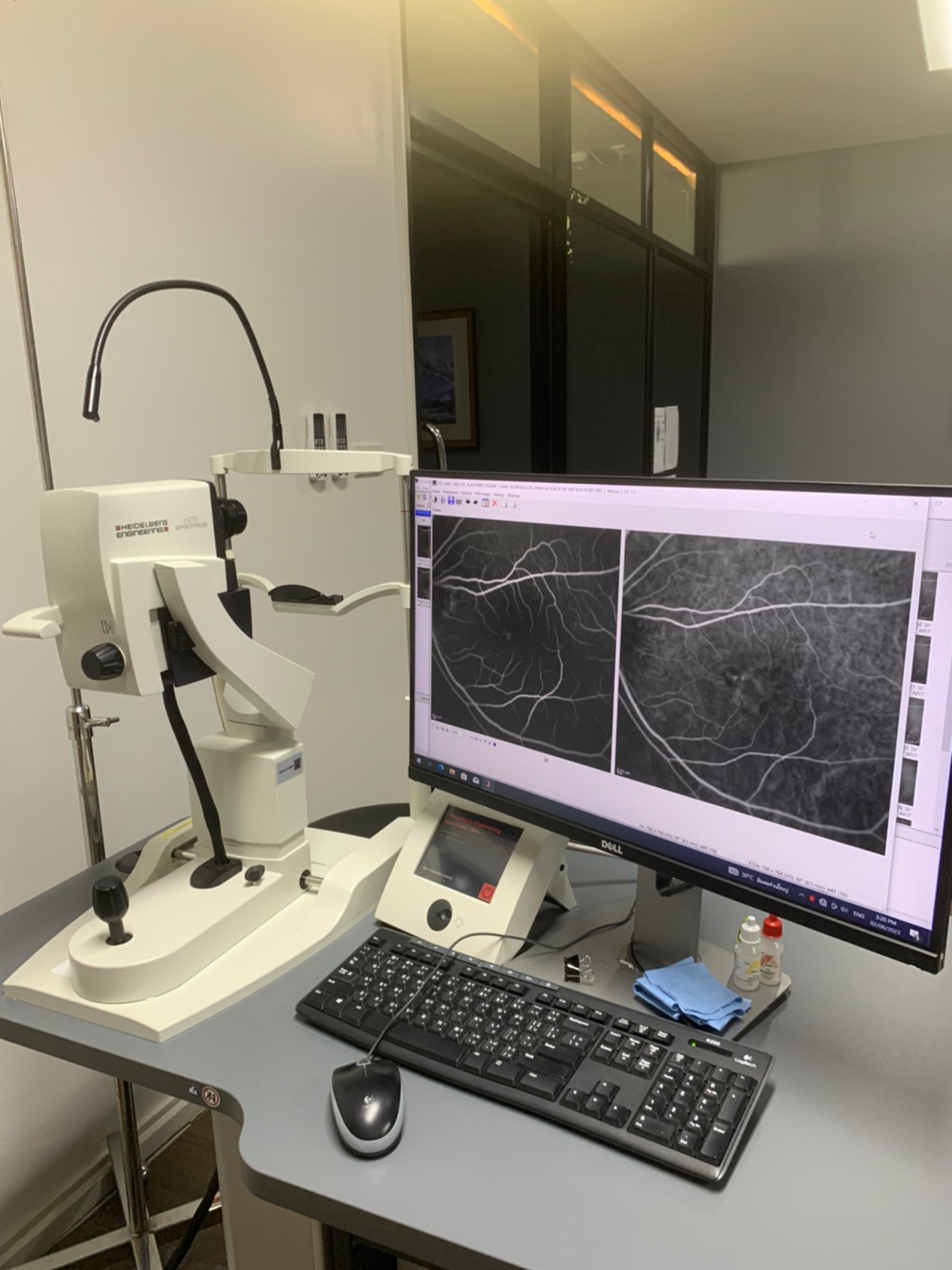
1. Optical coherence tomography [OCT]
– Heidelberg OCT Spectralis (Zeiss) version 1.10.4.0
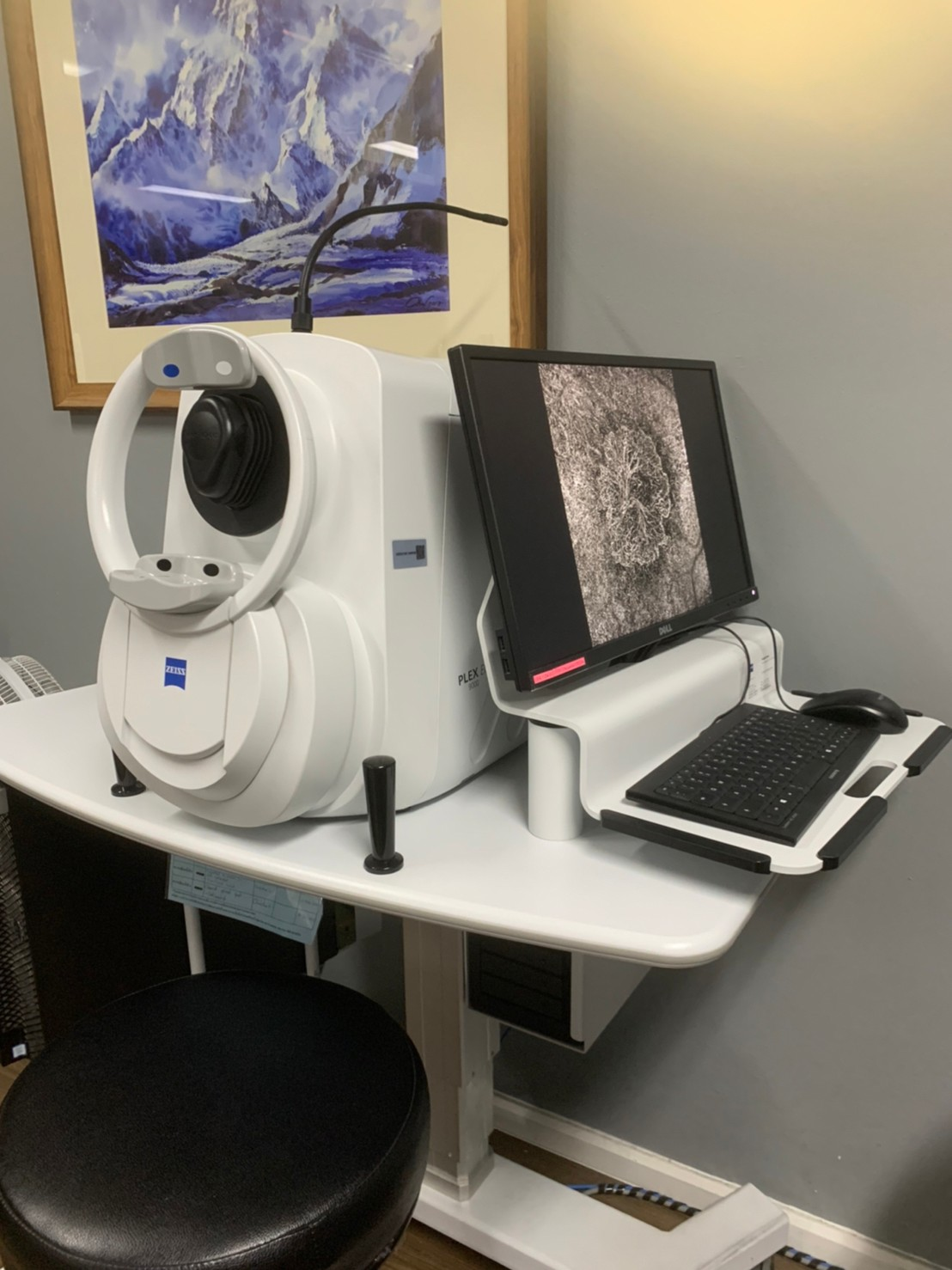
2. Optical coherence tomography angiography [OCTA]
– Heidelberg OCT Spectralis (Zeiss) version 1.10.4.0
– Plex Elite 9000 (Zeiss) version 2.1.0.55513
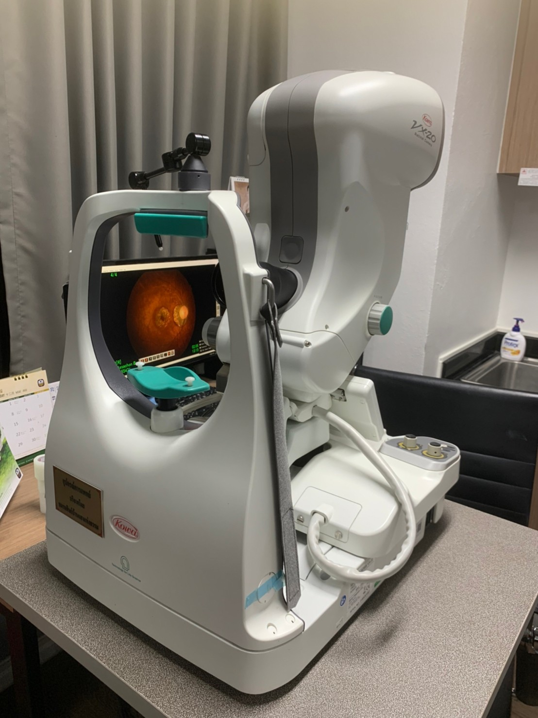
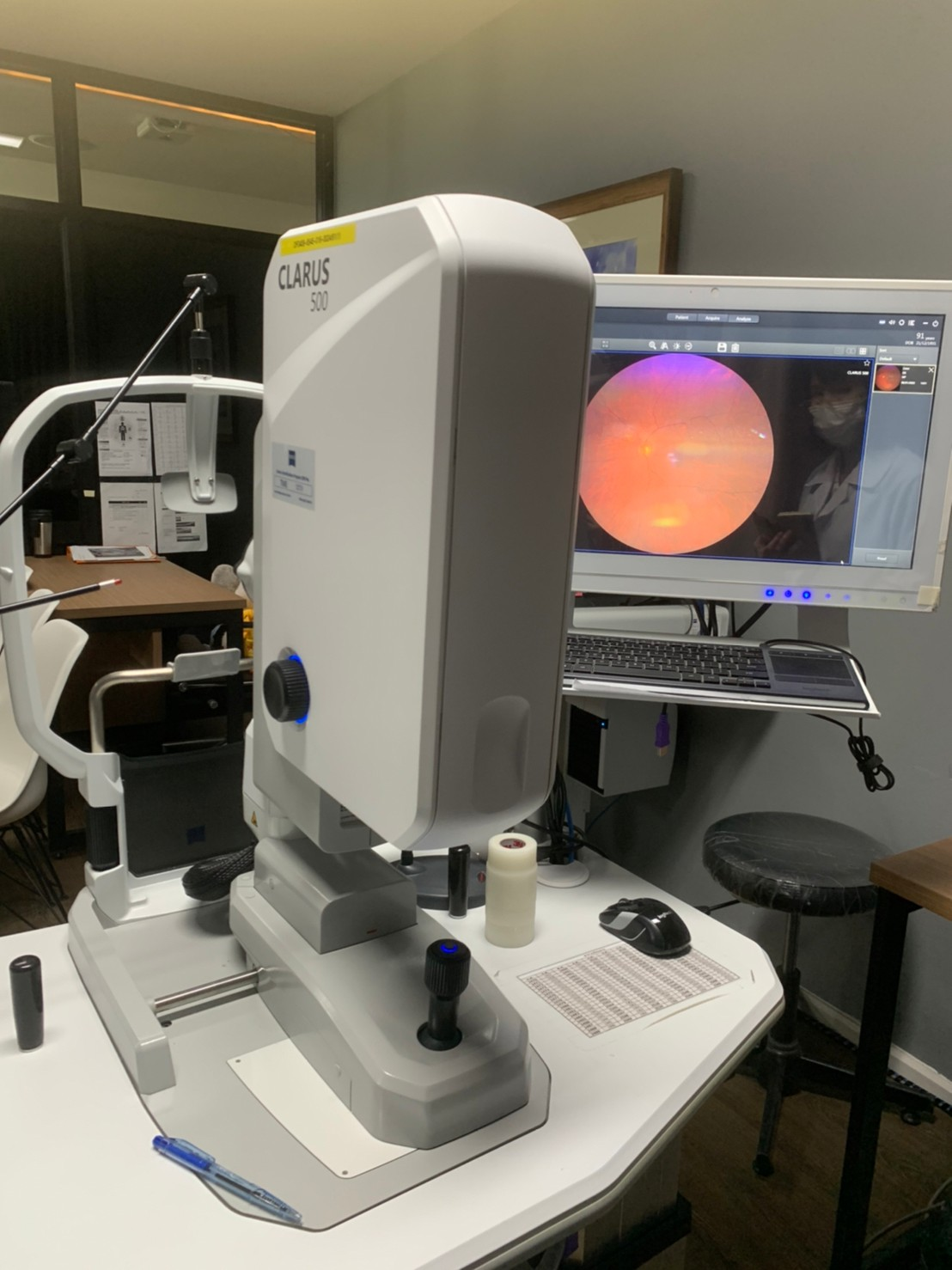
3. Color fundus photography and wide field fundus photography
– Kowa VX-20 version 5.8.0.0
– Clarus 500 (Zeiss) version 1.1.3.62215
4. fundus fluorescein angiography [FFA] and indocyanine green angiography [ICGA]
– Heidelberg HRA Spectralis (Zeiss) version 1.10.2.0
5. Fundus autofluorescence [FAF]
– Heidelberg HRA Spectralis (Zeiss) version 1.10.2.0
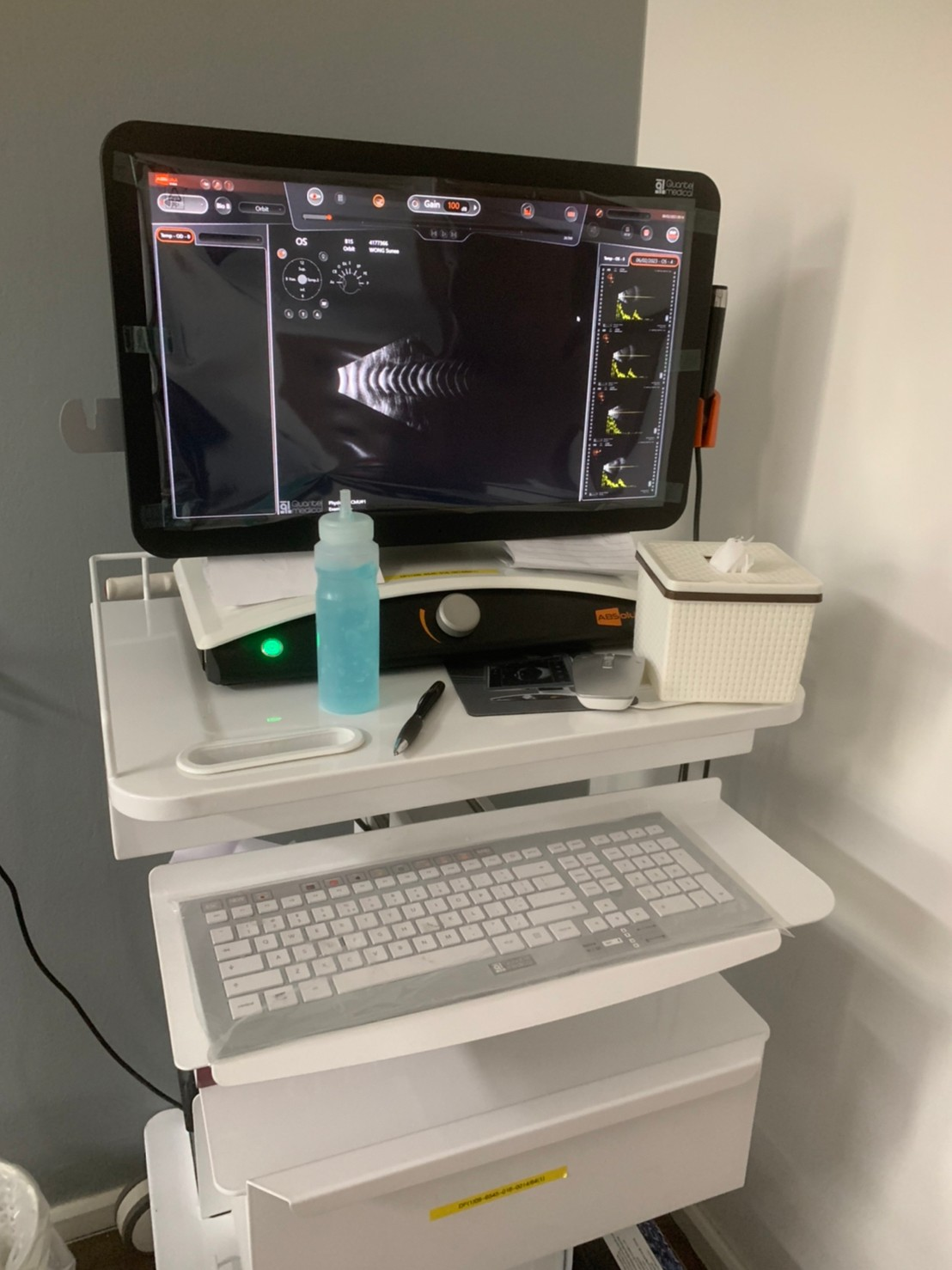
6. เครื่องตรวจลูกตาด้วยคลื่นความถี่สูง (B-scan ultrasonography)
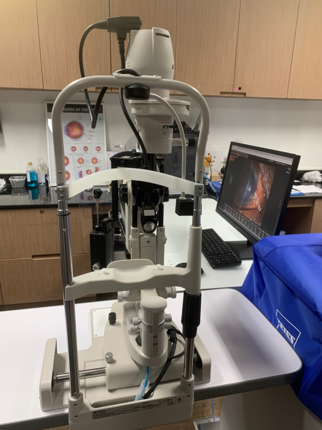
7. เครื่องถ่ายรูปตาส่วนหน้า (Anterior segment photography)
– Topcon DC-4 version 3.0.1.16534
Location
 ปัจจุบันคลินิกจอประสาทตาและน้ำวุ้นตาได้รองรับผู้ป่วยในจังหวัดเชียงใหม่และผู้ป่วยที่ได้รับการส่งต่อมาจากโรงพยาบาลต่าง ๆ ในเขตภาคเหนือตอนบน ผู้ป่วยที่จะเข้ารับบริการในคลินิกพิเศษนี้จะต้องผ่านการตรวจประเมินจากคลินิกตาทั่วไป (ชั้น 7 อาคารศรีพัฒน์) ก่อน เมื่อแพทย์พิจารณาแล้วจึงสามารถนัดเข้าคลินิกพิเศษนี้ได้